Monthly Archives: February 2021

Eradicating bacteria biofilm with nanocrystals

A January 8, 2021 news item on ScienceDaily announces new work from South Korea’s Pohang University of Science & Technology (POSTECH),

The COVID-19 pandemic is raising fears of new pathogens such as new viruses or drug-resistant bacteria. To this, a Korean research team has recently drawn attention for developing the technology for removing antibiotic-resistant bacteria by controlling the surface texture of nanomaterials.

A joint research team from POSTECH and UNIST [Ulsan National Institute of Science and Technology] has introduced mixed-FeCo-oxide-based surface-textured nanostructures (MTex) as highly efficient magneto-catalytic platform in the international journal Nano Letters. The team consisted of professors In Su Lee and Amit Kumar with Dr. Nitee Kumari of POSTECH’s Department of Chemistry and Professor Yoon-Kyung Cho and Dr. Sumit Kumar of UNIST’s Department of Biomedical Engineering.

Caption: Schematic diagram showing removal of bacterial biofilm via Mtex Credit: POSTECH

A January 8, 2021 POSTECH press release (also on EurkeAlert), which originated the news item, delves further,

First, the researchers synthesized smooth surface nanocrystals in which various metal ions were wrapped in an organic polymer shell and heated them at a very high temperature. While annealing the polymer shell, a high-temperature solid-state chemical reaction induced mixing of other metal ions on the nanocrystal surface, creating a number of few-nm-sized branches and holes on it. This unique surface texture was found to catalyze a chemical reaction that produced reactive oxygen species (ROS) that kills the bacteria. It was also confirmed to be highly magnetic and easily attracted toward the external magnetic field. The team had discovered a synthetic strategy for converting normal nanocrystals without surface features into highly functional mixed-metal-oxide nanocrystals.

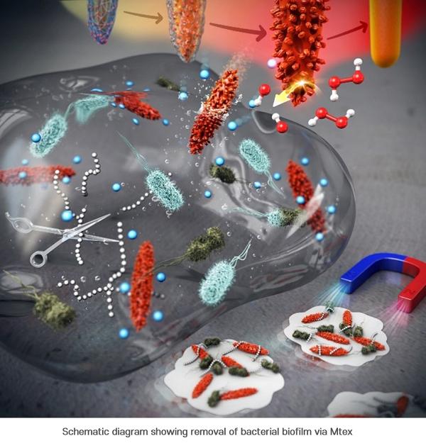
The research team named this surface topography – with branches and holes that resembles that of a ploughed field – “MTex.” This unique surface texture has been verified to increase the mobility of nanoparticles to allow efficient penetration into biofilm matrix while showing high activity in generating reactive oxygen species (ROS) that are lethal to bacteria.

This system produces ROS over a broad pH range and can effectively diffuse into the biofilm and kill the embedded bacteria resistant to antibiotics. And since the nanostructures are magnetic, biofilm debris can be scraped out even from the hard-to-reach microchannels.

“This newly developed MTex shows high catalytic activity, distinct from the stable smooth-surface of the conventional spinel forms,” explained Dr. Amit Kumar, one of the corresponding authors of the paper. “This characteristic is very useful in infiltrating biofilms even in small spaces and is effective in killing the bacteria and removing biofilms.”

“This research allows to regulate the surface nanotexturization, which opens up possibilities to augment and control the exposure of active sites,” remarked Professor In Su Lee who led the research. “We anticipate the nanoscale-textured surfaces to contribute significantly in developing a broad array of new enzyme-like properties at the nano-bio interface.”

Here’s a link to and a citation for the paper,

Surface-Textured Mixed-Metal-Oxide Nanocrystals as Efficient Catalysts for ROS Production and Biofilm Eradication by Nitee Kumari, Sumit Kumar, Mamata Karmacharya, Sateesh Dubbu, Taewan Kwon, Varsha Singh, Keun Hwa Chae, Amit Kumar, Yoon-Kyoung Cho, and In Su Lee. Nano Lett. 2021, 21, 1, 279–287 DOI: https://doi.org/10.1021/acs.nanolett.0c03639 Publication Date: December 11, 2020 Copyright © 2020 American Chemical Society

This paper is behind a paywall.

Beginner’s guide to folding DNA origami

I think this Aug. 6, 2010 post, Folding, origami, and shapeshifting and an article with over 50,000 authors is the first time I wrote about DNA (deoxyribonucleic acid) and origami (the Japanese art of paper folding).

Since then, the technique has become even more popular with the result that the US National Institute of Standards and Technology (NIST) has produced a beginner’s guide, according to a Jan. 8, 2021 news item on Nanowerk,

In a technique known as DNA origami, researchers fold long strands of DNA over and over again to construct a variety of tiny 3D structures, including miniature biosensors and drug-delivery containers. Pioneered at the California Institute of Technology in 2006, DNA origami has attracted hundreds of new researchers over the past decade, eager to build receptacles and sensors that could detect and treat disease in the human body, assess the environmental impact of pollutants, and assist in a host of other biological applications.

Although the principles of DNA origami are straightforward, the technique’s tools and methods for designing new structures are not always easy to grasp and have not been well documented. In addition, scientists new to the method have had no single reference they could turn to for the most efficient way of building DNA structures and how to avoid pitfalls that could waste months or even years of research.

That’s why Jacob Majikes and Alex Liddle, researchers at the National Institute of Standards and Technology (NIST) who have studied DNA origami for years, have compiled the first detailed tutorial on the technique. Their comprehensive report provides a step-by-step guide to designing DNA origami nanostructures, using state-of-the-art tools.

Here’s an image illustrating some of the techniques for DNA origami,

Caption: Collage shows some of the techniques and designs employed in DNA origami. Credit: K. Dill/NIST

A Jan. 8, 2021 US NIST news release (also on EurekAlert), which originated the news item, provide more detail as to the authors’ motivations, objectives, and future plans for their beginner’s guide,

“We wanted to take all the tools that people have developed and put them all in one place, and to explain things that you can’t say in a traditional journal article,” said Majikes. “Review papers might tell you everything that everyone’s done, but they don’t tell you how the people did it. “

DNA origami relies on the ability of complementary base pairs of the DNA molecule to bind to each other. Among DNA’s four bases — adenine (A), cytosine (C), guanine (G) and thymine (T) — A binds with T and G with C. This means that a specific sequence of As, Ts, Cs and Gs will find and bind to its complement.

The binding enables short strands of DNA to act as “staples,” keeping sections of long strands folded or joining separate strands. A typical origami design may require 250 staples. In this way, the DNA can self-assemble into a variety of shapes, forming a nanoscale framework to which an assortment of nanoparticles — many useful in medical treatment, biological research and environmental monitoring — can attach.

The challenges in using DNA origami are twofold, said Majikes. First, researchers are fabricating 3D structures using a foreign language — the base pairs A, G, T and C. In addition, they’re using those base-pair staples to twist and untwist the familiar double helix of DNA molecules so that the strands bend into specific shapes. That can be difficult to design and visualize. Majikes and Liddle urge researchers to strengthen their design intuition by building 3D mock-ups, such as sculptures made with bar magnets, before they start fabrication. These models, which can reveal which aspects of the folding process are critical and which ones are less important, should then be “flattened” into 2D to be compatible with computer-aided design tools for DNA origami, which typically use two-dimensional representations.

DNA folding can be accomplished in a variety of ways, some less efficient than others, noted Majikes. Some strategies, in fact, may be doomed to failure.

“Pointing out things like ‘You could do this, but it’s not a good idea’ — that type of perspective isn’t in a traditional journal article, but because NIST is focused on driving the state of technology in the nation, we’re able to publish this work in the NIST journal,” Majikes said. “I don’t think there’s anywhere else that would have given us the leeway and the time and the person hours to put all this together.”

Liddle and Majikes plan to follow up their work with several additional manuscripts detailing how to successfully fabricate nanoscale devices with DNA.

Here’s a link to and a citation for the beginner’s guide,

DNA Origami Design: A How-To Tutorial by Majikes, Jacob M. and Liddle, J. Alexander. Journal of Research of the National Institute of Standards and Technology Volume 126, Article No. 126001 (2021) Published online Jan. 8, 2021. DOI: 10.6028/jres.126.001

This is open access and it include such gems as this,

1.2 Education or Skill Level

Readers of this tutorial should be familiar with the physical properties of B-DNA, single-stranded DNA (ssDNA), and crossover junctions. In addition, once ready to create a structure for a specific application, the designer should determine the full list of functional requirements. This list includes answers to the following questions: What should the structure do? What specific properties are critical to the system’s performance?

1.3 Prerequisites

The designer should have either sufficient paper for manual design (not recommended) or a design program such as cadnano [1] (all versions sufficient), nanoengineer®, ParaboninSēquio®, or equivalent.1 A registered account with three-dimensional (3D) structure prediction servers such as CanDo [2, 3] is also recommended.

1.4Tools or Equipment

Equipment includes desktop or laptop computer equipment, craft supplies for macroscale models, and DNA nanotechnology computer-aided design (CAD) software.

Feel free to go forth and fold!

Stretching diamonds to improve electronic devices

On the last day of 2020, City University of Hong Kong (CityU) announced a technique for stretching diamonds that could result in a new generation of electronic devices. A December 31, 2020 news item on ScienceDaily makes the announcement,

Diamond is the hardest material in nature. It also has great potential as an excellent electronic material. A research team has demonstrated for the first time the large, uniform tensile elastic straining of microfabricated diamond arrays through the nanomechanical approach. Their findings have shown the potential of strained diamonds as prime candidates for advanced functional devices in microelectronics, photonics, and quantum information technologies.

A December 31, 2020 CityU press release on EurekAlert , which originated the news item, delves further into the research,

The research was co-led by Dr Lu Yang, Associate Professor in the Department of Mechanical Engineering (MNE) at CityU and researchers from Massachusetts Institute of Technology (MIT) and Harbin Institute of Technology (HIT). Their findings have been recently published in the prestigious scientific journal Science, titled “Achieving large uniform tensile elasticity in microfabricated diamond“.

“This is the first time showing the extremely large, uniform elasticity of diamond by tensile experiments. Our findings demonstrate the possibility of developing electronic devices through ‘deep elastic strain engineering’ of microfabricated diamond structures,” said Dr Lu.

Diamond: “Mount Everest” of electronic materials

Well known for its hardness, industrial applications of diamonds are usually cutting, drilling, or grinding. But diamond is also considered as a high-performance electronic and photonic material due to its ultra-high thermal conductivity, exceptional electric charge carrier mobility, high breakdown strength and ultra-wide bandgap. Bandgap is a key property in semi-conductor, and wide bandgap allows operation of high-power or high-frequency devices. “That’s why diamond can be considered as ‘Mount Everest’ of electronic materials, possessing all these excellent properties,” Dr Lu said.

However, the large bandgap and tight crystal structure of diamond make it difficult to “dope”, a common way to modulate the semi-conductors’ electronic properties during production, hence hampering the diamond’s industrial application in electronic and optoelectronic devices. A potential alternative is by “strain engineering”, that is to apply very large lattice strain, to change the electronic band structure and associated functional properties. But it was considered as “impossible” for diamond due to its extremely high hardness.

Then in 2018, Dr Lu and his collaborators discovered that, surprisingly, nanoscale diamond can be elastically bent with unexpected large local strain. This discovery suggests the change of physical properties in diamond through elastic strain engineering can be possible. Based on this, the latest study showed how this phenomenon can be utilized for developing functional diamond devices.

Uniform tensile straining across the sample

The team firstly microfabricated single-crystalline diamond samples from a solid diamond single crystals. The samples were in bridge-like shape – about one micrometre long and 300 nanometres wide, with both ends wider for gripping (See image: Tensile straining of diamond bridges). The diamond bridges were then uniaxially stretched in a well-controlled manner within an electron microscope. Under cycles of continuous and controllable loading-unloading of quantitative tensile tests, the diamond bridges demonstrated a highly uniform, large elastic deformation of about 7.5% strain across the whole gauge section of the specimen, rather than deforming at a localized area in bending. And they recovered their original shape after unloading.

By further optimizing the sample geometry using the American Society for Testing and Materials (ASTM) standard, they achieved a maximum uniform tensile strain of up to 9.7%, which even surpassed the maximum local value in the 2018 study, and was close to the theoretical elastic limit of diamond. More importantly, to demonstrate the strained diamond device concept, the team also realized elastic straining of microfabricated diamond arrays.

Tuning the bandgap by elastic strains

The team then performed density functional theory (DFT) calculations to estimate the impact of elastic straining from 0 to 12% on the diamond’s electronic properties. The simulation results indicated that the bandgap of diamond generally decreased as the tensile strain increased, with the largest bandgap reduction rate down from about 5 eV to 3 eV at around 9% strain along a specific crystalline orientation. The team performed an electron energy-loss spectroscopy analysis on a pre-strained diamond sample and verified this bandgap decreasing trend.

Their calculation results also showed that, interestingly, the bandgap could change from indirect to direct with the tensile strains larger than 9% along another crystalline orientation. Direct bandgap in semi-conductor means an electron can directly emit a photon, allowing many optoelectronic applications with higher efficiency.

These findings are an early step in achieving deep elastic strain engineering of microfabricated diamonds. By nanomechanical approach, the team demonstrated that the diamond’s band structure can be changed, and more importantly, these changes can be continuous and reversible, allowing different applications, from micro/nanoelectromechanical systems (MEMS/NEMS), strain-engineered transistors, to novel optoelectronic and quantum technologies. “I believe a new era for diamond is ahead of us,” said Dr Lu.

Here’s an illustration provided by the researchers,

Caption: Stretching of microfabricated diamonds pave ways for applications in next-generation microelectronics.. Credit: Dang Chaoqun / City University of Hong Kong

Here’s a link to and a citation for the paper,

Achieving large uniform tensile elasticity in microfabricated diamond by Chaoqun Dang, Jyh-Pin Chou, Bing Dai, Chang-Ti Chou, Yang Yang, Rong Fan, Weitong Lin, Fanling Meng, Alice Hu, Jiaqi Zhu, Jiecai Han, Andrew M. Minor, Ju Li, Yang Lu. Science 01 Jan 2021: Vol. 371, Issue 6524, pp. 76-78 DOI: 10.1126/science.abc4174

This paper is behind a paywall.

Mother-of-pearl self-assembles from disorder into perfection

Courtesy: Mother-of-pearl Courtesy: Technische Universitaet (TU) Dresden

Mother-of-pearl (also known as nacre) research has been featured here a few times (links at the end of this post). This time it touches on self-assembly, which is the source of much interest and, on occasion, much concern in the field of nanotechnology.

In any case, the latest mother-of-pearl work comes from the Technische Universität (TU) Dresden (Technical University of Dresden), located in Germany. From a January 4, 2021 news item on phys.org,

In a new study published in Nature Physics, researchers from the B CUBE—Center for Molecular Bioengineering at TU Dresden and European Synchrotron Radiation Facility (ESRF) in Grenoble [Grance] describe, for the first time, that structural defects in self-assembling nacre attract and cancel each other out, eventually leading to a perfect periodic structure.

A January 4, 2021 Technische Universität (TU) Dresden press release (also on EurekAlert), which originated the news item, explains the reason for the ongoing interest in mother-of-pearl and reveals an unexpected turn in the research,

Mollusks build shells to protect their soft tissues from predators. Nacre, also known as the mother of pearl, has an intricate, highly regular structure that makes it an incredibly strong material. Depending on the species, nacres can reach tens of centimeters in length. No matter the size, each nacre is built from materials deposited by a multitude of single cells at multiple different locations at the same time. How exactly this highly periodic and uniform structure emerges from the initial disorder was unknown until now.

Nacre formation starts uncoordinated with the cells depositing the material simultaneously at different locations. Not surprisingly, the early nacre structure is not very regular. At this point, it is full of defects. “In the very beginning, the layered mineral-organic tissue is full of structural faults that propagate through a number of layers like a helix. In fact, they look like a spiral staircase, having either right-handed or left-handed orientation,” says Dr. Igor Zlotnikov, research group leader at the B CUBE – Center for Molecular Bioengineering at TU Dresden. “The role of these defects in forming such a periodic tissue has never been established. On the other hand, the mature nacre is defect-free, with a regular, uniform structure. How could perfection emerge from such disorder?”

The researchers from the Zlotnikov group collaborated with the European Synchrotron Radiation Facility (ESRF) in Grenoble to take a very detailed look at the internal structure of the early and mature nacre. Using synchrotron-based holographic X-ray nano-tomography the researchers could capture the growth of nacre over time. “Nacre is an extremely fine structure, having organic features below 50 nm in size. Beamline ID16A at the ESRF provided us with an unprecedented capability to visualize nacre in three-dimensions,” explains Dr. Zlotnikov. “The combination of electron dense and highly periodical inorganic platelets with delicate and slender organic interfaces makes nacre a challenging structure to image. Cryogenic imaging helped us to obtain the resolving power we needed,” explains Dr. ‘Alexandra] Pacureanu from the X-ray Nanoprobe group at the ESRF.

The analysis of data was quite a challenge. The researchers developed a segmentation algorithm using neural networks and trained it to separate different layers of nacre. In this way, they were able to follow what happens to the structural defects as nacre grows.

The behavior of structural defects in a growing nacre was surprising. Defects of opposite screw direction were attracted to each other from vast distances. The right-handed and left-handed defects moved through the structure, until they met, and cancelled each other out. These events led to a tissue-wide synchronization. Over time, it allowed the structure to develop into a perfectly regular and defect-free.

Periodic structures similar to nacre are produced by many different animal species. The researchers think that the newly discovered mechanism could drive not only the formation of nacre but also other biogenic structures.

Here’s a link to and a citation for the paper,

Dynamics of topological defects and structural synchronization in a forming periodic tissue by Maksim Beliaev, Dana Zöllner, Alexandra Pacureanu, Paul Zaslansky & Igor Zlotnikov. Nature Physics (2021) First published online: 17 September 2020 Published: 04 January 2021

This paper is behind a paywall.

As promised here are the links for One tough mother, imitating mother-of-pearl for stronger ceramics (a March 14, 2014 posting) and Clues as to how mother of pearl is made (a December 15, 2015 posting).

CRISPR/Cas9 used successfully to edit SIV (simian immunodeficiency virus, which is similar to HIV) out of monkey genome

Before reading further please note, the research discussed in this posting is based on animal testing, which many people find highly disturbing.

CRISPR (clustered regularly interspaced short palindromic repeats)/Cas9 (CRISPR-associated protein 9), or more familiarly CRISPR/Cas9, has been been used to edit simian immunodeficiency virus from infected monkeys’ cells according to a December 2, 2020 article by Matthew Rozsa for Salon.com (Note: Links have been removed),

With multiple coronavirus vaccines being produced as we speak, the COVID-19 pandemic appears to have an end in sight, though the HIV pandemic continues after more than 40 years. That might seem like a head-scratcher: why is HIV, a virus we’ve known about for decades, so much harder to cure than a virus discovered just last year? Part of the reason is that HIV, as a retrovirus, is a more complex virus to vaccinate against than SARS-CoV-2 — hence why a vaccine or other cure has eluded scientists for decades. 

Now, a surprising new study on a related retrovirus shows incredible promise for the potential to develop a cure for HIV, or human immunodeficiency virus. In an article published in the scientific journal Nature Communications, scientists revealed that they had used CRISPR – a genetic technology that can alter DNA and whose developers won the 2020 Nobel Prize in Chemistry [specifically, Jennifer Doudna and Emanuelle Charpentier received the Nobel for developing CRISPR-cas9 or CRISPR/Cas9 not CRISPR alone) — to successfully edit SIV (simian immunodeficiency virus), a virus similar to HIV, out of the genomes of non-human primates.  Specifically, the scientists were able to edit out the SIV genome from rhesus macaque monkeys’ infected cells.

For anyone who’s interested in how CRISPR was developed and the many contributions which have led to the current state-of-the-art for CRISPR gene editing, see the History subsection of Wikipedia’s CRISPR entry.

Getting back to Rozsa’s December 2, 2020 article,

“This study used the CRISPR CaS9 system, which has been described as molecular scissors,” Andrew G. MacLean, PhD, wrote to Salon. MacLean is an associate professor at the Tulane National Primate Research Center and the Department of Microbiology and Immunology at Tulane University School of Medicine and was a senior co-investigator of the study. “It uses a highly specific targeting system to cut out a specific portion of DNA that is necessary for HIV to be able to produce more virus.”

He added, “Our collaborators at in the Khalili Lab at Temple University have developed a method of ‘packaging’ this within a single so-called vector. A vector is a non-disease causing virus that is used as a carrier for the CRISPR CaS9 scissors to get it into the tissues of interest.”

The experiments with SIV are considered to be a gateway to understanding HIV, as HIV is believed to have evolved from SIV, and is genetically similar.

“The rhesus macaque model of HIV/AIDS is the most valuable model to test efficacy of new interventions or approaches for preventing or treating HIV infection, prior to human clinical trials,” Binhua Ling, PhD, associate professor at the Southwest National Primate Research Center, Texas Biomedical Research Institute, wrote to Salon. “This first proof-of-principal [emphasis mine] study on the rhesus macaque model indicates that this virus-vehicle-delivered-CRISPR system can reach many tissue sites of the body, and is able to effectively delete virus DNA in infected cells. This paves the way for applying the same technology to the human body, which could lead to a cure for HIV infection.”

Tricia H. Burdo, PhD, another senior co-investigator on the new study who works at the Lewis Katz School of Medicine at Temple University, explained to Salon by email that “HIV is in a class of viruses (retroviruses) that inserts itself into the DNA of the host, so you can really think of this now as a genetic disease” — in other words, the kind of thing that would be ripe for CRISPR’s scissors-like ability to remove errant or unwanted genetic material. Burdo notes that the CRISPR technology discussed in the article “cuts out this foreign viral gene.”

The study was conducted on eight Rhesus macaque monkeys. That is a very small number to start with and not all of the monkeys received the CRISPR/Cas9 treatment. From the ‘Animals used in the study and ethical statement‘ subsection of the study, “Animals were sacrificed for tissue collection 3 weeks after … .” Leaving aside how anyone may feel about ‘sacrificing …’, three weeks is not a long time for observation.

Should you be interested, there is a November 30, 2020 Tulane University news release announcing the research.

If you want to read the whole study, here’s a link and a citation,

CRISPR based editing of SIV proviral DNA in ART treated non-human primates by Pietro Mancuso, Chen Chen, Rafal Kaminski, Jennifer Gordon, Shuren Liao, Jake A. Robinson, Mandy D. Smith, Hong Liu, Ilker K. Sariyer, Rahsan Sariyer, Tiffany A. Peterson, Martina Donadoni, Jaclyn B. Williams, Summer Siddiqui, Bruce A. Bunnell, Binhua Ling, Andrew G. MacLean, Tricia H. Burdo & Kamel Khalili. Nature Communications volume 11, Article number: 6065 (2020) DOI: https://doi.org/10.1038/s41467-020-19821-7 Published: 27 November 2020

This paper is open access.

As Rozsa notes in his December 2, 2020 article, the Joint United Nations Programme on HIV/AIDS estimates that 32.7 million [24.8 million–42.2 million] people have died from AIDS-related illnesses since the start (1981?) of the epidemic to the end of 2019.

Dancing with a robot

Dancing with Baryshnibot. Alice Williamson, Courtesy Merritt Moore

Dancing robots usually perform to pop music but every once in a while, there’s a move toward classical music and ballet, e.g., my June 8, 2011 posting was titled, Robot swan dances to Tchaikovsky’s Swan Lake. Unlike the dancing robot in the picture above, that robot swan danced alone. (You can still see the robot’s Swan Lake performance in the video embedded in the 2011 posting.)

I don’t usually associate dance magazines with robots but Chava Pearl Lansky’s Nov. 18, 2020 article about dancer/physicist Merritt Moore and her work with Baryshnibot is found in ballet magazine, Pointe (Note: Links have been removed),

When the world went into lockdown last March [2019], most dancers despaired. But not Merritt Moore. The Los Angeles native, who lives in London and has danced with Norwegian National Ballet, English National Ballet and Boston Ballet, holds a PhD in atomic and laser physics from the University of Oxford. A few weeks into the coronavirus pandemic, she came up with a solution for having to train and work alone: robots.

Moore had just come out of a six-week residency at Harvard ArtLab focused on the intersection between dance and robotics. “I knew I needed something to look forward to, and thought how bizarre I’d just been working with robots,” she says. “Who knew they’d be my only potential dance partners for a really long time?” She reached out to Universal Robotics and asked them to collaborate, and they agreed to send her a robot to experiment with.

Baryshnibot is an industrial robot normally used for automation and manufacturing. “It does not look impressive at all,” says Moore. “But there’s so much potential for different movement.” Creating dances for a robot, she says, is like an elaborate puzzle: “I have to figure out how to make this six-jointed rod emulate the dance moves of a head, two arms, a body and two legs.”

Moore started with the basics. She’d learn a simple TikTok dance, and then map the movements into a computer pad attached to the robot. “The 15-second-routine will take me five-hours-plus to program,” she says. Despite the arduous process, she’s built up to more advanced choreography, and is trying on different dance styles, from ballet to hip hop to salsa. For her newest pas de deux, titled Merritt + Robot, Moore worked with director Conor Gorman and cinematographer Howard Mills to beautifully capture her work with Baryshnibot on film. …

You can find Moore’s and Baryshnibot’s performance video embedded in Nov. 18, 2020 article.

Water and minerals have a nanoscale effect on bones

Courtesy: University of Arkansas

What a great image of bones! This December 3, 2020 University of Arkansas news release (also on EurekAlert) by Matt McGowan features research focused on bone material looks exciting. The date for the second study citation and link that I have listed (at the end of this posting) suggests the more recent study may have been initially overlooked in the deluge of COVID-19 research we are experiencing,

University of Arkansas researchers Marco Fielder and Arun Nair have conducted the first study of the combined nanoscale effects of water and mineral content on the deformation mechanisms and thermal properties of collagen, the essence of bone material.

The researchers also compared the results to the same properties of non-mineralized collagen reinforced with carbon nanotubes, which have shown promise as a reinforcing material for bio-composites. This research aids in the development of synthetic materials to mimic bone.

Using molecular dynamics — in this case a computer simulation of the physical movements of atoms and molecules — Nair and Fielder examined the mechanics and thermal properties of collagen-based bio-composites containing different weight percentages of minerals, water and carbon nanotubes when subjected to external loads.

They found that variations of water and mineral content had a strong impact on the mechanical behavior and properties of the bio-composites, the structure of which mimics nanoscale bone composition. With increased hydration, the bio-composites became more vulnerable to stress. Additionally, Nair and Fielder found that the presence of carbon nanotubes in non-mineralized collagen reduced the deformation of the gap regions.

The researchers also tested stiffness, which is the standard measurement of a material’s resistance to deformation. Both mineralized and non-mineralized collagen bio-composites demonstrated less stability with greater water content. Composites with 40% mineralization were twice as strong as those without minerals, regardless of the amount of water content. Stiffness of composites with carbon nanotubes was comparable to that of the mineralized collagen.

“As the degree of mineralization or carbon nanotube content of the collagenous bio-composites increased, the effect of water to change the magnitude of deformation decreased,” Fielder said.

The bio-composites made of collagen and carbon nanotubes were also found to have a higher specific heat than the studied mineralized collagen bio-composites, making them more likely to be resistant to thermal damage that could occur during implantation or functional use of the composite. Like most biological materials, bone is a hierarchical – with different structures at different length scales. At the microscale level, bone is made of collagen fibers, composed of smaller nanofibers called fibrils, which are a composite of collagen proteins, mineralized crystals called apatite and water. Collagen fibrils overlap each other in some areas and are separated by gaps in other areas.

“Though several studies have characterized the mechanics of fibrils, the effects of variation and distribution of water and mineral content in fibril gap and overlap regions are unexplored,” said Nair, who is an associate professor of mechanical engineering. “Exploring these regions builds an understanding of the structure of bone, which is important for uncovering its material properties. If we understand these properties, we can design and build better bio-inspired materials and bio-composites.”

Here are links and citations for both papers mentioned in the news release,

Effects of hydration and mineralization on the deformation mechanisms of collagen fibrils in bone at the nanoscale by Marco Fielder & Arun K. Nair. Biomechanics and Modeling in Mechanobiology volume 18, pages57–68 (2019) Biomech Model Mechanobiol 18, 57–68 (2019). DOI: https://doi.org/10.1007/s10237-018-1067-y First published: 07 August 2018 Issue Date: 15 February 2019

This paper is behind a paywall.

A computational study of mechanical properties of collagen-based bio-composites by Marco Fielder & Arun K. Nair. International Biomechanics Volume 7, 2020 – Issue 1 Pages 76-87 DOI: https://doi.org/10.1080/23335432.2020.1812428 Published online: 02 Sep 2020

This paper is open access.

Detecting COVID-19 in under five minutes with paper-based sensor made of graphene

A Dec. 7, 2020 news item on Nanowerk announced a new technology for rapid COVID-19 testing (Note: A link has been removed),

As the COVID-19 pandemic continues to spread across the world, testing remains a key strategy for tracking and containing the virus. Bioengineering graduate student, Maha Alafeef, has co-developed a rapid, ultrasensitive test using a paper-based electrochemical sensor that can detect the presence of the virus in less than five minutes.

The team led by professor Dipanjan Pan reported their findings in ACS Nano (“Rapid, Ultrasensitive, and Quantitative Detection of SARS-CoV-2 Using Antisense Oligonucleotides Directed Electrochemical Biosensor Chip”).

“Currently, we are experiencing a once-in-a-century life-changing event,” said Alafeef. “We are responding to this global need from a holistic approach by developing multidisciplinary tools for early detection and diagnosis and treatment for SARS-CoV-2.”

I wonder why they didn’t think to provide a caption for the graphene substrate (the square surface) underlying the gold electrode (the round thing) or provide a caption for the electrode. Maybe they assumed anyone knowledgeable about graphene would be able to identify it?

Caption: COVID-19 electrochemical sensing platform. Credit: University of Illinois

A Dec. 7, 2020 University of Illinois Grainger College of Engineering news release (also on EurekAlert) by Huan Song, which originated the news item, provides more technical detail including a description of the graphene substrate and the gold electrode, which make up the cheaper, faster COVID-19 sensing platform,

There are two broad categories of COVID-19 tests on the market. The first category uses reverse transcriptase real-time polymerase chain reaction (RT-PCR) and nucleic acid hybridization strategies to identify viral RNA. Current FDA [US Food and Drug Administration]-approved diagnostic tests use this technique. Some drawbacks include the amount of time it takes to complete the test, the need for specialized personnel and the availability of equipment and reagents.

The second category of tests focuses on the detection of antibodies. However, there could be a delay of a few days to a few weeks after a person has been exposed to the virus for them to produce detectable antibodies.

In recent years, researchers have had some success with creating point-of-care biosensors using 2D nanomaterials such as graphene to detect diseases. The main advantages of graphene-based biosensors are their sensitivity, low cost of production and rapid detection turnaround. “The discovery of graphene opened up a new era of sensor development due to its properties. Graphene exhibits unique mechanical and electrochemical properties that make it ideal for the development of sensitive electrochemical sensors,” said Alafeef. The team created a graphene-based electrochemical biosensor with an electrical read-out setup to selectively detect the presence of SARS-CoV-2 genetic material.

There are two components [emphasis mine] to this biosensor: a platform to measure an electrical read-out and probes to detect the presence of viral RNA. To create the platform, researchers first coated filter paper with a layer of graphene nanoplatelets to create a conductive film [emphasis mine]. Then, they placed a gold electrode with a predefined design on top of the graphene [emphasis mine] as a contact pad for electrical readout. Both gold and graphene have high sensitivity and conductivity which makes this platform ultrasensitive to detect changes in electrical signals.

Current RNA-based COVID-19 tests screen for the presence of the N-gene (nucleocapsid phosphoprotein) on the SARS-CoV-2 virus. In this research, the team designed antisense oligonucleotide (ASOs) probes to target two regions of the N-gene. Targeting two regions ensures the reliability of the senor in case one region undergoes gene mutation. Furthermore, gold nanoparticles (AuNP) are capped with these single-stranded nucleic acids (ssDNA), which represents an ultra-sensitive sensing probe for the SARS-CoV-2 RNA.

The researchers previously showed the sensitivity of the developed sensing probes in their earlier work published in ACS Nano. The hybridization of the viral RNA with these probes causes a change in the sensor electrical response. The AuNP caps accelerate the electron transfer and when broadcasted over the sensing platform, results in an increase in the output signal and indicates the presence of the virus.

The team tested the performance of this sensor by using COVID-19 positive and negative samples. The sensor showed a significant increase in the voltage of positive samples compared to the negative ones and confirmed the presence of viral genetic material in less than five minutes. Furthermore, the sensor was able to differentiate viral RNA loads in these samples. Viral load is an important quantitative indicator of the progress of infection and a challenge to measure using existing diagnostic methods.

This platform has far-reaching applications due to its portability and low cost. The sensor, when integrated with microcontrollers and LED screens or with a smartphone via Bluetooth or wifi, could be used at the point-of-care in a doctor’s office or even at home. Beyond COVID-19, the research team also foresees the system to be adaptable for the detection of many different diseases.

“The unlimited potential of bioengineering has always sparked my utmost interest with its innovative translational applications,” Alafeef said. “I am happy to see my research project has an impact on solving a real-world problem. Finally, I would like to thank my Ph.D. advisor professor Dipanjan Pan for his endless support, research scientist Dr. Parikshit Moitra, and research assistant Ketan Dighe for their help and contribution toward the success of this study.”

Here’s a link to and a citation for the paper,

Rapid, Ultrasensitive, and Quantitative Detection of SARS-CoV-2 Using Antisense Oligonucleotides Directed Electrochemical Biosensor Chip by Maha Alafeef, Ketan Dighe, Parikshit Moitra, and Dipanjan Pan. ACS Nano 2020, 14, 12, 17028–17045 DOI: https://doi.org/10.1021/acsnano.0c06392 Publication Date:October 20, 2020 Copyright © 2020 American Chemical Society

I’m not sure where I found this notice but it is most definitely from the American Chemical Society: “This paper is freely accessible, at this time, for unrestricted RESEARCH re-use and analyses in any form or by any means with acknowledgement of the original source. These permissions are granted for the duration of the World Health Organization (WHO) declaration of COVID-19 as a global pandemic.”

A better quality of cultivated meat from McMaster University?

This could be a bit stomach-churning for some folks.

Researchers at Canada’s McMaster University have developed and are commercializing a technique for cultivated meat (the first experiment involved mouse meat). (You could call it vat-grown meat.) A January 19, 2021 news item on phys.org makes the announcement (Note: Links have been removed),

McMaster researchers have developed a new form of cultivated meat using a method that promises more natural flavor and texture than other alternatives to traditional meat from animals.

Researchers Ravi Selvaganapathy and Alireza Shahin-Shamsabadi, both of the university’s School of Biomedical Engineering, have devised a way to make meat by stacking thin sheets of cultivated muscle and fat cells grown together in a lab setting. The technique is adapted from a method used to grow tissue for human transplants.

A January 19, 2021 McMaster University news release (also on EurekAlert) by Wade Hemsworth, which originated the news item, offers more details,

The sheets of living cells, each about the thickness of a sheet of printer paper, are first grown in culture and then concentrated on growth plates before being peeled off and stacked or folded together. The sheets naturally bond to one another before the cells die.

The layers can be stacked into a solid piece of any thickness, Selvaganapathy says, and “tuned” to replicate the fat content and marbling of any cut of meat – an advantage over other alternatives.

“We are creating slabs of meat,” he says. “Consumers will be able to buy meat with whatever percentage of fat they like – just like they do with milk.”

As they describe in the journal Cells Tissues Organs, the researchers proved the concept by making meat from available lines of mouse cells. Though they did not eat the mouse meat described in the research paper, they later made and cooked a sample of meat they created from rabbit cells.

“It felt and tasted just like meat,” says Selvaganapathy.

There is no reason to think the same technology would not work for growing beef, pork or chicken, and the model would lend itself well to large-scale production, Selvaganapathy says.

The researchers were inspired by the meat-supply crisis in which worldwide demand is growing while current meat consumption is straining land and water resources and generating troubling levels of greenhouse gases.

“Meat production right now is not sustainable,” Selvaganapathy says. “There has to be an alternative way of creating meat.”

Producing viable meat without raising and harvesting animals would be far more sustainable, more sanitary and far less wasteful, the researchers point out. While other forms of cultured meat have previously been developed, the McMaster researchers believe theirs has the best potential for creating products consumers will accept, enjoy and afford.

The researchers have formed a start-up company to begin commercializing the technology.

The researchers have included a picture of the ‘meat’,

Caption: A sample of meat cultivated by researchers at Canada’s McMaster University, using cells from mice. Credit: McMaster University

Here’s a link to and a citation for the paper,

Engineering Murine Adipocytes and Skeletal Muscle Cells in Meat-like Constructs Using Self-Assembled Layer-by-Layer Biofabrication: A Platform for Development of Cultivated Meat by Alireza Shahin-Shamsabadi and P. R. (Ravi) Selvaganapathy. Cells Tissues Organs (2021). DOI: 10.1159/000511764

This paper is behind a paywall.