Monthly Archives: August 2022

The reddest red and Schrödinger’s red pixel

Caption: Schrödinger’s red pixel by quasi-bound-states in-the-continuum Credit: 123RF

Science keeps moving. First, there was the June 2022 news and, then, there was the August 2022 news.

A June 8, 2022 Agency for Science, Technology and Research (A*STAR) press release (also on EurekAlert but published June 7, 2022 as an ‘article highlight’) announces more research into structural colour along with some colour theory from Erwin Schrödinger,

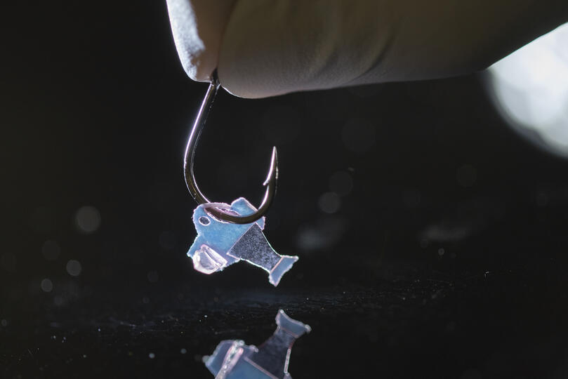
The brilliant and often iridescent colours that we see in some species of birds, beetles and butterflies arise from a regular arrangement of nanostructures that scatter selective wavelengths of light more strongly to generate colour. These colours are called structural colours, which usually range from blues to greens, and even magenta. However, vibrant or saturated reds are elusive and notably absent from the structural colour range in both natural and synthetic realms.

To achieve highly saturated reds, the material needs to absorb light from all wavelengths shorter than ~600 nm and reflect the remaining longer wavelengths, doing both as completely as possible. This sharp transition from absorption to reflection was prescribed theoretically by none other than Erwin Schrödinger of quantum theory fame. However, the physics of resonators tell us that high-order optical resonances in blue will also occur as soon as we have a fundamental resonance in red. This combination of blue and red thus results in the magenta observed in nature. It is therefore challenging to achieve the Schrödinger’s red pixel, which would produce the most saturated red in the world. Current nanoantenna-based approaches are insufficient to simultaneously satisfy the above conditions.

Researchers from the Agency for Science, Technology and Research’s (A*STAR) Institute of Materials Research and Engineering (IMRE), National University of Singapore (NUS) and Singapore University of Technology and Design (SUTD) have collaborated to design and realise reds at the ultimate limit of saturation as predicted by theory, where the team worked together on conceptualisation methodology, fabrications, characterisations and simulations. This research was published in Science Advances on 23 February 2022.

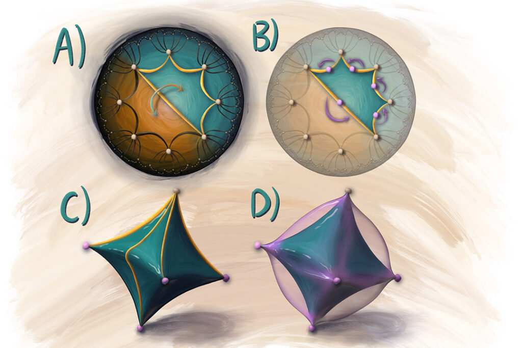
The design consists of regularly arranged silicon nanoantennas in the shape of ellipses. These produce possibly the most saturated and brightest reds with ~80% reflectance, exceeding the reds in the standard red, green and blue gamut (sRGB) and other well-known red pigments, e.g. cadmium red .

The nanoantennas support two partially overlapping quasi bound-states-in-the-continuum modes, where the optimal dimensions of the silicon nanoantenna arrays are derived by using a gradient descent algorithm to enable the antennas to achieve sharp spectral edges at red wavelengths. At the same time, high-order modes at blue or green wavelengths are suppressed via engineering the substrate‑induced diffraction channels and the absorption of amorphous silicon.

Potential uses for Schrödinger’s red include developing a polarisation dependent encryption method, with plans to scale up the Schrödinger’s red pixel for applications like functional nanofabrication devices such as optical spectrometers and reflective displays with high colour saturation.

“With this new design that can achieve the most saturated and brightest reds, we can exploit its sensitivity to polarisation and illumination angle on potential applications for information encryption. This proposed concept and design methodology could also be generalised to other Schrödinger’s colour pixels. The highly-saturated red achieved could be potentially scaled up through methods such as deep ultraviolet and nano-imprint lithography, to reach the dimensions of reflective displays based on multilayer film configuration, which could lead to potential applications like compact red filters, highly saturated reflective displays, nonlocal metasurfaces, and miniaturised spectrometers”, said Dr. Dong Zhaogang, Deputy Department Head of Nanofabrication at A*STAR’s IMRE.

“The creation of the record-high saturation and brightness in red opens up possibilities for a plethora of applications related to anti-counterfeiting technologies, high-calibre colour display and more, which were previously perceived as unachievable with structural colour. It showcases a wonderful synergy between conceptual breakthrough, powerful algorithm and advanced nanofabrication”, said Prof. Cheng-Wei Qiu, Dean’s Chair Professor at NUS.

“This work in structural colours goes to show that we can sometimes outdo evolution through clever use of the tools in nanofabrication and accurate optical simulations”, said Prof. Joel Yang, Provost Chair Professor and Associate Professor in Engineering Product Development at SUTD.

Here’s a link to and a citation for the paper,

Schrödinger’s red pixel by quasi-bound-states-in-the-continuum by Zhaogang Dong, Lei Jin, Soroosh Daqiqeh Rezaei, Hao Wang, Yang Chen, Febiana Tjiptoharsono, Jinfa Ho, Sergey Gorelik, Ray Jia Hong Ng, Qifeng Ruan, Cheng-Wei Qiu and Joel K. W. Yang. Science Advances Vol 8, Issue 8 DOI: 10.1126/sciadv.abm4512 Published 23 Feb 2022

This paper is open access.

Math error, colour theory, and perception

An August 10, 2022 news item on phys.org announced a math error made by Erwin Schrödinger and others,

A new study corrects an important error in the 3D mathematical space developed by the Nobel Prize-winning physicist Erwin Schrödinger and others, and used by scientists and industry for more than 100 years to describe how your eye distinguishes one color from another. The research has the potential to boost scientific data visualizations, improve TVs and recalibrate the textile and paint industries.

“The assumed shape of color space requires a paradigm shift,” said Roxana Bujack, a computer scientist with a background in mathematics who creates scientific visualizations at Los Alamos National Laboratory. Bujack is lead author of the paper by a Los Alamos team in the Proceedings of the National Academy of Sciences on the mathematics of color perception.

“Our research shows that the current mathematical model of how the eye perceives color differences is incorrect. That model was suggested by Bernhard Riemann and developed by Hermann von Helmholtz and Erwin Schrödinger—all giants in mathematics and physics—and proving one of them wrong is pretty much the dream of a scientist,” said Bujack.

While the Los Alamos National Laboratory work was published in April 2022 (online) and May 2022 (in print), their news announcement doesn’t seem to have been made until August. I can’t be certain but I believe this should have an impact on the work from A*STAR as that team’s paper cites: E. Schrödinger, Theorie der Pigmente von größter Leuchtkraft. Ann. Phys. 367, 603–622 (1920).

An August 10, 2022 Los Alamos National Laboratory (LANL) news release (also on EurekAlert) provides more information about the discovery,

Modeling human color perception enables automation of image processing, computer graphics and visualization tasks.

“Our original idea was to develop algorithms to automatically improve color maps for data visualization, to make them easier to understand and interpret,” Bujack said. So the team was surprised when they discovered they were the first to determine that the longstanding application of Riemannian geometry, which allows generalizing straight lines to curved surfaces, didn’t work.

To create industry standards, a precise mathematical model of perceived color space is needed. First attempts used Euclidean spaces—the familiar geometry taught in many high schools; more advanced models used Riemannian geometry. The models plot red, green and blue in the 3D space. Those are the colors registered most strongly by light-detecting cones on our retinas, and—not surprisingly—the colors that blend to create all the images on your RGB computer screen.

In the study, which blends psychology, biology and mathematics, Bujack and her colleagues discovered that using Riemannian geometry overestimates the perception of large color differences. That’s because people perceive a big difference in color to be less than the sum you would get if you added up small differences in color that lie between two widely separated shades.

Riemannian geometry cannot account for this effect.

“We didn’t expect this, and we don’t know the exact geometry of this new color space yet,” Bujack said. “We might be able to think of it normally but with an added dampening or weighing function that pulls long distances in, making them shorter. But we can’t prove it yet.”

Here’s a link to and a citation for the paper,

The non-Riemannian nature of perceptual color space by Roxana Bujack, Emily Teti, Jonah Miller, Elektra Caffrey, and Terece L. Turton. Proceedings of the National Academy of Sciences (PNAS) 119 (18) e2119753119 DOI: https://doi.org/10.1073/pnas.2119753119 Published: April 29, 2022

This paper is behind a paywall.

Mildred Dresselhaus (Queen of Carbon) gets a book

She died in 2017 and left behind a legacy many would envy. From a March 8, 2022 book review by Jess Wade for Physics World (Note: Links have been removed),

Mildred Dresselhaus, materials-science pioneer and nanotechnology trailblazer, should be a household name. Her contributions to science were immense: unravelling the electronic structure of carbon and paving the way for the discovery of fullerenes, carbon nanotubes and graphene. She was the first woman to be appointed Institute Professor at the Massachusetts Institute of Technology (MIT), which is the highest title that is awarded there. She was also the first woman to win a National Medal of Science in the category of engineering (awarded by the US president) and the first individual winner of the Kavli Prize in Nanoscience.

Dresselhaus’ resilience and determination meant that she succeeded in a world that was not welcoming to her. At the time, a lot of people still believed that “a woman’s place is in the home”. Her contributions to nanoscience were nothing short of incredible. She studied thermoelectric materials, as well as the magnetic, optical and electrical properties of semimetals, creating novel nanomaterials that provided the foundation for lithium-ion batteries, fullerenes and carbon nanotubes. Her attention to detail and creativity allowed her to formulate the design rules for nanomaterials, with a focus on sustainability.

Now, there is a book, “Carbon Queen: The Remarkable Life of Nanoscience Pioneer Mildred Dresselhaus” (2022) by Maia Weinstock. Slate.com features a March 13, 2022 posting of an excerpt from the book,

The late 1940s encompassed a unique period for women in science in the United States. After scores of women had entered scientific, technological, engineering, and mathematical fields for the first time to support the war effort, American women were routinely discouraged from pursuing STEM [science, technology, engineering, and mathematics] careers in the postwar era. Many top colleges and universities refused to admit women as students until the late 1960s or early 1970s. Women of color were particularly hard to find in labs and in scientific journals during the mid-twentieth century.

This was the climate in which Mildred “Millie” Dresselhaus found herself when she first enrolled as an undergraduate at Hunter College in New York City in 1948. Dresselhaus would eventually become a decorated MIT physicist, making highly influential discoveries about the properties of materials. Based on her far-reaching foundational research, scientists and engineers have made enormous advances at the nanoscale—discovering structures like spherical carbon “buckyballs,” cylindrical carbon nanotubes, and 2D carbon sheets known as graphene that have made products from aircraft to cellphones stronger, lighter, and more efficient. …

There are postings here about Mildred Dresselhaus and her work with the last in 2017 being an RIP posting.

Biohybrid fish made from human cardiac cells could lead to artificial hearts

Biohybrid fish on a hook (Photo credit to Michael Rosnach, Keel Yong Lee, Sung-Jin Park, Kevin Kit Parker)

A February 10, 2022 news item on ScienceDaily announces research on a biohybrid fish,

Harvard University researchers, in collaboration with colleagues from Emory University, have developed the first fully autonomous biohybrid fish from human stem-cell derived cardiac muscle cells. The artificial fish swims by recreating the muscle contractions of a pumping heart, bringing researchers one step closer to developing a more complex artificial muscular pump and providing a platform to study heart disease like arrhythmia.

A February 10, 2022 Harvard University John A. Paulson School of Engineering and Applied Sciences news release (also on EurekAlert) by Leah Burrows explains how this research could lead to an artificial heart (Note: Links have been removed),

“Our ultimate goal is to build an artificial heart to replace a malformed heart in a child,” said Kit Parker, the Tarr Family Professor of Bioengineering and Applied Physics at the Harvard John A. Paulson School of Engineering and Applied Sciences (SEAS) and senior author of the paper.  “Most of the work in building heart tissue or hearts, including some work we have done, is focused on replicating the anatomical features or replicating the simple beating of the heart in the engineered tissues. But here, we are drawing design inspiration from the biophysics of the heart, which is harder to do. Now, rather than using heart imaging as a blueprint, we are identifying the key biophysical principles that make the heart work, using them as design criteria, and replicating them in a system, a living, swimming fish, where it is much easier to see if we are successful.”

The research is published in Science

The biohybrid fish developed by the team builds off previous research from Parker’s Disease Biophysics Group. In 2012, the lab used cardiac muscle cells from rats to build a jellyfish-like biohybrid pump and in 2016 the researchers developed a swimming, artificial stingray also from rat heart muscle cells.

In this research, the team built the first autonomous biohybrid device made from human stem-cell derived cardiomyocytes. This device was inspired by the shape and swimming motion of a zebrafish. Unlike previous devices, the biohybrid zebrafish has two layers of muscle cells, one on each side of the tail fin. When one side contracts, the other stretches. That stretch triggers the opening of a mechanosensitive protein channel, which causes a contraction, which triggers a stretch and so on and so forth, leading to a closed loop system that can propel the fish for more than 100 days. 

“By leveraging cardiac mechano-electrical signaling between two layers of muscle, we recreated the cycle where each contraction results automatically as a response to the stretching on the opposite side,” said Keel Yong Lee, a postdoctoral fellow at SEAS and co-first author of the study. “The results highlight the role of feedback mechanisms in muscular pumps such as the heart.”

The researchers also engineered an autonomous pacing node, like a pacemaker, which controls the frequency and rhythm of these spontaneous contractions. Together, the two layers of muscle and the autonomous pacing node enabled the generation of continuous, spontaneous, and coordinated, back-and-forth fin movements.

“Because of the two internal pacing mechanisms, our fish can live longer, move faster and swim more efficiently than previous work,” said Sung-Jin Park, a former postdoctoral fellow in the Disease Biophysics Group at SEAS and co-first author of the study. “This new research provides a model to investigate mechano-electrical signaling as a therapeutic target of heart rhythm management and for understanding pathophysiology in sinoatrial node dysfunctions and cardiac arrhythmia.”

Park is currently an Assistant Professor at the Coulter Department of Biomedical Engineering at Georgia Institute of Technology and Emory University School of Medicine.

Unlike a fish in your refrigerator, this biohybrid fish improves with age. Its muscle contraction amplitude, maximum swimming speed, and muscle coordination all increased for the first month as the cardiomyocyte cells matured.  Eventually, the biohybrid fish reached speeds and swimming efficacy similar to zebrafish in the wild. 

Next, the team aims to build even more complex biohybrid devices from human heart cells. 

“I could build a model heart out of Play-Doh, it doesn’t mean I can build a heart,” said Parker. “You can grow some random tumor cells in a dish until they curdle into a throbbing lump and call it a cardiac organoid. Neither of those efforts is going to, by design, recapitulate the physics of a system that beats over a billion times during your lifetime while simultaneously rebuilding its cells on the fly. That is the challenge. That is where we go to work.”

The research was co-authored by David G. Matthews, Sean L. Kim, Carlos Antonio Marquez, John F. Zimmerman, Herdeline Ann M. Ardona, Andre G. Kleber and George V. Lauder. 

It was supported in part by National Institutes of Health National Center for Advancing Translational Sciences grant UH3TR000522, and National Science Foundation Materials Research Science and Engineering Center grant DMR-142057.

Before giving you a link and a citation for the paper, here’s a little more information about the work from a February 10, 2022 American Association for the Advancement of Science (AAAS) news release on EurekAlert announcing publication of the paper in their journal Science, Note: A link has been removed,

An autonomously swimming biohybrid fish, designed with a focus on two key regulatory features of the human heart, has revealed the importance of feedback mechanisms in muscular pumps (such as the heart). The findings could one day help inform the development of an artificial heart made from living muscle cells. Biohybrid systems – devices containing both biological and artificial components – are an effective way to investigate the physiological control mechanisms in biological organisms and to discover bio-inspired robotic solutions to a host of pressing concerns, including those related to human health. When it comes to natural fluid transport pumps, like those that circulate blood, the performance of biohybrid systems has been lacking, however.  Here, researchers considered whether two functional regulatory features of the heart — mechanoelectrical signaling and automaticity — could be transferred to a synthetic analog of another fluid transport system: a swimming fish. Lee et al. developed an autonomously swimming fish constructed from a bilayer of human cardiac cells; the muscular bilayer was integrated using tissue engineering techniques. Lee and team were able to control muscle contractions in the biohybrid fish using external optogenetic stimulation, allowing the fish analog to swim. In tests, the biohybrid fish outperformed the locomotory speed of previous biohybrid muscular systems, the authors say. It maintained spontaneous activity for 108 days. By contrast, say the authors, biohybrid fish equipped with single-layered muscle showed deteriorating activity within the first month. The data in this study demonstrate the potential of muscular bilayer systems and mechanoelectrical signaling as a means to promote maturation of in vitro muscle tissues, write Lee and colleagues. “Taken together,” the authors conclude, “the technology described here may represent foundational work toward the goal of creating autonomous systems capable of homeostatic regulation and adaptive behavioral control.”

For reporters interested in trends, this work builds upon previous work published in a July 2016 study in Science, in which Sung-jin Park et al. used cardiac cells from rats to develop a self-propelling ray fish analog.

Here’s a link to and a citation for the paper,

An autonomously swimming biohybrid fish designed with human cardiac biophysics by Keel Yong Lee, Sung-Jin Park, David G. Matthews. Sean L. Kim, Carlos Antonio Marquez, John F. Zimmerman, Herdeline Ann M. Ardoña, Andre G. Kleber, George V. Lauder and Kevin Kit Parker. Science • 10 Feb 2022 • Vol 375, Issue 6581 • pp. 639-647 • DOI: 10.1126/science.abh0474

This paper is behind a paywall.

Illustrating math at the University of Saskatchewan (Canada)

Art and math intersect in Dr. Steven Rayan’s work on quantum materials at the University of Saskatchewan (USask).

An illustration by Elliot Kienzle (undergraduate research assistant, quanTA Centre, USask) of a hyperbolic crystal in action

A May 2, 2022 USask news release (also received via email) describes Rayan’s work in more detail,

Art and mathematics may go hand-in-hand when building new and better materials for use in quantum computing and other quantum applications, according to University of Saskatchewan (USask) mathematician Dr. Steven Rayan (PhD).

Quantum materials are what futuristic dreams are made of. Such materials are able to efficiently conduct and insulate electric currents – the everyday equivalent of never having a lightbulb flicker. Quantum materials may be the fabric of tomorrow’s supercomputers, ones that can quickly and accurately analyze and solve problems to a degree far beyond what was previously thought possible.

“Before the 1700s, people were amazed that metals could be melted down and reshaped to suit their needs, be it the need for building materials or for tools. There was no thought that, perhaps, metals were capable of something much more — such as conducting electricity,” said Rayan, an associate professor of mathematics and statistics in the USask College of Arts and Science who also serves as the director of the USask Centre for Quantum Topology and its Applications (quanTA).

“Today, we’re at a similar juncture. We may be impressed with what materials are capable of right now, but tomorrow’s materials will redefine our expectations. We are standing at a doorway and on the other side of it is a whole new world of materials capable of things that we previously could not imagine.”

Many conducting materials exhibit a crystal-like structure that consists of tiny cells repeating over and over. Previous research published in Science Advances had highlighted Rayan and University of Alberta physicist Dr. Joseph Maciejko’s (PhD) success in defining a new type of quantum material that does not follow a typical crystal structure but instead consists of “hyperbolic” crystals that are warped and curved. 

“This is an immense paradigm shift in the understanding of what it means to be a ‘material’,” said Rayan.

It is expected that hyperbolic materials will exhibit the perfect conductivity of current quantum materials, but at slightly higher temperatures. Today’s quantum materials often need to be supercooled to extremely low temperatures to reach their full potential. Maintaining such temperatures is an obstacle to implementing widespread quantum computing, which has the potential to impact information security, drug design, vaccine development, and other crucial tasks. Hyperbolic materials may be part of the solution to this problem.

Hyperbolic materials may also be the key to new types of sensors and medical imaging devices, such as magnetic resonance imaging (MRI) machines that take advantage of quantum effects in order to be more lightweight for use in rural or remote environments.

USask recently named Quantum Innovation as one of its three new signature areas of research [Note: Link removed] to respond to emerging questions and needs in the pursuit of new knowledge.

“All of this comes at the right time, as new technologies like quantum computers, quantum sensors, and next-generation fuel cells are putting new demands on materials and exposing the limits of existing components,” said Rayan.

This year has seen two new articles by Rayan together with co-authors extending previous research of hyperbolic materials. The first is written with Maciejko and appears in the prestigious journal Proceedings of the National Academy of Sciences (PNAS). The second has been written with University of Maryland undergraduate student Elliot Kienzle, who served as a USask quanTA research assistant under Rayan’s supervision in summer of 2021.

In these two articles, the power of mathematics used to study quantum and hyperbolic crystals is significantly extended through the use of tools from geometry. These tools have not typically been applied to the study of materials. The results will make it much easier for scientists experimenting with hyperbolic materials to make accurate predictions about how they will behave as electrical conductors.

Reflecting on the initial breakthrough of considering hyperbolic geometry rather than ordinary geometry, Rayan said, “What is interesting is that these warped crystals have appeared in mathematics for over 100 years as well as in art – for instance, in the beautiful woodcuts of M.C. Escher – and it is very satisfying to see these ideas practically applied in science.”

The work also intersects with art in another way. The article with Kienzle, which was released in pre-publication form on February 1, 2022 [sic], was accompanied by exclusive hand drawings provided by Kienzle. With concepts in mathematics and physics often being difficult to visualize, the artwork helps the work to come to life and invites everyone to learn about the function and power of quantum materials. 

The artwork, which is unusual for mathematics or physics papers, has garnered a lot of positive attention on social media.

“Elliot is tremendously talented not only as an emerging researcher in mathematics and physics, but also as an artist,” said Rayan. “His illustrations have added a new dimension to our work, and I hope that this is the start of a new trend in these types of papers where the quality and creativity of illustrations are as important as the correctness of equations.”

Here are links to and citations for both of Rayan’s most recent papers,

Hyperbolic band theory through Higgs bundles by Elliot Kienzle and Steven Rayan. arXiv:2201.12689 (or arXiv:2201.12689v1 [math-ph] for this version) DOI: https://doi.org/10.48550/arXiv.2201.1268 Submitted on 30 Jan 2022

This paper is open access and open for peer review.

Automorphic Bloch theorems for hyperbolic lattices by Joseph Maciejko and Steven Rayan. PNAS February 25, 2022 | 119 (9) e2116869119 DOI: https://doi.org/10.1073/pnas.2116869119

This peer-reviewed paper is behind a paywall.

AI & creativity events for August and September 2022 (mostly)

This information about these events and papers comes courtesy of the Metacreation Lab for Creative AI (artificial intelligence) at Simon Fraser University and, as usual for the lab, the emphasis is on music.

Music + AI Reading Group @ Mila x Vector Institute

Philippe Pasquier, Metacreation Lab director and professor, is giving a presentation on Friday, August 12, 2022 at 11 am PST (2 pm EST). Here’s more from the August 10, 2022 Metacreation Lab announcement (received via email),

Metacreaton Lab director Philippe Pasquier and PhD researcher Jeff Enns will be presenting next week [tomorrow on August 12 ,2022] at the Music + AI Reading Group hosted by Mila. The presentation will be available as a Zoom meeting. 

Mila is a community of more than 900 researchers specializing in machine learning and dedicated to scientific excellence and innovation. The institute is recognized for its expertise and significant contributions in areas such as modelling language, machine translation, object recognition and generative models.

I believe it’s also possible to view the presentation from the Music + AI Reading Group at MILA: presentation by Dr. Philippe Pasquier webpage on the Simon Fraser University website.

For anyone curious about Mila – Québec Artificial Intelligence Institute (based in Montréal) and the Vector Institute for Artificial Intelligence (based in Toronto), both are part of the Pan-Canadian Artificial Intelligence Strategy (a Canadian federal government funding initiative).

Getting back to the Music + AI Reading Group @ Mila x Vector Institute, there is an invitation to join the group which meets every Friday at 2 pm EST, from the Google group page,

unread,Feb 24, 2022, 2:47:23 PMto Community Announcements🎹🧠🚨Online Music + AI Reading Group @ Mila x Vector Institute 🎹🧠🚨

Dear members of the ISMIR [International Society for Music Information Retrieval] Community,

Together with fellow researchers at Mila (the Québec AI Institute) in Montréal, canada [sic], we have the pleasure of inviting you to join the Music + AI Reading Group @ Mila x Vector Institute. Our reading group gathers every Friday at 2pm Eastern Time. Our purpose is to build an interdisciplinary forum of researchers, students and professors alike, across industry and academia, working at the intersection of Music and Machine Learning. 

During each meeting, a speaker presents a research paper of their choice during 45’, leaving 15 minutes for questions and discussion. The purpose of the reading group is to :
– Gather a group of Music+AI/HCI [human-computer interface]/others people to share their research, build collaborations, and meet peer students. We are not constrained to any specific research directions, and all people are welcome to contribute.
– People share research ideas and brainstorm with others.
– Researchers not actively working on music-related topics but interested in the field can join and keep up with the latest research in the area, sharing their thoughts and bringing in their own backgrounds.

Our topics of interest cover (beware : the list is not exhaustive !) :
🎹 Music Generation
🧠 Music Understanding
📇 Music Recommendation
🗣  Source Separation and Instrument Recognition
🎛  Acoustics
🗿 Digital Humanities …
🙌  … and more (we are waiting for you :]) !


If you wish to attend one of our upcoming meetings, simply join our Google Group : https://groups.google.com/g/music_reading_group. You will automatically subscribe to our weekly mailing list and be able to contact other members of the group.

Here is the link to our Youtube Channel where you’ll find recordings of our past meetings : https://www.youtube.com/channel/UCdrzCFRsIFGw2fiItAk5_Og.
Here are general information about the reading group (presentation slides) : https://docs.google.com/presentation/d/1zkqooIksXDuD4rI2wVXiXZQmXXiAedtsAqcicgiNYLY/edit?usp=sharing.

Finally, if you would like to contribute and give a talk about your own research, feel free to fill in the following spreadhseet in the slot of your choice ! —> https://docs.google.com/spreadsheets/d/1skb83P8I30XHmjnmyEbPAboy3Lrtavt_jHrD-9Q5U44/edit?usp=sharing

Bravo to the two student organizers for putting this together!

Calliope Composition Environment for music makers

From the August 10, 2022 Metacreation Lab announcement,

Calling all music makers! We’d like to share some exciting news on one of the latest music creation tools from its creators, and   .

Calliope is an interactive environment based on MMM for symbolic music generation in computer-assisted composition. Using this environment, the user can generate or regenerate symbolic music from a “seed” MIDI file by using a practical and easy-to-use graphical user interface (GUI). Through MIDI streaming, the  system can interface with your favourite DAW (Digital Audio Workstation) such as Ableton Live, allowing creators to combine the possibilities of generative composition with their preferred virtual instruments sound design environments.

The project has now entered an open beta-testing phase, and inviting music creators to try the compositional system on their own! Head to the metacreation website to learn more and register for the beta testing.

Learn More About Calliope Here

You can also listen to a Calliope piece “the synthrider,” an Italo-disco fantasy of a machine, by Philippe Pasquier and Renaud Bougueng Tchemeube for the 2022 AI Song Contest.

3rd Conference on AI Music Creativity (AIMC 2022)

This in an online conference and it’s free but you do have to register. From the August 10, 2022 Metacreation Lab announcement,

Registration has opened  for the 3rd Conference on AI Music Creativity (AIMC 2022), which will be held 13-15 September, 2022. The conference features 22 accepted papers, 14 music works, and 2 workshops. Registered participants will get full access to the scientific and artistic program, as well as conference workshops and virtual social events. 

The full conference program is now available online

Registration, free but mandatory, is available here:

Free Registration for AIMC 2022 

The conference theme is “The Sound of Future Past — Colliding AI with Music Tradition” and I noticed that a number of the organizers are based in Japan. Often, the organizers’ home country gets some extra time in the spotlight, which is what makes these international conferences so interesting and valuable.

Autolume Live

This concerns generative adversarial networks (GANs) and a paper proposing “… Autolume-Live, the first GAN-based live VJing-system for controllable video generation.”

Here’s more from the August 10, 2022 Metacreation Lab announcement,

Jonas Kraasch & Phiippe Pasquier recently presented their latest work on the Autolume system at xCoAx, the 10th annual Conference on Computation, Communication, Aesthetics & X. Their paper is an in-depth exploration of the ways that creative artificial intelligence is increasingly used to generate static and animated visuals. 

While there are a host of systems to generate images, videos and music videos, there is a lack of real-time video synthesisers for live music performances. To address this gap, Kraasch and Pasquier propose Autolume-Live, the first GAN-based live VJing-system for controllable video generation.

Autolume Live on xCoAx proceedings  

As these things go, the paper is readable even by nonexperts (assuming you have some tolerance for being out of your depth from time to time). Here’s an example of the text and an installation (in Kelowna, BC) from the paper, Autolume-Live: Turning GANsinto a Live VJing tool,

Due to the 2020-2022 situation surrounding COVID-19, we were unable to use
our system to accompany live performances. We have used different iterations
of Autolume-Live to create two installations. We recorded some curated sessions
and displayed them at the Distopya sound art festival in Istanbul 2021 (Dystopia
Sound and Art Festival 2021) and Light-Up Kelowna 2022 (ARTSCO 2022) [emphasis mine]. In both iterations, we let the audio mapping automatically generate the video without using any of the additional image manipulations. These installations show
that the system on its own is already able to generate interesting and responsive
visuals for a musical piece.

For the installation at the Distopya sound art festival we trained a Style-GAN2 (-ada) model on abstract paintings and rendered a video using the de-scribed Latent Space Traversal mapping. For this particular piece we ran a super-resolution model on the final video as the original video output was in 512×512 and the wanted resolution was 4k. For our piece at Light-Up Kelowna [emphasis mine] we ran Autolume-Live with the Latent Space Interpolation mapping. The display included three urban screens, which allowed us to showcase three renders at the same time. We composed a video triptych using a dataset of figure drawings, a dataset of medical sketches and to tie the two videos together a model trained on a mixture of both datasets.

I found some additional information about the installation in Kelowna (from a February 7, 2022 article in The Daily Courier),

The artwork is called ‘Autolume Acedia’.

“(It) is a hallucinatory meditation on the ancient emotion called acedia. Acedia describes a mixture of contemplative apathy, nervous nostalgia, and paralyzed angst,” the release states. “Greek monks first described this emotion two millennia ago, and it captures the paradoxical state of being simultaneously bored and anxious.”

Algorithms created the set-to-music artwork but a team of humans associated with Simon Fraser University, including Jonas Kraasch and Philippe Pasquier, was behind the project.

These are among the artistic images generated by a form of artificial intelligence now showing nightly on the exterior of the Rotary Centre for the Arts in downtown Kelowna. [downloaded from https://www.kelownadailycourier.ca/news/article_6f3cefea-886c-11ec-b239-db72e804c7d6.html]

You can find the videos used in the installation and more information on the Metacreation Lab’s Autolume Acedia webpage.

Movement and the Metacreation Lab

Here’s a walk down memory lane: Tom Calvert, a professor at Simon Fraser University (SFU) and deceased September 28, 2021, laid the groundwork for SFU’s School of Interactive Arts & Technology (SIAT) and, in particular studies in movement. From SFU’s In memory of Tom Calvert webpage,

As a researcher, Tom was most interested in computer-based tools for user interaction with multimedia systems, human figure animation, software for dance, and human-computer interaction. He made significant contributions to research in these areas resulting in the Life Forms system for human figure animation and the DanceForms system for dance choreography. These are now developed and marketed by Credo Interactive Inc., a software company of which he was CEO.

While the Metacreation Lab is largely focused on music, other fields of creativity are also studied, from the August 10, 2022 Metacreation Lab announcement,

MITACS Accelerate award – partnership with Kinetyx

We are excited to announce that the Metacreation Lab researchers will be expanding their work on motion capture and movement data thanks to a new MITACS Accelerate research award. 

The project will focus on ​​body pose estimation using Motion Capture data acquisition through a partnership with Kinetyx, a Calgary-based innovative technology firm that develops in-shoe sensor-based solutions for a broad range of sports and performance applications.

Movement Database – MoDa

On the subject of motion data and its many uses in conjunction with machine learning and AI, we invite you to check out the extensive Movement Database (MoDa), led by transdisciplinary artist and scholar Shannon Cyukendall, and AI Researcher Omid Alemi. 

Spanning a wide range of categories such as dance, affect-expressive movements, gestures, eye movements, and more, this database offers a wealth of experiments and captured data available in a variety of formats.

Explore the MoDa Database

MITACS (originally a federal government mathematics-focused Network Centre for Excellence) is now a funding agency (most of the funds they distribute come from the federal government) for innovation.

As for the Calgary-based company (in the province of Alberta for those unfamiliar with Canadian geography), here they are in their own words (from the Kinetyx About webpage),

Kinetyx® is a diverse group of talented engineers, designers, scientists, biomechanists, communicators, and creators, along with an energy trader, and a medical doctor that all bring a unique perspective to our team. A love of movement and the science within is the norm for the team, and we’re encouraged to put our sensory insoles to good use. We work closely together to make movement mean something.

We’re working towards a future where movement is imperceptibly quantified and indispensably communicated with insights that inspire action. We’re developing sensory insoles that collect high-fidelity data where the foot and ground intersect. Capturing laboratory quality data, out in the real world, unlocking entirely new ways to train, study, compete, and play. The insights we provide will unlock unparalleled performance, increase athletic longevity, and provide a clear path to return from injury. We transform lives by empowering our growing community to remain moved.

We believe that high quality data is essential for us to have a meaningful place in the Movement Metaverse [1]. Our team of engineers, sport scientists, and developers work incredibly hard to ensure that our insoles and the insights we gather from them will meet or exceed customer expectations. The forces that are created and experienced while standing, walking, running, and jumping are inferred by many wearables, but our sensory insoles allow us to measure, in real-time, what’s happening at the foot-ground intersection. Measurements of force and power in addition to other traditional gait metrics, will provide a clear picture of a part of the Kinesome [2] that has been inaccessible for too long. Our user interface will distill enormous amounts of data into meaningful insights that will lead to positive behavioral change. 

[1] The Movement Metaverse is the collection of ever-evolving immersive experiences that seamlessly span both the physical and virtual worlds with unprecedented interoperability.

[2] Kinesome is the dynamic characterization and quantification encoded in an individual’s movement and activity. Broadly; an individual’s unique and dynamic movement profile. View the kinesome nft. [Note: Was not able to successfully open link as of August 11, 2022)

“… make movement mean something … .” Really?

The reference to “… energy trader …” had me puzzled but an August 11, 2022 Google search at 11:53 am PST unearthed this,

An energy trader is a finance professional who manages the sales of valuable energy resources like gas, oil, or petroleum. An energy trader is expected to handle energy production and financial matters in such a fast-paced workplace.May 16, 2022

Perhaps a new meaning for the term is emerging?

AI and visual art show in Vancouver (Canada)

The Vancouver Art Gallery’s (VAG) latest exhibition, “The Imitation Game: Visual Culture in the Age of Artificial Intelligence” is running March 5, 2022 – October 23, 2022. Should you be interested in an exhaustive examination of the exhibit and more, I have a two-part commentary: Mad, bad, and dangerous to know? Artificial Intelligence at the Vancouver (Canada) Art Gallery (1 of 2): The Objects and Mad, bad, and dangerous to know? Artificial Intelligence at the Vancouver (Canada) Art Gallery (2 of 2): Meditations.

Enjoy the show and/or the commentary, as well as, any other of the events and opportunities listed in this post.

We have math neurons and singing neurons?

According to the two items I have here, the answer is: yes, we have neurons that are specific to math and to the sound of singing.

Math neurons

A February 14, 2022 news item on ScienceDaily explains how specific the math neurons are,

The brain has neurons that fire specifically during certain mathematical operations. This is shown by a recent study conducted by the Universities of Tübingen and Bonn [both in Germany]. The findings indicate that some of the neurons detected are active exclusively during additions, while others are active during subtractions. They do not care whether the calculation instruction is written down as a word or a symbol. The results have now been published in the journal Current Biology.

Using ultrafine electrodes – implanted in the temporal lobes of epilepsy patients, researchers can visualize the activity of brain regions. © Photo: Christian Burkert/Volkswagen-Stiftung/University of Bonn

A February 14, 2022 University of Bonn press release (also on EurekAlert), which originated the news item, delves further,

Most elementary school children probably already know that three apples plus two apples add up to five apples. However, what happens in the brain during such calculations is still largely unknown. The current study by the Universities of Bonn and Tübingen now sheds light on this issue.

The researchers benefited from a special feature of the Department of Epileptology at the University Hospital Bonn. It specializes in surgical procedures on the brains of people with epilepsy. In some patients, seizures always originate from the same area of the brain. In order to precisely localize this defective area, the doctors implant several electrodes into the patients. The probes can be used to precisely determine the origin of the spasm. In addition, the activity of individual neurons can be measured via the wiring.

Some neurons fire only when summing up

Five women and four men participated in the current study. They had electrodes implanted in the so-called temporal lobe of the brain to record the activity of nerve cells. Meanwhile, the participants had to perform simple arithmetic tasks. “We found that different neurons fired during additions than during subtractions,” explains Prof. Florian Mormann from the Department of Epileptology at the University Hospital Bonn.

It was not the case that some neurons responded only to a “+” sign and others only to a “-” sign: “Even when we replaced the mathematical symbols with words, the effect remained the same,” explains Esther Kutter, who is doing her doctorate in Prof. Mormann’s research group. “For example, when subjects were asked to calculate ‘5 and 3’, their addition neurons sprang back into action; whereas for ‘7 less 4,’ their subtraction neurons did.”

This shows that the cells discovered actually encode a mathematical instruction for action. The brain activity thus showed with great accuracy what kind of tasks the test subjects were currently calculating: The researchers fed the cells’ activity patterns into a self-learning computer program. At the same time, they told the software whether the subjects were currently calculating a sum or a difference. When the algorithm was confronted with new activity data after this training phase, it was able to accurately identify during which computational operation it had been recorded.

Prof. Andreas Nieder from the University of Tübingen supervised the study together with Prof. Mormann. “We know from experiments with monkeys that neurons specific to certain computational rules also exist in their brains,” he says. “In humans, however, there is hardly any data in this regard.” During their analysis, the two working groups came across an interesting phenomenon: One of the brain regions studied was the so-called parahippocampal cortex. There, too, the researchers found nerve cells that fired specifically during addition or subtraction. However, when summing up, different addition neurons became alternately active during one and the same arithmetic task. Figuratively speaking, it is as if the plus key on the calculator were constantly changing its location. It was the same with subtraction. Researchers also refer to this as “dynamic coding.”

“This study marks an important step towards a better understanding of one of our most important symbolic abilities, namely calculating with numbers,” stresses Mormann. The two teams from Bonn and Tübingen now want to investigate exactly what role the nerve cells found play in this.

Funding:

The study was funded by the German Research Foundation (DFG) and the Volkswagen Foundation.

Here’s a link to and a citation for the paper,

Neuronal codes for arithmetic rule processing in the human brain by Esther F. Kutter, Jan Boström, Christian E. Elger, Andreas Nieder, Florian Mormann. Current Biology, 2022; DOI: 10.1016/j.cub.2022.01.054 Published February 14, 2022

This paper appears to be open access.

Neurons for the sounds of singing

This work from the Massachusetts Institute of Technology (MIT) according to a February 22, 2022 news item on ScienceDaily,

For the first time, MIT neuroscientists have identified a population of neurons in the human brain that lights up when we hear singing, but not other types of music.

Pretty nifty, eh? As is the news release headline with its nod to a classic Hollywood musical and song, from a February 22, 2022 MIT news release (also on EurekAlert),

Singing in the brain

These neurons, found in the auditory cortex, appear to respond to the specific combination of voice and music, but not to either regular speech or instrumental music. Exactly what they are doing is unknown and will require more work to uncover, the researchers say.

“The work provides evidence for relatively fine-grained segregation of function within the auditory cortex, in a way that aligns with an intuitive distinction within music,” says Sam Norman-Haignere, a former MIT postdoc who is now an assistant professor of neuroscience at the University of Rochester Medical Center.

The work builds on a 2015 study in which the same research team used functional magnetic resonance imaging (fMRI) to identify a population of neurons in the brain’s auditory cortex that responds specifically to music. In the new work, the researchers used recordings of electrical activity taken at the surface of the brain, which gave them much more precise information than fMRI.

“There’s one population of neurons that responds to singing, and then very nearby is another population of neurons that responds broadly to lots of music. At the scale of fMRI, they’re so close that you can’t disentangle them, but with intracranial recordings, we get additional resolution, and that’s what we believe allowed us to pick them apart,” says Norman-Haignere.

Norman-Haignere is the lead author of the study, which appears today in the journal Current Biology. Josh McDermott, an associate professor of brain and cognitive sciences, and Nancy Kanwisher, the Walter A. Rosenblith Professor of Cognitive Neuroscience, both members of MIT’s McGovern Institute for Brain Research and Center for Brains, Minds and Machines (CBMM), are the senior authors of the study.

Neural recordings

In their 2015 study, the researchers used fMRI to scan the brains of participants as they listened to a collection of 165 sounds, including different types of speech and music, as well as everyday sounds such as finger tapping or a dog barking. For that study, the researchers devised a novel method of analyzing the fMRI data, which allowed them to identify six neural populations with different response patterns, including the music-selective population and another population that responds selectively to speech.

In the new study, the researchers hoped to obtain higher-resolution data using a technique known as electrocorticography (ECoG), which allows electrical activity to be recorded by electrodes placed inside the skull. This offers a much more precise picture of electrical activity in the brain compared to fMRI, which measures blood flow in the brain as a proxy of neuron activity.

“With most of the methods in human cognitive neuroscience, you can’t see the neural representations,” Kanwisher says. “Most of the kind of data we can collect can tell us that here’s a piece of brain that does something, but that’s pretty limited. We want to know what’s represented in there.”

Electrocorticography cannot be typically be performed in humans because it is an invasive procedure, but it is often used to monitor patients with epilepsy who are about to undergo surgery to treat their seizures. Patients are monitored over several days so that doctors can determine where their seizures are originating before operating. During that time, if patients agree, they can participate in studies that involve measuring their brain activity while performing certain tasks. For this study, the MIT team was able to gather data from 15 participants over several years.

For those participants, the researchers played the same set of 165 sounds that they used in the earlier fMRI study. The location of each patient’s electrodes was determined by their surgeons, so some did not pick up any responses to auditory input, but many did. Using a novel statistical analysis that they developed, the researchers were able to infer the types of neural populations that produced the data that were recorded by each electrode.

“When we applied this method to this data set, this neural response pattern popped out that only responded to singing,” Norman-Haignere says. “This was a finding we really didn’t expect, so it very much justifies the whole point of the approach, which is to reveal potentially novel things you might not think to look for.”

That song-specific population of neurons had very weak responses to either speech or instrumental music, and therefore is distinct from the music- and speech-selective populations identified in their 2015 study.

Music in the brain

In the second part of their study, the researchers devised a mathematical method to combine the data from the intracranial recordings with the fMRI data from their 2015 study. Because fMRI can cover a much larger portion of the brain, this allowed them to determine more precisely the locations of the neural populations that respond to singing.

“This way of combining ECoG and fMRI is a significant methodological advance,” McDermott says. “A lot of people have been doing ECoG over the past 10 or 15 years, but it’s always been limited by this issue of the sparsity of the recordings. Sam is really the first person who figured out how to combine the improved resolution of the electrode recordings with fMRI data to get better localization of the overall responses.”

The song-specific hotspot that they found is located at the top of the temporal lobe, near regions that are selective for language and music. That location suggests that the song-specific population may be responding to features such as the perceived pitch, or the interaction between words and perceived pitch, before sending information to other parts of the brain for further processing, the researchers say.

The researchers now hope to learn more about what aspects of singing drive the responses of these neurons. They are also working with MIT Professor Rebecca Saxe’s lab to study whether infants have music-selective areas, in hopes of learning more about when and how these brain regions develop.

Here’s a link to and a citation for the paper,

A neural population selective for song in human auditory cortex by Sam V. Norman-Haignere, Jenelle Feather, Dana Boebinger, Peter Brunner, Anthony Ritaccio, Josh H. Mcdermott, Gerwin Schalk, Nancy Kanwisher. Current Biology, 2022 DOI: 10.1016/j.cub.2022.01.069 Published February 22, 2022.

This paper appears to be open access.

I couldn’t resist,

Protein wires for nanoelectronics

A February 24, 2022 news item on phys.org describes research into using proteins as electrical conductors,

Proteins are among the most versatile and ubiquitous biomolecules on earth. Nature uses them for everything from building tissues to regulating metabolism to defending the body against disease.

Now, a new study shows that proteins have other, largely unexplored capabilities. Under the right conditions, they can act as tiny, current-carrying wires, useful for a range human-designed nanoelectronics.

….

A February 25, 2022 Arizona State University (ASU) news release (also on EurekAlert but published February 24, 2022), which originated the news item, delves further into the intricacies of nanoelectronics (Note: Links have been removed),

In new research appearing in the journal ACS Nano, Stuart Lindsay and his colleagues show that certain proteins can act as efficient electrical conductors. In fact, these tiny protein wires may have better conductance properties than similar nanowires composed of DNA [deoxyribonucleic acid], which have already met with considerable success for a host of human applications. 

Professor Lindsay directs the Biodesign Center for Single-Molecule Biophysics. He is also professor with ASU’s Department of Physics and the School of Molecular Sciences.

Just as in the case of DNA, proteins offer many attractive properties for nanoscale electronics including stability, tunable conductance and vast information storage capacity. Although proteins had traditionally been regarded as poor conductors of electricity, all that recently changed when Lindsay and his colleagues demonstrated that a protein poised between a pair of electrodes could act as an efficient conductor of electrons.

The new research examines the phenomenon of electron transport through proteins in greater detail. The study results establish that over long distances, protein nanowires display better conductance properties than chemically-synthesized nanowires specifically designed to be conductors. In addition, proteins are self-organizing and allow for atomic-scale control of their constituent parts.

Synthetically designed protein nanowires could give rise to new ultra-tiny electronics, with potential applications for medical sensing and diagnostics, nanorobots to carry out search and destroy missions against diseases or in a new breed of ultra-tiny computer transistors. Lindsay is particularly interested in the potential of protein nanowires for use in new devices to carry out ultra-fast DNA and protein sequencing, an area in which he has already made significant strides.

In addition to their role in nanoelectronic devices, charge transport reactions are crucial in living systems for processes including respiration, metabolism and photosynthesis. Hence, research into transport properties through designed proteins may shed new light on how such processes operate within living organisms.

While proteins have many of the benefits of DNA for nanoelectronics in terms of electrical conductance and self-assembly, the expanded alphabet of 20 amino acids used to construct them offers an enhanced toolkit for nanoarchitects like Lindsay, when compared with just four nucleotides making up DNA.

Transit Authority

Though electron transport has been a focus of considerable research, the nature of the flow of electrons through proteins has remained something of a mystery. Broadly speaking, the process can occur through electron tunneling, a quantum effect occurring over very short distances or through the hopping of electrons along a peptide chain—in the case of proteins, a chain of amino acids.

One objective of the study was to determine which of these regimes seemed to be operating by making quantitative measurements of electrical conductance over different lengths of protein nanowire. The study also describes a mathematical model that can be used to calculate the molecular-electronic properties of proteins.

For the experiments, the researchers used protein segments in four nanometer increments, ranging from 4-20 nanometers in length. A gene was designed to produce these amino acid sequences from a DNA template, with the protein lengths then bonded together into longer molecules. A highly sensitive instrument known as a scanning tunneling microscope was used to make precise measurements of conductance as electron transport progressed through the protein nanowire.

The data show that conductance decreases over nanowire length in a manner consistent with hopping rather than tunneling behavior of the electrons. Specific aromatic amino acid residues, (six tyrosines and one tryptophan in each corkscrew twist of the protein), help guide the electrons along their path from point to point like successive stations along a train route. “The electron transport is sort of like skipping stone across water—the stone hasn’t got time to sink on each skip,” Lindsay says.

Wire wonders

While the conductance values of the protein nanowires decreased over distance, they did so more gradually than with conventional molecular wires specifically designed to be efficient conductors.

When the protein nanowires exceeded six nanometers in length, their conductance outperformed molecular nanowires, opening the door to their use in many new applications. The fact that they can be subtly designed and altered with atomic scale control and self-assembled from a gene template permits fine-tuned manipulations that far exceed what can currently be achieved with conventional transistor design.

One exciting possibility is using such protein nanowires to connect other components in a new suite of nanomachines. For example, nanowires could be used to connect an enzyme known as a DNA polymerase to electrodes, resulting in a device that could potentially sequence an entire human genome at low cost in under an hour. A similar approach could allow the integration of proteosomes into nanoelectronic devices able to read amino acids for protein sequencing.

“We are beginning now to understand the electron transport in these proteins. Once you have quantitative calculations, not only do you have great molecular electronic components, but you have a recipe for designing them,” Lindsay says. “If you think of the SPICE program that electrical engineers use to design circuits, there’s a glimmer now that you could get this for protein electronics.”

Here’s a link to and a citation for the paper,

Electronic Transport in Molecular Wires of Precisely Controlled Length Built from Modular Proteins by Bintian Zhang, Eathen Ryan, Xu Wang, Weisi Song, and Stuart Lindsay. ACS Nano 2022, 16, 1, 1671–1680 DOI: https://doi.org/10.1021/acsnano.1c10830 Publication Date:January 14, 2022 Copyright © 2022 American Chemical Society

This paper is behind a paywall.

Documentary “NNI Retrospective Video: Creating a National Initiative” celebrates the US National Nanotechnology Initiative (NNI) and a lipid nanoparticle question

i stumbled across an August 4, 2022 tvworldwide.com news release about a video celbrating the US National Nanotechnology Initiative’s (NNI) over 20 years of operation, (Note: A link has been removed),

TV Worldwide, since 1999, a pioneering web-based global TV network, announced that it was releasing a video trailer highlighting a previously released documentary on NNI over the past 20 years, entitled, ‘NNI Retrospective Video: Creating a National Initiative’.

The video and its trailer were produced in cooperation with the National Nanotechnology Initiative (NNI), the National Science Foundation and the University of North Carolina Greensboro.

Video Documentary Synopsis

Nanotechnology is a megatrend in science and technology at the beginning of the 21 Century. The National Nanotechnology Initiative (NNI) has played a key role in advancing the field after it was announced by President Clinton in January 2000. Neil Lane was Presidential Science Advisor. Mike Roco proposed the initiative at the White House in March 1999 on behalf of the Interagency Working Group on Nanotechnology and was named the founding Chair of NSET to implement NNI beginning with Oct. 2000. NSF led the preparation of this initiative together with other agencies including NIH, DoD, DOE, NASA, and EPA. Jim Murday was named the first Director of NNCO to support NSET. The scientific and societal success of NNI has been recognized in the professional communities, National Academies, PCAST, and Congress. Nanoscale science, engineering and technology are strongly connected and collectively called Nanotechnology.

This video documentary was made after the 20th NNI grantees conference at NSF. It is focused on creating and implementing NNI, through video interviews. The interviews focused on three questions: (a) Motivation and how NNI started; (b) The process and reason for the success in creating NNI; (c) Outcomes of NNI after 20 years, and how the initial vision has been realized.

About the National Nanotechnology Initiative (NNI)

The National Nanotechnology Initiative (NNI) is a U.S. Government research and development (R&D) initiative. Over thirty Federal departments, independent agencies, and commissions work together toward the shared vision of a future in which the ability to understand and control matter at the nanoscale leads to ongoing revolutions in technology and industry that benefit society. The NNI enhances interagency coordination of nanotechnology R&D, supports a shared infrastructure, enables leveraging of resources while avoiding duplication, and establishes shared goals, priorities, and strategies that complement agency-specific missions and activities.

The NNI participating agencies work together to advance discovery and innovation across the nanotechnology R&D enterprise. The NNI portfolio encompasses efforts along the entire technology development pathway, from early-stage fundamental science through applications-driven activities. Nanoscience and nanotechnology are prevalent across the R&D landscape, with an ever-growing list of applications that includes nanomedicine, nanoelectronics, water treatment, precision agriculture, transportation, and energy generation and storage. The NNI brings together representatives from multiple agencies to leverage knowledge and resources and to collaborate with academia and the private sector, as appropriate, to promote technology transfer and facilitate commercialization. The breadth of NNI-supported infrastructure enables not only the nanotechnology community but also researchers from related disciplines.

In addition to R&D efforts, the NNI is helping to build the nanotechnology workforce of the future, with focused efforts from K–12 through postgraduate research training. The responsible development of nanotechnology has been an integral pillar of the NNI since its inception, and the initiative proactively considers potential implications and technology applications at the same time. Collectively, these activities ensure that the United States remains not only the place where nanoscience discoveries are made, but also where these discoveries are translated and manufactured into products to benefit society.

I’m embedding the trailer here and a lipid nanoparticle question follows (The origin story told in Vancouver [Canada] is that the work was started at the University of British Columbia by Pieter Quilty.),

I was curious about what involvement the US NNI had with the development of lipid nanoparticles (LNPs) and found a possible answer to that question on Wikipedia The LNP Wikipedia entry certainly gives the bulk of the credit to Quilty but there was work done prior to his involvement (Note: Links have been removed),

A significant obstacle to using LNPs as a delivery vehicle for nucleic acids is that in nature, lipids and nucleic acids both carry a negative electric charge—meaning they do not easily mix with each other.[19] While working at Syntex in the mid-1980s,[20] Philip Felgner [emphasis mine] pioneered the use of artificially-created cationic lipids (positively-charged lipids) to bind lipids to nucleic acids in order to transfect the latter into cells.[21] However, by the late 1990s, it was known from in vitro experiments that this use of cationic lipids had undesired side effects on cell membranes.[22]

During the late 1990s and 2000s, Pieter Cullis of the University of British Columbia [emphasis mine] developed ionizable cationic lipids which are “positively charged at an acidic pH but neutral in the blood.”[8] Cullis also led the development of a technique involving careful adjustments to pH during the process of mixing ingredients in order to create LNPs which could safely pass through the cell membranes of living organisms.[19][23] As of 2021, the current understanding of LNPs formulated with such ionizable cationic lipids is that they enter cells through receptor-mediated endocytosis and end up inside endosomes.[8] The acidity inside the endosomes causes LNPs’ ionizable cationic lipids to acquire a positive charge, and this is thought to allow LNPs to escape from endosomes and release their RNA payloads.[8]

From 2005 into the early 2010s, LNPs were investigated as a drug delivery system for small interfering RNA (siRNA) drugs.[8] In 2009, Cullis co-founded a company called Acuitas Therapeutics to commercialize his LNP research [emphasis mine]; Acuitas worked on developing LNPs for Alnylam Pharmaceuticals’s siRNA drugs.[24] In 2018, the FDA approved Alnylam’s siRNA drug Onpattro (patisiran), the first drug to use LNPs as the drug delivery system.[3][8]

By that point in time, siRNA drug developers like Alnylam were already looking at other options for future drugs like chemical conjugate systems, but during the 2010s, the earlier research into using LNPs for siRNA became a foundation for new research into using LNPs for mRNA.[8] Lipids intended for short siRNA strands did not work well for much longer mRNA strands, which led to extensive research during the mid-2010s into the creation of novel ionizable cationic lipids appropriate for mRNA.[8] As of late 2020, several mRNA vaccines for SARS-CoV-2 use LNPs as their drug delivery system, including both the Moderna COVID-19 vaccine and the Pfizer–BioNTech COVID-19 vaccines.[3] Moderna uses its own proprietary ionizable cationic lipid called SM-102, while Pfizer and BioNTech licensed an ionizable cationic lipid called ALC-0315 from Acuitas.[8] [emphases mine]

You can find out more about Philip Felgner here on his University of California at Irvine (UCI) profile page.

I wish they had been a little more careful about some of the claims that Thomas Kalil made about lipid nanoparticles in both the trailer and video but, getting back to the trailer (approx. 3 mins.) and the full video (approx. 25 mins.), either provides insight into a quite extraordinary effort.

Bravo to the US NNI!