Tag Archives: Aalto University

Emergence in Toronto and Ottawa and brains in Vancouver (Canada): three April 2018 events

April 2018 is shaping up to be quite the month where art/sci events are concerned. I just published a March 27, 2018 posting titled ‘Curiosity collides with the quantum and with the Science Writers and Communicators of Canada in Vancouver (Canada)‘ and I’ve now received news about more happenings in Toronto and Ottawa.  Plus, there’s a science-themed meeting organized by ARPICO (Society of Italian Researchers &; Professionals in Western Canada) featuring brains and brain imaging in Vancouver.

Toronto’s and Ottawa’s Emergence

There’s an art/sci exhibit opening, from a March 27, 2018 Art/Sci Salon announcement (received via email),

You are invited!

FaceBook event:

The Oakwood Village Library and Arts Centre event:

341 Oakwood Avenue, Toronto, ON  M6E 2W1

I check the library webpage listed in the above and found this artist’s statement,

Artist / Scientist Statement [Stephen Morris]

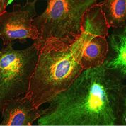
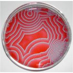
I am interested in self-organized, emergent patterns and textures. I make images of patterns both from the natural world and of experiments in my laboratory in the Department of Physics at the University of Toronto. Patterns naturally attract casual attention but are also the subject of serious scientific research. Some things just evolve all by themselves into strikingly regular shapes and textures. Why? These shapes emerge spontaneously from a dynamic process of growing, folding, cracking, wrinkling, branching, flowing and other kinds of morphological development. My photos are informed by the scientific aesthetic of nonlinear physics, and celebrate the subtle interplay of order and complexity in emergent patterns. They are a kind of “Scientific Folk Art” of the science of Emergence.

While the official opening is April 5, 2018, the event itself runs from April 1 – 30, 2018.

Next, there’s another March 27, 2018 announcement (received via email) from the Art/Sci Salon but this one concerns a series of talks about ’emergence’, Note: Some of the event information was a little difficult to decipher so I’ve added a note to the relevant section).

What is Emergent Form?

Nature teems with self-organized forms that seem to spring spontaneously from the smooth background of things, by mechanisms that are not always apparent. Think of rippled sand on a beach or regular stripes in the clouds.  Plants, insects and animals exhibit spirals and spots and stripes in an exuberant riot of colours.  Fluid flows in amazingly regular swirls and eddies.  The emergence of form is ubiquitous, and presents a challenge and an inspiration to both artists and scientists. In mathematics, patterns appear as solutions of the nonlinear partial differential equations in the continuum limit of classical physics, chemistry and biology. In the arts and humanities, “emergent form” addresses the entangled ways in which humans, plants animals, microorganisms inevitably co-exist in the universe; the way that human intervention and natural transformation can generate new landscapes and new forms of life.

With Emergent Form, we want to question the idea of a fixed world.

For us, Emergent Form is not just a series of natural and human phenomena too complicated to understand, measure or predict, but also a concept to help us identify ways in which we can come to term with, and embrace their complexity as a source of inspiration.

Join us in Toronto and Ottawa for a series of interdisciplinary discussions, performances and exhibitions on Emergent Form on Apr 10, 11, 12 (Toronto) and Apr. 14 [2018] (Ottawa).

This series is the result of a collaboration among several parties. Each event of the series is different and has its dedicated RSVP 

Tue. Apr 10 The Fields Institute, 222 College Street

Emergent form: an interdisciplinary concept 6:00-8:00 pm Pier Luigi Capucci, Accademia di Belle Arti Urbino. Founder and director, Noemalab*, Charles Sowers, Independent artist and exhibit designer, the Exploratorium, Stephen Morris, Professor of of Physics University of Toronto, Ron Wild, smART Maps

CLICK HERE FOR MORE AND TO RSVP

Wed. Apr 11 The Fields Institute6:00-8:00 pm

Anatomy of an Interconnected SystemA Performative Lecture with Margherita Pevere, Aalto University, Helsinki

CLICK HERE FOR MORE AND TO RSVP

Thu. Apr 12 (Note: I believe that from 5 – 6 pm, you’re invited to see Pevere’s exhibit and then proceed to Luella Massey Studio Theatre for performances)

5:00 pm  Cabinets in the Koffler Student Centre [I believe this is at the University of Toronto] Anatomy of an Interconnected System An exhibition by Margherita Pevere

6:00 pm Luella Massey Studio Theatre, 4 Glen Morris Ave., Toronto biopoetriX – conFiGURing AI

6:00-8:00 pm Performance: 

6:00pm Performance “Corpus Nil. A Ritual of Birth for a Modified Body” conceived and performed by Marco Donnarumma

6.30pm LAB dance: Blitz media posters on labs in the arts, sciences and engineering

7.10pm Panel: Performing AI, hybrid media and humans in/as technologyMarco Donnarumma, Doug van Nort (Dispersion Lab, York U.), Jane Tingley (Stratford User Research & Gameful Experiences Lab –SURGE-, U of Waterloo), Angela Schoellig (Dynamic Systems Lab, U of T)

Panel animators: Antje Budde (Digital Dramaturgy Lab) and Roberta Buiani (ArtSci Salon)

8.15pm Reception at the Italian Cultural Institute, 496 Huron St, Toronto

CLICK HERE FOR MORE AND TO RSVP

Ottawa. Sat. Apr. 14 National Arts Centre, 1 Elgin Street11:00 am-1:00 pm

Emergent Form and complex phenomenaA creative panel discussion and surprise demonstrationsWith Pier Luigi Capucci, Margherita Pevere, Marco Donnarumma, Stephen Morris

CLICK HERE FOR MORE AND TO RSVP

This event would not be possible without the support of The Fields Institute for Research in Mathematical Science, The Italian Embassy, the Centre for Drama, Theatre and Performance Studies at the University of Toronto, the Digital Dramaturgy Lab, and the Istituto Italiano di Cultura. Many thanks to our community partner BYOR (Bring your own Robot)

I wonder if some of the funding from Italy is in support of Italian Research in World Day. This is the inaugural year for the event, which will be held annually on April 15.

Vancouver’s brains

The Society of Italian Researchers and Professionals in Western Canada (ARPICO) is hosting an event in Vancouver (from a March 22, 2018 ARICO announcement received via email),

Our second speaking event of the year, in collaboration with the Consulate General of Italy in Vancouver, has been scheduled for Wednesday, April 11th, 2018 at the Roundhouse Community Centre. Professor Vesna Sossi’s talk will be examining how positron emission tomography (PET) imaging has contributed to better understanding of the brain function and disease with particular focus on Parkinson’s disease. You can read a summary of Prof. Sossi’s lecture as well as her short professional biography at the bottom of this message.

This event is organized in collaboration with the Consulate General of Italy in Vancouver to celebrate the newly instituted Italian Research in the World Day, as part of the Piano Straordinario “Vivere all’Italiana” – Giornata della ricerca Italiana nel mondo. You can read more on our website event page.

We look forward to seeing everyone there.

Please register for the event by visiting the EventBrite link or RSVPing to info@arpico.ca.

The evening agenda is as follows:

  • 6:45 pm – Doors Open
  • 7:00 pm – Lecture by Prof. Vesna Sossi
  • ~8:00 pm – Q & A Period
  • Mingling & Refreshments until about 9:30 pm

If you have not yet RSVP’d, please do so on our EventBrite page.

Further details are also available at arpico.ca, our facebook page, and Eventbrite.


Imaging: A Window into the Brain

Brain illness, comprising neurological disorders, mental illness and addiction, is considered the major health challenge in the 21st century with a socio-economic cost greater than cancer and cardiovascular disease combined. There are at least three unique challenges hampering brain disease management: relative inaccessibility, disease onset often preceding the onset of clinical symptoms by many years and overlap between clinical and pathological symptoms that makes accurate disease identification often difficult. This talk will give examples of how positron emission tomography (PET) imaging has contributed to better understanding of the brain function and disease with particular focus on Parkinson’s disease. Emphasis will be placed on the interplay between scientific discoveries and instrumentation and data analysis development as exemplified by the current understanding of the brain function as comprised by interactions between connectivity networks and neurochemistry and advancement in multi-modal imaging such as simultaneous PET and magnetic resonance imaging (MRI).

Vesna Sossi is a Professor in the University of British Columbia (UBC) Physics and Astronomy Department and at the UBC Djavad Mowafaghian Center for Brain Health. She directs the UBC Positron Emission Tomography (PET) imaging centre, which is known for its use of imaging as applied to neurodegeneration with emphasis on Parkinson’s disease. Her main areas of interest comprise development of imaging methods to enhance the investigation of neurochemical mechanisms that lead to an increased risk of Parkinson’s disease (PD) and mechanisms that contribute to treatment-related complications. She uses PET imaging to explore how alterations of the different neurotransmitter systems contribute to different trajectories of disease progression. Her other areas of interest are PET image analysis, instrumentation and multi-modal, multi-parameter data analysis. She published more than 180 peer review papers, is funded by several granting agencies, including the Michael J Fox Foundation, and sits on several national and international review panels.


WHEN: Wednesday, April 11th, 2018 at 7:00pm (doors open at 6:45pm)
WHERE: Roundhouse Community Centre, Room B – 181 Roundhouse Mews, Vancouver, BC, V6Z 2W3
RSVP: Please RSVP at EventBrite (https://imaging-a-window-into-the-brain.eventbrite.ca) or email info@arpico.ca


Tickets are Needed

  • Tickets are FREE, but all individuals are requested to obtain “free-admission” tickets on EventBrite site due to limited seating at the venue. Organizers need accurate registration numbers to manage wait lists and prepare name tags.
  • All ARPICO events are 100% staffed by volunteer organizers and helpers, however, room rental, stationery, and guest refreshments are costs incurred and underwritten by members of ARPICO. Therefore to be fair, all audience participants are asked to donate to the best of their ability at the door or via EventBrite to “help” defray costs of the event.

You can find directions for the Roundhouse Community Centre here

I have one idle question. What’s going to happen these groups if Canadians change their use of  Facebook or abandon the platform as they are threatening to do in the face of Cambridge Analytica’s use of their data? A March 25, 2018 article on huffingtonpost.ca outlines the latest about Canadians’ reaction to the Cambridge Analytical news according to an Angus Reid poll,

A survey by Angus Reid Institute suggests 73 per cent of Canadian Facebook users say they will make changes, while 27 per cent say it will be “business as usual.”

Nearly a quarter (23 per cent) said they would use Facebook less in the future, and 41 per cent of users said they would check and/or change their privacy settings.

The survey also found that one in 10 say they plan to abandon the platform, at least temporarily.

Facebook has been under fire for its ability to protect user privacy after Cambridge Analytica was accused of lifting the Facebook profiles of more than 50 million users without their permission.

There you have it.

*Well, a bit more information about one of the “Emergent’ speakers was received in an April 4, 2018 ArtSci Salon email announcement,

Do make sure to check out Pier Luigi Capucci’s EU-based (but with international breadth) Noemalab platform. https://noemalab.eu/ since the mid-nineties, this platform has been an important node of information for New Media Art and the relation between the arts and science.

noemalab’s blog regularly hosts reviews of events and conferences occurring around the world, including  the Subtle Technologies Festival between 2007 and 2014. you can search its archives here http://blogs.noemalab.eu/

Capucci has been writing several reflections on emergent forms of Life and theorized what he called the “third life”. See a recent essay https://noemalab.eu/memo/events/evolutionary-creativity-the-inner-life-and-meaning-of-art/ here is a picture which I would love him to explain during Emergent Form. Intrigued? come listen to him!

Novel self-assembly at 102 atoms

A Jan. 13, 2017 news item on ScienceDaily announces a discovery about self-assembly of 102-atom gold nanoclusters,

Self-assembly of matter is one of the fundamental principles of nature, directing the growth of larger ordered and functional systems from smaller building blocks. Self-assembly can be observed in all length scales from molecules to galaxies. Now, researchers at the Nanoscience Centre of the University of Jyväskylä and the HYBER Centre of Excellence of Aalto University in Finland report a novel discovery of self-assembling two- and three-dimensional materials that are formed by tiny gold nanoclusters of just a couple of nanometres in size, each having 102 gold atoms and a surface layer of 44 thiol molecules. The study, conducted with funding from the Academy of Finland and the European Research Council, has been published in Angewandte Chemie.

A Jan. 13, 2017 Academy of Finland press release, which originated the news item, provides more technical information about the work,

The atomic structure of the 102-atom gold nanocluster was first resolved by the group of Roger D Kornberg at Stanford University in 2007 (2). Since then, several further studies of its properties have been conducted in the Jyväskylä Nanoscience Centre, where it has also been used for electron microscopy imaging of virus structures (3). The thiol surface of the nanocluster has a large number of acidic groups that can form directed hydrogen bonds to neighbouring nanoclusters and initiate directed self-assembly.

The self-assembly of gold nanoclusters took place in a water-methanol mixture and produced two distinctly different superstructures that were imaged in a high-resolution electron microscope at Aalto University. In one of the structures, two-dimensional hexagonally ordered layers of gold nanoclusters were stacked together, each layer being just one nanocluster thick. Modifying the synthesis conditions, also three-dimensional spherical, hollow capsid structures were observed, where the thickness of the capsid wall corresponds again to just one nanocluster size (see figure).

While the details of the formation mechanisms of these superstructures warrant further systemic investigations, the initial observations open several new views into synthetically made self-assembling nanomaterials.

“Today, we know of several tens of different types of atomistically precise gold nanoclusters, and I believe they can exhibit a wide variety of self-assembling growth patterns that could produce a range of new meta-materials,” said Academy Professor Hannu Häkkinen, who coordinated the research at the Nanoscience Centre. “In biology, typical examples of self-assembling functional systems are viruses and vesicles. Biological self-assembled structures can also be de-assembled by gentle changes in the surrounding biochemical conditions. It’ll be of great interest to see whether these gold-based materials can be de-assembled and then re-assembled to different structures by changing something in the chemistry of the surrounding solvent.”

“The free-standing two-dimensional nanosheets will bring opportunities towards new-generation functional materials, and the hollow capsids will pave the way for highly lightweight colloidal framework materials,” Postdoctoral Researcher Nonappa (Aalto University) said.

Professor Olli Ikkala of Aalto University said: “In a broader framework, it has remained as a grand challenge to master the self-assemblies through all length scales to tune the functional properties of materials in a rational way. So far, it has been commonly considered sufficient to achieve sufficiently narrow size distributions of the constituent nanoscale structural units to achieve well-defined structures. The present findings suggest a paradigm change to pursue strictly defined nanoscale units for self-assemblies.”

References:

(1)    Nonappa, T. Lahtinen, J.S. Haataja, T.-R. Tero, H. Häkkinen and O. Ikkala, “Template-Free Supracolloidal Self-Assembly of Atomically Precise Gold Nanoclusters: From 2D Colloidal Crystals to Spherical Capsids”, Angewandte Chemie International Edition, published online 23 November 2016, DOI: 10.1002/anie.201609036

(2)    P. Jadzinsky et al., “Structure of a thiol-monolayer protected gold nanoparticle at 1.1Å resolution”, Science 318, 430 (2007)

(3)    V. Marjomäki et al., “Site-specific targeting of enterovirus capsid by functionalized monodispersed gold nanoclusters”, PNAS 111, 1277 (2014)

Here’s the figure mentioned in the news release,

Figure: 2D hexagonal sheet-like and 3D capsid structures based on atomically precise gold nanoclusters as guided by hydrogen bonding between the ligands. The inset in the top left corner shows the atomic structure of one gold nanocluster.

Here’s a link to and a citation for the paper,

Template-Free Supracolloidal Self-Assembly of Atomically Precise Gold Nanoclusters: From 2D Colloidal Crystals to Spherical Capsids by Dr. Nonappa, Dr. Tanja Lahtinen, M. Sc. Johannes. S. Haataja, Dr. Tiia-Riikka Tero, Prof. Hannu Häkkinen, and Prof. Olli Ikkala. Angewandte Chemie International Edition Volume 55, Issue 52, pages 16035–16038, December 23, 2016 Version of Record online: 23 NOV 2016 DOI: 10.1002/anie.201609036

© 2016 Wiley-VCH Verlag GmbH & Co. KGaA, Weinheim

This paper is behind a paywall.

A plasmonic nanolaser operating at visible light frequencies using ‘dark lattice’ modes

Finnish scientists have created lasers made of nanoparticles according to a Jan. 3, 2017 news item on ScienceDaily,

Researchers at Aalto University, Finland are the first to develop a plasmonic nanolaser that operates at visible light frequencies and uses so-called dark lattice modes.

The laser works at length scales 1000 times smaller than the thickness of a human hair. The lifetimes of light captured in such small dimensions are so short that the light wave has time to wiggle up and down only a few tens or hundreds of times. The results open new prospects for on-chip coherent light sources, such as lasers, that are extremely small and ultrafast.

The laser operation in this work is based on silver nanoparticles arranged in a periodic array.

A Jan. 3, 2017 Aalto University press release (also on EurekAlert), which originated the news item, describes the work in more detail,

 In contrast to conventional lasers, where the feedback of the lasing signal is provided by ordinary mirrors, this nanolaser utilizes radiative coupling between silver nanoparticles. These 100-nanometer-sized particles act as tiny antennas. To produce high intensity laser light, the interparticle distance was matched with the lasing wavelength so that all particles of the array radiate in unison. Organic fluorescent molecules were used to provide the input energy (the gain) that is needed for lasing.

Light from the dark

A major challenge in achieving lasing this way was that light may not exist long enough in such small dimensions to be helpful. The researchers found a smart way around this potential problem: they produced lasing in dark modes.

“A dark mode can be intuitively understood by considering regular antennas: A single antenna, when driven by a current, radiates strongly, whereas two antennas — if driven by opposite currents and positioned very close to each other — radiate very little,” explains Academy Professor Päivi Törmä.

“A dark mode in a nanoparticle array induces similar opposite-phase currents in each nanoparticle, but now with visible light frequencies”, she continues.

“Dark modes are attractive for applications where low power consumption is needed. But without any tricks, dark mode lasing would be quite useless because the light is essentially trapped at the nanoparticle array and cannot leave”, adds staff scientist Tommi Hakala.

“But by utilizing the small size of the array, we found an escape route for the light. Towards the edges of the array, the nanoparticles start to behave more and more like regular antennas that radiate to the outer world”, tells Ph.D. student Heikki Rekola.

The research team used the nanofabrication facilities and cleanrooms of the national OtaNano research infrastructure.

The researchers have produced a video elucidating their research,

A revelatory soundtrack by Kevin MacLeod has been added to this video.

Finally, here’s a link to and a citation for the paper,

Lasing in dark and bright modes of a finite-sized plasmonic lattice by T. K. Hakala, H. T. Rekola, A. I. Väkeväinen, J.-P. Martikainen, M. Nečada, A. J. Moilanen & P. Törmä. Nature Communications  8, Article number: 13687 doi:10.1038/ncomms13687 Published 03 January 2017

This is an open access paper.

Electrochemical measurements of biomolecules

This work comes from Finland and features some new nano shapes. From a Nov. 10, 2016 news item on phys.org,

Tomi Laurila’s research topic has many quirky names.

“Nanodiamond, nanohorn, nano-onion…,” lists off the Aalto University Professor, recounting the many nano-shapes of carbon. Laurila is using these shapes to build new materials: tiny sensors, only a few hundred nanometres across, that can achieve great things due to their special characteristics.

For one, the sensors can be used to enhance the treatment of neurological conditions. That is why Laurila, University of Helsinki Professor Tomi Taira and experts from HUS (the Hospital District of Helsinki and Uusimaa) are looking for ways to use the sensors for taking electrochemical measurements of biomolecules. Biomolecules are e.g. neurotransmitters such as glutamate, dopamine and opioids, which are used by nerve cells to communicate with each other.

A Nov. 10, 2016 Aalto University press release, which originated the news item, expands on the theme,

Most of the drugs meant for treating neurological diseases change the communication between nerve cells that is based on neurotransmitters. If we had real time and individual information on the operation of the neurotransmitter system, it would make it much easier to for example plan precise treatments’, explains Taira.

Due to their small size, carbon sensors can be taken directly next to a nerve cell, where the sensors will report what kind of neurotransmitter the cell is emitting and what kind of reaction it is inducing in other cells.

‘In practice, we are measuring the electrons that are moving in oxidation and reduction reactions’, Laurila explains the operating principle of the sensors.

‘The advantage of the sensors developed by Tomi and the others is their speed and small size. The probes used in current measurement methods can be compared to logs on a cellular scale – it’s impossible to use them and get an idea of the brain’s dynamic’, summarizes Taira.

Feedback system and memory traces

For the sensors, the journey from in vitro tests conducted in glass dishes and test tubes to in vivo tests and clinical use is long. However, the researchers are highly motivated.

‘About 165 million people are suffering from various neurological diseases in Europe alone. And because they are so expensive to treat, neurological diseases make up as much as 80 per cent of health care costs’, tells Taira.

Tomi Laurila believes that carbon sensors will have applications in fields such as optogenetics. Optogenetics is a recently developed method where a light-sensitive molecule is brought into a nerve cell so that the cell’s electric operation can then be turned on or off by stimulating it with light. A few years ago, a group of scientists proved in the scientific journal Nature that they had managed to use optogenetics to activate a memory trace that had been created previously due to learning. Using the same technique, researchers were able to demonstrate that with a certain type of Alzheimer’s, the problem is not that there are no memory traces being created, but that the brain cannot read the traces.

‘So the traces exist, and they can be activated by boosting them with light stimuli’, explains Taira but stresses that a clinical application is not yet a reality. However, clinical applications for other conditions may be closer by. One example is Parkinson’s disease. In Parkinson’s disease, the amount of dopamine starts to decrease in the cells of a particular brain section, which causes the typical symptoms such as tremors, rigidity and slowness of movement. With the sensors, the level of dopamine could be monitored in real time.

‘A sort of feedback system could be connected to it, so that it would react by giving an electric or optical stimulus to the cells, which would in turn release more dopamine’, envisions Taira.

‘Another application that would have an immediate clinical use is monitoring unconscious and comatose patients. With these patients, the level of glutamate fluctuates very much, and too much glutamate damages the nerve cell – online monitoring would therefore improve their treatment significantly.

Atom by atom

Manufacturing carbon sensors is definitely not a mass production process; it is slow and meticulous handiwork.

‘At this stage, the sensors are practically being built atom by atom’, summarises Tomi Laurila.

‘Luckily, we have many experts on carbon materials of our own. For example, the nanobuds of Professor Esko Kauppinen and the carbon films of Professor Jari Koskinen help with the manufacturing of the sensors. Carbon-based materials are mainly very compatible with the human body, but there is still little information about them. That’s why a big part of the work is to go through the electrochemical characterisation that has been done on different forms of carbon.’

The sensors are being developed and tested by experts from various fields, such as chemistry, materials science, modelling, medicine and imaging. Twenty or so articles have been published on the basic properties of the materials. Now, the challenge is to build them into geometries that are functional in a physiological environment. And taking measurements is not simple, either.

‘Brain tissue is delicate and doesn’t appreciate having objects being inserted in it. But if this were easy, someone would’ve already done it’, conclude the two.

I wish the researchers good luck.

Move objects by playing a melody

At this point, moving objects by playing a melody is a laboratory experiment but who knows, perhaps one day you’ll be able to sing your front door open. A Sept. 9, 2016 news item on ScienceDaily announces the research on acoustic waves,

Researchers of Aalto University have made a breakthrough in controlling the motion of multiple objects on a vibrating plate with a single acoustic source. By playing carefully constructed melodies, the scientists can simultaneously and independently move multiple objects on the plate towards desired targets. This has enabled scientists, for instance, writing words consisting of separate letters with loose metal pieces on the plate by playing a melody.

A Sept. 9, 2016 Aalto University press release (also on EurekAlert), which originated the news item, describes the research in more detail,

Already in 1878, the first studies of sand moving on a vibrating plate were done by Ernst Chladni, known as the father of acoustics. Chladni discovered that when a plate is vibrating at a frequency, objects move towards a few positions, called the nodal lines, specific to that frequency. Since then, the prevailing view has been that the particle motion is random on the plate before they reached the nodal line. “We have shown that the motion is also predictable away from the nodal lines. Now that the object does not have to be at a nodal line, we have much more freedom in controlling its motion and have achieved independent control of up to six objects simultaneously using just one single actuator. We are very excited about the results, because this probably is a new world record of how many independent motions can be controlled by a single acoustic actuator,” says Professor Quan Zhou.

The objects to be controlled have been placed on top of a manipulation plate, and imaged by a tracking camera. Based on the detected positions, the computer goes through a list of music notes to find a note that is most likely to move the objects towards the desired directions. After playing the note, the new positions of the objects are detected, and the control cycle is restarted. This cycle is repeated until the objects have reached their desired target locations. The notes played during the control cycles form a sequence, a bit like music.

The new method has been applied to manipulate a wide range of miniature objects including electronic components, water droplets, plant seeds, candy balls and metal parts. “Some of the practical applications we foresee include conveying and sorting microelectronic chips, delivering drug-loaded particles for pharmaceutical applications or handling small liquid volumes for lab on chips,” says Zhou. “Also, the basic idea should be transferrable to other kinds of systems with vibration phenomena. For example, it should be possible to use waves and ripples to control floating objects in a pond using our technique.”

Here’s a link to and a citation for the paper,

Controlling the motion of multiple objects on a Chladni plate by Quan Zhou, Veikko Sariola, Kourosh Latifi, Ville Liimatainen. Nature Communications 7, Article number: 12764 doi:10.1038/ncomms12764 Published 09 September 2016

This article is open access.

Could your photo be a solar cell?

Scientists at Aalto University (Finland) have found a way to print photographs that produce energy (like a solar cell does) according to a July 25, 2016 news item on Nanowerk,

Solar cells have been manufactured already for a long from inexpensive materials with different printing techniques. Especially organic solar cells and dye-sensitized solar cells are suitable for printing.

“We wanted to take the idea of printed solar cells even further, and see if their materials could be inkjet-printed as pictures and text like traditional printing inks,” tells University Lecturer Janne Halme.

A semi-transparent dye-sensitized solar cell with inkjet-printed photovoltaic portraits of the Aalto researchers (Ghufran Hashmi, Merve Özkan, Janne Halme) and a QR code that links to the original research paper. Courtesy: Aalto University

A semi-transparent dye-sensitized solar cell with inkjet-printed photovoltaic portraits of the Aalto researchers (Ghufran Hashmi, Merve Özkan, Janne Halme) and a QR code that links to the original research paper. Courtesy: Aalto University

A July 26, 2016 Aalto University press release, which originated the news item, describes the innovation in more detail,

When light is absorbed in an ordinary ink, it generates heat. A photovoltaic ink, however, coverts part of that energy to electricity. The darker the color, the more electricity is produced, because the human eye is most sensitive to that part of the solar radiation spectrum which has highest energy density. The most efficient solar cell is therefore pitch-black.

The idea of a colorful, patterned solar cell is to combine also other properties that take advantage of light on the same surface, such as visual information and graphics.

– For example, installed on a sufficiently low-power electrical device, this kind of solar cell could be part of its visual design, and at the same time produce energy for its needs, ponders Halme.

With inkjet printing, the photovoltaic dye could be printed to a shape determined by a selected image file, and the darkness and transparency of the different parts of the image could be adjusted accurately.

– The inkjet-dyed solar cells were as efficient and durable as the corresponding solar cells prepared in a traditional way. They endured more than one thousand hours of continuous light and heat stress without any signs of performance degradation, says Postdoctoral Researcher Ghufran Hashmi.

The dye and electrolyte that turned out to be best were obtained from the research group in the Swiss École Polytechnique Fédérale de Lausanne, where Dr. Hashmi worked as a visiting researcher.

– The most challenging thing was to find suitable solvent for the dye and the right jetting parameters that gave precise and uniform print quality, tells Doctoral Candidate Merve Özkan.

This puts solar cells (pun alert) in a whole new light.

Here’s a link to and a citation for the paper,

Dye-sensitized solar cells with inkjet-printed dyes by Syed Ghufran Hashmi, Merve Özkan, Janne Halme, Shaik Mohammed Zakeeruddin, Jouni Paltakari, Michael Grätzel, and Peter D. Lund. Energy Environ. Sci., 2016,9, 2453-2462 DOI: 10.1039/C6EE00826G First published online 09 Jun 2016

This paper is behind a paywall.

Constructing an autonomous Maxwell’s demon as a self-contained information-powered refrigerator

Aalto University (Finland) was the lead research institution for  INFERNOS, a European Union-funded project concerning Maxwell’s demon. Here’s an excerpt from an Oct. 14, 2013 post featuring the project,

An Oct. 9, 2013 news item on Nanowerk ties together INFERNOS and the ‘demon’,

Maxwell’s Demon is an imaginary creature that the mathematician James Clerk Maxwell created in 1897. The creature could turn heat into work without causing any other change, which violates the second law of thermodynamics. The primary goal of the European project INFERNOS (Information, fluctuations, and energy control in small systems) is to realize experimentally Maxwell’s Demon; in other words, to develop the electronic and biomolecular nanodevices that support this principle.

I like the INFERNOS logo, demon and all,

Logo of the European project INFERNOS (Information, fluctuations, and energy control in small systems).

A Jan. 11, 2016 news item on Nanowerk seems to be highlighting a paper resulting from the INFERNOS project (Note: A link has been removed),

On [a] theoretical level, the thought experiment has been an object of consideration for nearly 150 years, but testing it experimentally has been impossible until the last few years. Making use of nanotechnology, scientists from Aalto University have now succeeded in constructing an autonomous Maxwell’s demon that makes it possible to analyse the microscopic changes in thermodynamics. The research results were recently published in Physical Review Letters (“On-Chip Maxwell’s Demon as an Information-Powered Refrigerator”). The work is part of the forthcoming PhD thesis of MSc Jonne Koski at Aalto University.

An image illustrating the theory underlying the proposed device has been made available,

An autonomous Maxwell's demon. When the demon sees the electron enter the island (1.), it traps the electron with a positive charge (2.). When the electron leaves the island (3.), the demon switches back a negative charge (4.). Image: Jonne Koski.

An autonomous Maxwell’s demon. When the demon sees the electron enter the island (1.), it traps the electron with a positive charge (2.). When the electron leaves the island (3.), the demon switches back a negative charge (4.). Image: Jonne Koski.

A Jan. 11, 2016 Aalto University press release, which originated the news item, provides more technical details,

The system we constructed is a single-electron transistor that is formed by a small metallic island connected to two leads by tunnel junctions made of superconducting materials. The demon connected to the system is also a single-electron transistor that monitors the movement of electrons in the system. When an electron tunnels to the island, the demon traps it with a positive charge. Conversely, when an electron leaves the island, the demon repels it with a negative charge and forces it to move uphill contrary to its potential, which lowers the temperature of the system,’ explains Professor Jukka Pekola.

What makes the demon autonomous or self-contained is that it performs the measurement and feedback operation without outside help. Changes in temperature are indicative of correlation between the demon and the system, or, in simple terms, of how much the demon ‘knows’ about the system. According to Pekola, the research would not have been possible without the Low Temperature Laboratory conditions.

‘We work at extremely low temperatures, so the system is so well isolated that it is possible to register extremely small temperature changes,’ he says.

‘An electronic demon also enables a very large number of repetitions of the measurement and feedback operation in a very short time, whereas those who, elsewhere in the world, used molecules to construct their demons had to contend with not more than a few hundred repetitions.’

The work of the team led by Pekola remains, for the time being, basic research, but in the future, the results obtained may, among other things, pave the way towards reversible computing.

‘As we work with superconducting circuits, it is also possible for us to create qubits of quantum computers. Next, we would like to examine these same phenomena on the quantum level,’ Pekola reveals.

Here’s a link to and a citation for the paper,

On-Chip Maxwell’s Demon as an Information-Powered Refrigerator by J.V. Koski, A. Kutvonen, I.M. Khaymovich, T. Ala-Nissila, and J.P. Pekola. Phys. Rev. Lett. 115, 260602 DOI: http://dx.doi.org/10.1103/PhysRevLett.115.260602 Published 30 December 2015

This paper is behind a paywall.

One final comment, this is the 150th anniversary of Maxwell’s publication of a series of equations explaining the relationships between electric charges and electric and magnetic fields (featured here in a Nov. 27, 2015 posting).

Europe’s search for raw materials and hopes for nanotechnology-enabled solutions

A Feb. 27, 2015 news item on Nanowerk highlights the concerns over the availability of raw materials and European efforts to address those concerns,

Critical raw materials’ are crucial to many European industries but they are vulnerable to scarcity and supply disruption. As such, it is vital that Europe develops strategies for meeting the demand for raw materials. One such strategy is finding methods or substances that can replace the raw materials that we currently use. With this in mind, four EU projects working on substitution in catalysis, electronics and photonics presented their work at the Third Innovation Network Workshop on substitution of Critical Raw Materials hosted by the CRM_INNONET project in Brussels earlier this month [February 2015].

A Feb. 26, 2015 CORDIS press release, which originated the news item, goes on to describe four European Union projects working on nanotechnology-enabled solutions,

NOVACAM

NOVACAM, a coordinated Japan-EU project, aims to develop catalysts using non-critical elements designed to unlock the potential of biomass into a viable energy and chemical feedstock source.

The project is using a ‘catalyst by design’ approach for the development of next generation catalysts (nanoscale inorganic catalysts), as NOVACAM project coordinator Prof. Emiel Hensen from Eindhoven University of Technology in the Netherlands explained. Launched in September 2013, the project is developing catalysts which incorporate non-critical metals to catalyse the conversion of lignocellulose into industrial chemical feedstocks and bio-fuels. The first part of the project has been to develop the principle chemistry while the second part is to demonstrate proof of process. Prof. Hensen predicts that perhaps only two of three concepts will survive to this phase.

The project has already made significant progress in glucose and ethanol conversion, according to Prof. Hensen, and has produced some important scientific publications. The consortium is working with and industrial advisory board comprising Shell in the EU and Nippon Shokubai in Japan.

FREECATS

The FREECATS project, presented by project coordinator Prof. Magnus Rønning from the Norwegian University of Science and Technology, has been working over the past three years to develop new metal-free catalysts. These would be either in the form of bulk nanomaterials or in hierarchically organised structures – both of which would be capable of replacing traditional noble metal-based catalysts in catalytic transformations of strategic importance.

Prof. Magnus Rønning explained that the application of the new materials could eliminate the need for the use for platinum group metals (PGM) and rare earth metals – in both cases Europe is very reliant on other countries for these materials. Over the course of its research, FREECATS targeted three areas in particular – fuel cells, the production of light olefins and water and wastewater purification.

By working to replace the platinum in fuel cells, the project is supporting the EU’s aim of replacing the internal combustion engine by 2050. However, as Prof. Rønning noted, while platinum has been optimized for use over several decades, the materials FREECATS are using are new and thus come with their new challenges which the project is addressing.

HARFIR

Prof. Atsufumi Hirohata of the University of York in the United Kingdom, project coordinator of HARFIR, described how the project aims to discover an antiferromagnetic alloy that does not contain the rare metal Iridium. Iridium is becoming more and more widely used in numerous spin electronic storage devices, including read heads in hard disk drives. The world supply depends on Platinum ore that comes mainly from South Africa. The situation is much worse than for other rare earth elements as the price has been shooting up over recent years, according to Prof. Hirohata.

The HARFIR team, divided between Europe and Japan, aims to replace Iridium alloys with Heusler alloys. The EU team, led by Prof. Hirohata, has been working on the preparation of polycrystalline and epitaxial thin films of Heusler Alloys, with the material design led by theoretical calculations. The Japanese team, led by Prof. Koki Takanashi at Tohoku University, is meanwhile working on the preparation of epitaxial thin films, measurements of fundamental properties and structural/magnetic characterisation by neutron and synchrotron x-ray beams.

One of the biggest challenges has been that Heusler alloys have a relatively complicated atomic structure. In terms of HARFIR’s work, if any atomic disordering at the edge of nanopillar devices, the magnetic properties that are needed are lost. The team is exploring solutions to this challenge.

IRENA

Prof. of Esko Kauppinen Aalto University in Finland closed off the first session of the morning with his presentation of the IRENA project. Launched in September 2013, the project will run until mid 2017 working towards the aim of developing high performance materials, specifically metallic and semiconducting single-walled carbon nanotube (SWCNT) thin films to completely eliminate the use of the critical metals in electron devices. The ultimate aim is to replace Indium in transparent conducting films, and Indium and Gallium as a semiconductor in thin film field effect transistors (TFTs).

The IRENA team is developing an alternative that is flexible, transparent and stretchable so that it can meet the demands of the electronics of the future – including the possibility to print electronics.

IRENA involves three partners from Europe and three from Japan. The team has expertise in nanotube synthesis, thin film manufacturing and flexible device manufacturing, modelling of nanotube growth and thin film charge transport processes, and the project has benefitted from exchanges of team members between institutions. One of the key achievements so far is that the project has succeeded in using a nanotube thin film for the first time as the both the electrode and hole blocking layer in an organic solar cell.

You’ll note that Japan is a partner in all of these projects. In all probability, these initiatives have something to do with rare earths which are used in much of today’s electronics technology and Japan is sorely lacking in those materials. China, by comparison, has dominated the rare earths export industry and here’s an excerpt from my Nov. 1, 2013 posting where I outline the situation (which I suspect hasn’t changed much since),

As for the short supply mentioned in the first line of the news item, the world’s largest exporter of rare earth elements at 90% of the market, China, recently announced a cap according to a Sept. 6, 2013 article by David Stanway for Reuters. The Chinese government appears to be curtailing exports as part of an ongoing, multi-year strategy. Here’s how Cientifica‘s (an emerging technologies consultancy, etc.) white paper (Simply No Substitute?) about critical materials published in 2012 (?), described the situation,

Despite their name, REE are not that rare in the Earth’s crust. What has happened in the past decade is that REE exports from China undercut prices elsewhere, leading to the closure of mines such as the Mountain Pass REE mine in California. Once China had acquired a dominant market position, prices began to rise. But this situation will likely ease. The US will probably begin REE production from the Mountain Pass mine later in 2012, and mines in other countries are expected to start operation soon as well.

Nevertheless, owing to their broad range of uses REE will continue to exert pressures on their supply – especially for countries without notable REE deposits. This highlights two aspects of importance for strategic materials: actual rarity and strategic supply issues such as these seen for REE. Although strategic and diplomatic supply issues may have easier solutions, their consideration for manufacturing industries will almost be the same – a shortage of crucial supply lines.

Furthermore, as the example of REE shows, the identification of long-term supply problems can often be difficult, and not every government has the same strategic foresight that the Chinese demonstrated. And as new technologies emerge, new elements may see an unexpected, sudden demand in supply. (pp. 16-17)

Meanwhile, in response to China’s decision to cap its 2013 REE exports, the Russian government announced a $1B investment to 2018 in rare earth production,, according to a Sept. 10, 2013 article by Polina Devitt for Reuters.

I’m not sure you’ll be able to access Tim Harper’s white paper as he is now an independent, serial entrepreneur. I most recently mentioned him in relation to his articles (on Azonano) about the nanotechnology scene in a Feb. 12, 2015 posting where you’ll also find contact details for him.

Inhibiting viruses with nanocrystalline cellulose (NCC) in Finland

Research and interest in cellulose nanomaterials of one kind or another seems to be reaching new heights. That’s my experience since this is my third posting on the topic in one week.

The latest research features NCC (nanocrystalline cellulose [NCC] or, as it’s sometimes known, cellulose nanocrystals [CNC]) ,as a ‘viral inhibitor’ and is described in an April 15, 2014 news item on Nanowerk,

Researchers from Aalto University [Finland] and and the University of Eastern Finland have succeeded in creating a surface on nano-sized cellulose crystals that imitates a biological structure. The surface adsorbs viruses and disables them. The results can prove useful in the development of antiviral ointments and surfaces, for instance.

There are many viral diseases in the world for which no pharmaceutical treatment exists. These include, among others, dengue fever, which is spread by mosquitoes in the tropics, as well as a type of diarrhea, which is more familiar in Finland and is easily spread by the hands and can be dangerous especially for small children and the elderly.

An April 15, 2014 Aalto University news release, which originated the news item, provides more detail,

Researchers at Aalto University and the University of Eastern Finland have now succeeded in preliminary tests to prevent the spread of one type of virus into cells with the help of a new type of nanocrystalline cellulose. Nano-sized cellulose crystals were manufactured out of cotton fibre or filter paper with the help of sulphuric acid, causing sulphate ions with negative charges to attach to their surfaces. The ions then attached to alphaviruses used in the test and neutralised them. When the researchers replaced the sulphate ions with cellulose derivatives that imitate tyrosine sulphates, the activity of the viruses was further reduced. The experiments succeeded in preventing viral infection in 88-100 percent of the time with no noticeable effect on the viability of the cells by the nanoparticles. The research findings were published in the journal Biomacromolecules.

Here’s a diagram illustrating how the new type of NCC works,

Courtesy of Aalto University

Courtesy of Aalto University

The news release includes perspectives from the researchers,

’Certain cellulose derivatives had been seen to have an impact on viruses before. The nano scale increases the proportion of the surface area to that of the number of grams to a very high level, which is an advantage, because viruses specifically attach themselves to surfaces. Making the cellulose crystals biomimetic, which means that they mimic biological structures, was an important step, as we know that in nature viruses often interact specifically with tyrosine structures,’ he [Jukka Seppälä, Professor of Polymer Technology at Aalto University] says.

Both Jukka Seppälä and Ari Hinkkanen, Professor of Gene Transfer Technology at the University of Eastern Finland, emphasise that the research is still in the early stages.

‘Now we know that the attachment of a certain alphavirus can be effectively prevented when we use large amounts of nanocrystalline cellulose.  Next we need to experiment with other alpha viruses and learn to better understand the mechanisms that prevent viral infection. In addition, it is necessary to ascertain if cellulose can also block other viruses and in what conditions, and to investigate whether or not the sulphates have a deleterious effects on an organism,’ Ari Hinkkanen explains.

According to Kristiina Järvinen, Professor of Pharmaceutical Technology at the University of Eastern Finland, there are many routes that can be taken in the commercialisation of the results. The development of an antiviral medicine is the most distant of these; the idea could be sooner applied in disinfectant ointments and coatings, for instance.

‘It would be possible to provide protection against viruses, spread by mosquitoes, by applying ointment containing nanocrystalline cellulose onto the skin. Nanocrystalline cellulose applied on hospital door handles could kill viruses and prevent them from spreading.  However, we first need to ascertain if the compounds will remain effective in a non-liquid form and how they work in animal tests,’ she ponders.

For the curious, here’s a link to and a citation for the paper,

Synthesis of Cellulose Nanocrystals Carrying Tyrosine Sulfate Mimetic Ligands and Inhibition of Alphavirus Infection by Justin O. Zoppe, Ville Ruottinen, Janne Ruotsalainen, Seppo Rönkkö, Leena-Sisko Johansson, Ari Hinkkanen, Kristiina Järvinen, and Jukka Seppälä. Biomacromolecules, 2014, 15 (4), pp 1534–1542 DOI: 10.1021/bm500229d Publication Date (Web): March 14, 2014

Copyright © 2014 American Chemical Society

This paper is behind a paywall.

As for my other recent postings on cellulose nanomaterials, there’s this April 14, 2014 piece titled: Preparing nanocellulose for eventual use in dressings for wounds and this from April 10, 2014 titled: US Dept. of Agriculture wants to commercialize cellulose nanomaterials.

Cancer as a fashion statement at the University of British Columbia (Canada) and a Marimekko dress made of birch in Finland

The ‘Fashioning Cancer Project’ at the University of British Columbia (UBC) bears some resemblance to the types of outreach projects supported by the UK’s Wellcome Trust (for an example see my June 21, 2011 posting) where fashion designers are inspired by some aspect of science. Here’s more about the ‘Fashioning Cancer Project’ and its upcoming fashion show (on March 25, 2014). From the March 12, 2014 UBC news release (Note: Links have been removed),

A UBC costume design professor has created a collection of ball gowns inspired by microscopic photos of cancer cells and cellular systems to get people talking about the disease, beauty and body image.

The project aims to create alternative imagery for discussions of cancer, to complement existing examples such as the pink ribbon, which is an important symbol of cancer awareness, but may not accurately represent women’s experience with the disease.

“Many women who have battled cancer express a disconnect with the fashion imagery that commonly represents the disease,” says Jacqueline Firkins, an assistant professor in UBC’s Dept. of Theatre and Film, who designed the collection of 10 dresses and dubbed the work ‘Fashioning Cancer: The Correlation between Destruction and Beauty.’

Inspired by cellular images captured by researchers in the lab of UBC scientist Christian Naus, a Peter Wall Distinguished Scholar in Residence, the project seeks to create artistic imagery based on the disease itself.

“My hope is that somehow through fashion, I more closely tap into what a woman might be feeling about her body as she undergoes the disease, but simultaneously reflect a strength, beauty, and resilience,” says Firkins, who will use the collection to raise money for cancer research, patients and survivors.

“This will be an opportunity for people to share their thoughts about the gowns,” says Firkins. “Are they too pretty to reflect something as destructive as cancer? Do they encourage you to tell your own story? Do they evoke any emotions related to your own experience?”

Before giving you where and when, here are two images (a cell and a dress based on the cell),

http://news.ubc.ca/2014/03/12/prof-challenges-cancer-fashion/

Cell7_brain_cells_in_a_dish; Astrocytes from the brain growing in a culture dish. Green colour indicates the cytoskeleton of these cells, red colour shows specific membrance [sic] channels (gap junctions), blue colour indicates the cell nuclei (DNA). The ability to grow cells in a dish has contributed to our understand of the changes these cells undergo when they become channels. Photo credit: John Bechberger, MSc., Christian Naus, PhD.

Cell7_Mercedes_de_la_Zerda: Dress modeled by BFA Acting student Mercedes de la Zerda.Black organza cap sleeve w/ sheer top and multicolour organza diagonal trim. Photo credit: Tim Matheson

Cell7_Mercedes_de_la_Zerda: Dress modeled by BFA Acting student Mercedes de la Zerda.Black organza cap sleeve w/ sheer top and multicolour organza diagonal trim. Photo credit: Tim Matheson

Details about the show (from the UBC event description webpage where you can also find a slide show more pictures),

  • Event: Fashioning Cancer: The Correlation between Destruction and Beauty
  • Date: Tue. March 25, 2014 | Time: 12-1pm
  • Location: UBC’s Frederic Wood Theatre, 6354 Crescent Rd.
  • MAP: http://bit.ly/1fZ4bC8

On a more or less related note, Aalto University (Finland) has announced a dress made of birch cellulose fibre, from a March 13, 2014 news item on ScienceDaily,

The first garment made out of birch cellulose fibre using the Ioncell method is displayed at a fashion show in Finland on 13 March [2014]. The Ioncell method, which was developed by researchers at Aalto University, is an environmentally friendly alternative to cotton in textile production. The dress produced for Marimekko is a significant step forward in the development of fibre for industrial production.

Researchers were looking for new alternatives to cotton, because demand for textile fibres is expected to nearly double by 2030. The raw material for the Ioncell fibre is a birch-based pulp from Finnish pulp mills. Growing birch wood does not require artificial irrigation in its native habitat, for instance.

The Aalto University March 12, 2014 news release, which originated the news item, describes the new Ioncell fibre and its relationship with Finnish clothing company Marimekko,

The production method for Ioncell has been developed by Professor Herbert Sixta’s research group. The method is based on a liquid salt (ionic liquid) developed under the guidance of Professor Ilkka Kilpeläinen which is a very efficient cellulose solvent. The fibres derived from it are carded and spun to yarns at the Textile University of Börås in Sweden.

‒ We made a breakthrough in the development of the method about a year ago. Progress has been rapid since then. [see my Oct. 3, 2013 posting for another Finnish team’s work with wood cellulose to create fabric]  Production of the fibre and the thread is still a cumbersome process, but we have managed to triple the amount of fibre that is produced in six months. The quality has also improved: the fibers are stronger and of more even quality, Professor Sixta says with satisfaction.

The surface of the ready textile has a dim glow and it is pleasing to the touch. According to Sixta, because of its strength, the strength properties of the Ioncell fibre are equal or even better than other pulp-based fibres on the market. The fibres are even stronger than cotton and viscose.

The Finnish textile and clothing design company Marimekko became inspired by the new fibre at an event organised by the Finnish Bioeconomy Cluster FIBIC, which coordinates bioeconomy research, and immediately got in touch with Professor Herbert Sixta at Aalto University.

‒ We monitor product development for materials closely in order to be able to offer our customers new and more ecological alternatives. It was a wonderful opportunity to be able to join this Aalto University development project at such an early stage. Fibre made from birch pulp seems to be a promising material by virtue of its durability and other characteristics, and we hope that we will soon be able to utilise this new material in our collections, says Noora Niinikoski, Head of Fashion at Marimekko.

Here’s the birch cellulose dress,

Marimekko Birch Dress Courtesy: Aalto University

Let’s all have a fashionable day!