Tag Archives: Abraham Vazquez-Guardado

Cortical spheroids (like mini-brains) could unlock (larger) brain’s mysteries

A March 19, 2021 Northwestern University news release on EurekAlert announces the creation of a device designed to monitor brain organoids (for anyone unfamiliar with brain organoids there’s more information after the news),

A team of scientists, led by researchers at Northwestern University, Shirley Ryan AbilityLab and the University of Illinois at Chicago (UIC), has developed novel technology promising to increase understanding of how brains develop, and offer answers on repairing brains in the wake of neurotrauma and neurodegenerative diseases.

Their research is the first to combine the most sophisticated 3-D bioelectronic systems with highly advanced 3-D human neural cultures. The goal is to enable precise studies of how human brain circuits develop and repair themselves in vitro. The study is the cover story for the March 19 [March 17, 2021 according to the citation] issue of Science Advances.

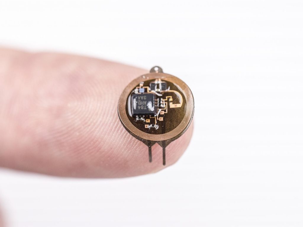
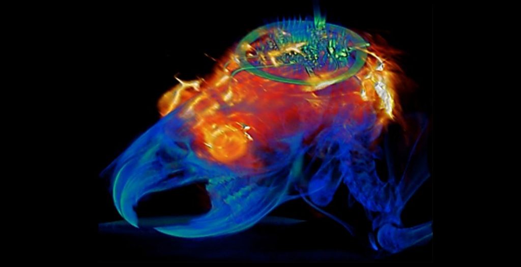
The cortical spheroids used in the study, akin to “mini-brains,” were derived from human-induced pluripotent stem cells. Leveraging a 3-D neural interface system that the team developed, scientists were able to create a “mini laboratory in a dish” specifically tailored to study the mini-brains and collect different types of data simultaneously. Scientists incorporated electrodes to record electrical activity. They added tiny heating elements to either keep the brain cultures warm or, in some cases, intentionally overheated the cultures to stress them. They also incorporated tiny probes — such as oxygen sensors and small LED lights — to perform optogenetic experiments. For instance, they introduced genes into the cells that allowed them to control the neural activity using different-colored light pulses.

This platform then enabled scientists to perform complex studies of human tissue without directly involving humans or performing invasive testing. In theory, any person could donate a limited number of their cells (e.g., blood sample, skin biopsy). Scientists can then reprogram these cells to produce a tiny brain spheroid that shares the person’s genetic identity. The authors believe that, by combining this technology with a personalized medicine approach using human stem cell-derived brain cultures, they will be able to glean insights faster and generate better, novel interventions.

“The advances spurred by this research will offer a new frontier in the way we study and understand the brain,” said Shirley Ryan AbilityLab’s Dr. Colin Franz, co-lead author on the paper who led the testing of the cortical spheroids. “Now that the 3-D platform has been developed and validated, we will be able to perform more targeted studies on our patients recovering from neurological injury or battling a neurodegenerative disease.”

Yoonseok Park, postdoctoral fellow at Northwestern University and co-lead author, added, “This is just the beginning of an entirely new class of miniaturized, 3-D bioelectronic systems that we can construct to expand the capacity of the regenerative medicine field. For example, our next generation of device will support the formation of even more complex neural circuits from brain to muscle, and increasingly dynamic tissues like a beating heart.”

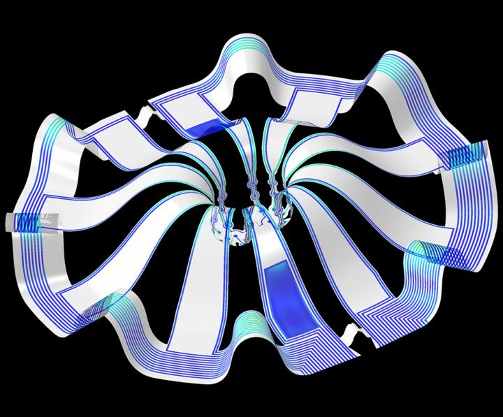
Current electrode arrays for tissue cultures are 2-D, flat and unable to match the complex structural designs found throughout nature, such as those found in the human brain. Moreover, even when a system is 3-D, it is extremely challenging to incorporate more than one type of material into a small 3-D structure. With this advance, however, an entire class of 3-D bioelectronics devices has been tailored for the field of regenerative medicine.

“Now, with our small, soft 3-D electronics, the capacity to build devices that mimic the complex biological shapes found in the human body is finally possible, providing a much more holistic understanding of a culture,” said Northwestern’s John Rogers, who led the technology development using technology similar to that found in phones and computers. “We no longer have to compromise function to achieve the optimal form for interfacing with our biology.”

As a next step, scientists will use the devices to better understand neurological disease, test drugs and therapies that have clinical potential, and compare different patient-derived cell models. This understanding will then enable a better grasp of individual differences that may account for the wide variation of outcomes seen in neurological rehabilitation.

“As scientists, our goal is to make laboratory research as clinically relevant as possible,” said Kristen Cotton, research assistant in Dr. Franz’s lab. “This 3-D platform opens the door to new experiments, discovery and scientific advances in regenerative neurorehabilitation medicine that have never been possible.”

Caption: Three dimensional multifunctional neural interfaces for cortical spheroids and engineered assembloids Credit: Northwestern University

As for what brain ogranoids might be, Carl Zimmer in an Aug. 29, 2019 article for the New York Times provides an explanation,

Organoids Are Not Brains. How Are They Making Brain Waves?

Two hundred and fifty miles over Alysson Muotri’s head, a thousand tiny spheres of brain cells were sailing through space.

The clusters, called brain organoids, had been grown a few weeks earlier in the biologist’s lab here at the University of California, San Diego. He and his colleagues altered human skin cells into stem cells, then coaxed them to develop as brain cells do in an embryo.

The organoids grew into balls about the size of a pinhead, each containing hundreds of thousands of cells in a variety of types, each type producing the same chemicals and electrical signals as those cells do in our own brains.

In July, NASA packed the organoids aboard a rocket and sent them to the International Space Station to see how they develop in zero gravity.

Now the organoids were stowed inside a metal box, fed by bags of nutritious broth. “I think they are replicating like crazy at this stage, and so we’re going to have bigger organoids,” Dr. Muotri said in a recent interview in his office overlooking the Pacific.

What, exactly, are they growing into? That’s a question that has scientists and philosophers alike scratching their heads.

On Thursday, Dr. Muotri and his colleagues reported that they  have recorded simple brain waves in these organoids. In mature human brains, such waves are produced by widespread networks of neurons firing in synchrony. Particular wave patterns are linked to particular forms of brain activity, like retrieving memories and dreaming.

As the organoids mature, the researchers also found, the waves change in ways that resemble the changes in the developing brains of premature babies.

“It’s pretty amazing,” said Giorgia Quadrato, a neurobiologist at the University of Southern California who was not involved in the new study. “No one really knew if that was possible.”

But Dr. Quadrato stressed it was important not to read too much into the parallels. What she, Dr. Muotri and other brain organoid experts build are clusters of replicating brain cells, not actual brains.

If you have the time, I recommend reading Zimmer’s article in its entirety. Perhaps not coincidentally, Zimmer has an excerpt titled “Lab-Grown Brain Organoids Aren’t Alive. But They’re Not Not Alive, Either.” published in Slate.com,

From Life’s Edge: The Search For What It Means To Be Alive by Carl Zimmer, published by Dutton, an imprint of Penguin Publishing Group, a division of Penguin Random House, LLC. Copyright © 2021 by Carl Zimmer.

Cleber Trujillo led me to a windowless room banked with refrigerators, incubators, and microscopes. He extended his blue-gloved hands to either side and nearly touched the walls. “This is where we spend half our day,” he said.

In that room Trujillo and a team of graduate students raised a special kind of life. He opened an incubator and picked out a clear plastic box. Raising it above his head, he had me look up at it through its base. Inside the box were six circular wells, each the width of a cookie and filled with what looked like watered-down grape juice. In each well 100 pale globes floated, each the size of a housefly head.

Getting back to the research about monitoring brain organoids, here’s a link to and a citation for the paper about cortical spheroids,

Three-dimensional, multifunctional neural interfaces for cortical spheroids and engineered assembloids by Yoonseok Park, Colin K. Franz, Hanjun Ryu, Haiwen Luan, Kristen Y. Cotton, Jong Uk Kim, Ted S. Chung, Shiwei Zhao, Abraham Vazquez-Guardado, Da Som Yang, Kan Li, Raudel Avila, Jack K. Phillips, Maria J. Quezada, Hokyung Jang, Sung Soo Kwak, Sang Min Won, Kyeongha Kwon, Hyoyoung Jeong, Amay J. Bandodkar, Mengdi Han, Hangbo Zhao, Gabrielle R. Osher, Heling Wang, KunHyuck Lee, Yihui Zhang, Yonggang Huang, John D. Finan and John A. Rogers. Science Advances 17 Mar 2021: Vol. 7, no. 12, eabf9153 DOI: 10.1126/sciadv.abf9153

This paper appears to be open access.

According to a March 22, 2021 posting on the Shirley Riley AbilityLab website, the paper is featured on the front cover of Science Advances (vol. 7 no. 12).

Controlling neurons with light: no batteries or wires needed

Caption: Wireless and battery-free implant with advanced control over targeted neuron groups. Credit: Philipp Gutruf

This January 2, 2019 news item on ScienceDaily describes the object seen in the above and describes the problem it’s designed to solve,

University of Arizona biomedical engineering professor Philipp Gutruf is first author on the paper Fully implantable, optoelectronic systems for battery-free, multimodal operation in neuroscience research, published in Nature Electronics.

Optogenetics is a biological technique that uses light to turn specific neuron groups in the brain on or off. For example, researchers might use optogenetic stimulation to restore movement in case of paralysis or, in the future, to turn off the areas of the brain or spine that cause pain, eliminating the need for — and the increasing dependence on — opioids and other painkillers.

“We’re making these tools to understand how different parts of the brain work,” Gutruf said. “The advantage with optogenetics is that you have cell specificity: You can target specific groups of neurons and investigate their function and relation in the context of the whole brain.”

In optogenetics, researchers load specific neurons with proteins called opsins, which convert light to electrical potentials that make up the function of a neuron. When a researcher shines light on an area of the brain, it activates only the opsin-loaded neurons.

The first iterations of optogenetics involved sending light to the brain through optical fibers, which meant that test subjects were physically tethered to a control station. Researchers went on to develop a battery-free technique using wireless electronics, which meant subjects could move freely.

But these devices still came with their own limitations — they were bulky and often attached visibly outside the skull, they didn’t allow for precise control of the light’s frequency or intensity, and they could only stimulate one area of the brain at a time.

A Dec. 21, 2018 University of Azrizona news release (published Jan. 2, 2019 on EurekAlert), which originated the news item, discusses the work in more detail,

“With this research, we went two to three steps further,” Gutruf said. “We were able to implement digital control over intensity and frequency of the light being emitted, and the devices are very miniaturized, so they can be implanted under the scalp. We can also independently stimulate multiple places in the brain of the same subject, which also wasn’t possible before.”

The ability to control the light’s intensity is critical because it allows researchers to control exactly how much of the brain the light is affecting — the brighter the light, the farther it will reach. In addition, controlling the light’s intensity means controlling the heat generated by the light sources, and avoiding the accidental activation of neurons that are activated by heat.

The wireless, battery-free implants are powered by external oscillating magnetic fields, and, despite their advanced capabilities, are not significantly larger or heavier than past versions. In addition, a new antenna design has eliminated a problem faced by past versions of optogenetic devices, in which the strength of the signal being transmitted to the device varied depending on the angle of the brain: A subject would turn its head and the signal would weaken.

“This system has two antennas in one enclosure, which we switch the signal back and forth very rapidly so we can power the implant at any orientation,” Gutruf said. “In the future, this technique could provide battery-free implants that provide uninterrupted stimulation without the need to remove or replace the device, resulting in less invasive procedures than current pacemaker or stimulation techniques.”

Devices are implanted with a simple surgical procedure similar to surgeries in which humans are fitted with neurostimulators, or “brain pacemakers.” They cause no adverse effects to subjects, and their functionality doesn’t degrade in the body over time. This could have implications for medical devices like pacemakers, which currently need to be replaced every five to 15 years.

The paper also demonstrated that animals implanted with these devices can be safely imaged with computer tomography, or CT, and magnetic resonance imaging, or MRI, which allow for advanced insights into clinically relevant parameters such as the state of bone and tissue and the placement of the device.

This image of a combined MRI (magnetic resonance image) and CT (computer tomography) scan bookends, more or less, the picture of the device which headed this piece,

Combined image analysis with MRI and CT results superimposed on a 3D rendering of the animal implanted with the programmable bilateral multi µ-ILED device. Courtesy: University of Arizona

Here’s a link to and a citation for the paper,

Fully implantable optoelectronic systems for battery-free, multimodal operation in neuroscience research by Philipp Gutruf, Vaishnavi Krishnamurthi, Abraham Vázquez-Guardado, Zhaoqian Xie, Anthony Banks, Chun-Ju Su, Yeshou Xu, Chad R. Haney, Emily A. Waters, Irawati Kandela, Siddharth R. Krishnan, Tyler Ray, John P. Leshock, Yonggang Huang, Debashis Chanda, & John A. Rogers. Nature Electronics volume 1, pages652–660 (2018) DOI: https://doi.org/10.1038/s41928-018-0175-0 Published 13 December 2018

This paper is behind a paywall.

World’s first full-color, flexible thin-film reflective display: a step forward for camouflage?

Caption: Dr. Chanda used an iconic National Geographic photographic of an Afghan girl to demonstrate the color-changing abilities of the nanostructured reflective display developed by his team. Credit: University of Central Florida, used with permission from National Geographic

Caption: Dr. Chanda used an iconic National Geographic photographic of an Afghan girl to demonstrate the color-changing abilities of the nanostructured reflective display developed by his team. Credit: University of Central Florida, used with permission from National Geographic

This has gotten a lot of attention. A June 25, 2015 news item on Azonano describes a couple of possible applications,

Imagine a soldier who can change the color and pattern of his camouflage uniform from woodland green to desert tan at will. Or an office worker who could do the same with his necktie. Is someone at the wedding reception wearing the same dress as you? No problem – switch yours to a different color in the blink of an eye.

A June 24, 2015 University of Central Florida news release on EurekAlert, which originated the news item, provides some insight into the research along with some technical details,

Chanda’s [Professor Debashis Chanda] research was inspired by nature. Traditional displays like those on a mobile phone require a light source, filters and a glass plates. But animals like chameleons, octopuses and squids are born with thin, flexible, color-changing displays that don’t need a light source – their skin.

“All manmade displays – LCD, LED, CRT – are rigid, brittle and bulky. But you look at an octopus, they can create color on the skin itself covering a complex body contour, and it’s stretchable and flexible,” Chanda said. “That was the motivation: Can we take some inspiration from biology and create a skin-like display?”

As detailed in the cover article of the June issue of the journal Nature Communications, Chanda is able to change the color on an ultrathin nanostructured surface by applying voltage. The new method doesn’t need its own light source. Rather, it reflects the ambient light around it.

A thin liquid crystal layer is sandwiched over a metallic nanostructure shaped like a microscopic egg carton that absorbs some light wavelengths and reflects others. The colors reflected can be controlled by the voltage applied to the liquid crystal layer. The interaction between liquid crystal molecules and plasmon waves on the nanostructured metallic surface played the key role in generating the polarization-independent, full-color tunable display.

His method is groundbreaking. It’s a leap ahead of previous research that could produce only a limited color palette. And the display is only about few microns thick, compared to a 100-micron-thick human hair. Such an ultrathin display can be applied to flexible materials like plastics and synthetic fabrics.

The research has major implications for existing electronics like televisions, computers and mobile devices that have displays considered thin by today’s standards but monstrously bulky in comparison. But the potentially bigger impact could be whole new categories of displays that have never been thought of.

“Your camouflage, your clothing, your fashion items – all of that could change,” Chanda said. “Why would I need 50 shirts in my closet if I could change the color and pattern?”

Researchers used a simple and inexpensive nano-imprinting technique that can produce the reflective nanostructured surface over a large area.

“This is a cheap way of making displays on a flexible substrate with full-color generation,” Chanda said. “That’s a unique combination.”

Here’s a link to and a citation for the paper,

Polarization-independent actively tunable colour generation on imprinted plasmonic surfaces by Daniel Franklin, Yuan Chen, Abraham Vazquez-Guardado, Sushrut Modak, Javaneh Boroumand, Daming Xu, Shin-Tson Wu & Debashis Chanda. Nature Communications 6, Article number: 7337 doi:10.1038/ncomms8337 Published 11 June 2015

This paper is open access.