Tag Archives: Alzheimer’s Disease

Cornell University researchers breach blood-brain barrier

There are other teams working on ways to breach the blood-brain barrier (my March 26, 2015 post highlights work from a team at the University of Montréal) but this team from  Cornell is working with a drug that has already been approved by the US Food and Drug Administration (FDA) according to an April 8, 2016 news item on ScienceDaily,

Cornell researchers have discovered a way to penetrate the blood brain barrier (BBB) that may soon permit delivery of drugs directly into the brain to treat disorders such as Alzheimer’s disease and chemotherapy-resistant cancers.

The BBB is a layer of endothelial cells that selectively allow entry of molecules needed for brain function, such as amino acids, oxygen, glucose and water, while keeping others out.

Cornell researchers report that an FDA-approved drug called Lexiscan activates receptors — called adenosine receptors — that are expressed on these BBB cells.

An April 4, 2016 Cornell University news release by Krishna Ramanujan, which originated the news item, expands on the theme,

“We can open the BBB for a brief window of time, long enough to deliver therapies to the brain, but not too long so as to harm the brain. We hope in the future, this will be used to treat many types of neurological disorders,” said Margaret Bynoe, associate professor in the Department of Microbiology and Immunology in Cornell’s College of Veterinary Medicine. …

The researchers were able to deliver chemotherapy drugs into the brains of mice, as well as large molecules, like an antibody that binds to Alzheimer’s disease plaques, according to the paper.

To test whether this drug delivery system has application to the human BBB, the lab engineered a BBB model using human primary brain endothelial cells. They observed that Lexiscan opened the engineered BBB in a manner similar to its actions in mice.

Bynoe and Kim discovered that a protein called P-glycoprotein is highly expressed on brain endothelial cells and blocks the entry of most drugs delivered to the brain. Lexiscan acts on one of the adenosine receptors expressed on BBB endothelial cells specifically activating them. They showed that Lexiscan down-regulates P-glycoprotein expression and function on the BBB endothelial cells. It acts like a switch that can be turned on and off in a time dependent manner, which provides a measure of safety for the patient.

“We demonstrated that down-modulation of P-glycoprotein function coincides exquisitely with chemotherapeutic drug accumulation” in the brains of mice and across an engineered BBB using human endothelial cells, Bynoe said. “The amount of chemotherapeutic drugs that accumulated in the brain was significant.”

In addition to P-glycoprotein’s role in inhibiting foreign substances from penetrating the BBB, the protein is also expressed by many different types of cancers and makes these cancers resistant to chemotherapy.

“This finding has significant implications beyond modulation of the BBB,” Bynoe said. “It suggests that in the future, we may be able to modulate adenosine receptors to regulate P-glycoprotein in the treatment of cancer cells resistant to chemotherapy.”

Because Lexiscan is an FDA-approved drug, ”the potential for a breakthrough in drug delivery systems for diseases such as Alzheimer’s disease, Parkinson’s disease, autism, brain tumors and chemotherapy-resistant cancers is not far off,” Bynoe said.

Another advantage is that these molecules (adenosine receptors  and P-glycoprotein are naturally expressed in mammals. “We don’t have to knock out a gene or insert one for a therapy to work,” Bynoe said.

The study was funded by the National Institutes of Health and the Kwanjung Educational Foundation.

Here’s a link to and a citation for the paper,

A2A adenosine receptor modulates drug efflux transporter P-glycoprotein at the blood-brain barrier by Do-Geun Kim and Margaret S. Bynoe. J Clin Invest. doi:10.1172/JCI76207 First published April 4, 2016

Copyright © 2016, The American Society for Clinical Investigation.

This paper appears to be open access.
Nanotechnology delivery system for skin disease therapies

A Feb. 29, 2016 news item on ScienceDaily announces a new development concerning free radicals that may be helpful with skin diseases and pathologies,

Researchers at The Hebrew University of Jerusalem have developed a nanotechnology-based delivery system containing a protective cellular pathway inducer that activates the body’s natural defense against free radicals efficiently, a development that could control a variety of skin pathologies and disorders.

A Feb. 29, 2016 Hebrew University of Jerusalem press release on EurekAlert, which originated the news item, expands on the theme,

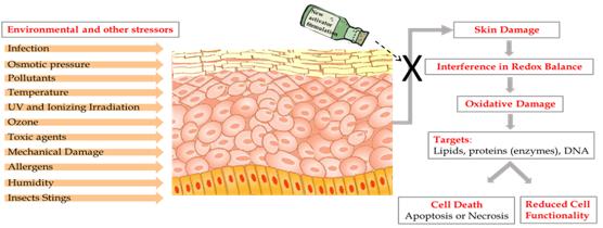
The human skin is constantly exposed to various pollutants, UV rays, radiation and other stressors that exist in our day-to-day environment. When they filter into the body they can create Reactive Oxygen Species (ROS) – oxygen molecules known as Free Radicals, which are able to damage and destroy cells, including lipids, proteins and DNA.

In the skin – the largest organ of the body – an excess of ROS can lead to various skin conditions, including inflammatory diseases, pigmenting disorders, wrinkles and some types of skin cancer, and can also affect internal organs. This damage is known as Oxidative Stress.

The body is naturally equipped with defense mechanisms to counter oxidative stress. It has anti-oxidants and, more importantly, anti-oxidant enzymes that attack the ROS before they cause damage.

In a review article published in the journal Cosmetics, a PhD student from The Hebrew University of Jerusalem, working in collaboration with researchers at the Technion – Israel Institute of Technology, suggested an innovative way to invigorate the body to produce antioxidant enzymes, while maintaining skin cell redox balance – a gentle equilibrium between Reactive Oxygen Species and their detoxification.

“The approach of using the body’s own defense system is very effective. We showed that activation of the body’s defense system with the aid of a unique delivery system is feasible, and may leverage dermal cure,” said Hebrew University researcher Maya Ben-Yehuda Greenwald.

Ben-Yehuda Greenwald showed that applying nano-size droplets of microemulsion liquids containing a cellular protective pathway inducer into the skin activates the natural skin defense systems.

“Currently, there are many scientific studies supporting the activation of the body’s defense mechanisms. However, none of these studies has demonstrated the use of a nanotechnology-based delivery system to do so,” Ben-Yehuda Greenwald said.

Production of antioxidant enzymes in the body is signaled in the DNA by activation of Nrf2 – a powerful protein that exists in every cell in our body. This Nrf2 cellular-protective signaling pathway is a major intersection of many other signaling pathways affecting each other and determining cell functionality and fate. Nrf2 is capable of coordinating the cellular response to internal as well as external stressors by tight regulation of phase-II protective enzymes, such as the antioxidant enzymes.

Ben-Yehuda Greenwald has also discovered a new family of compounds capable of activating the Nrf2 pathway. Moreover, by incorporating them into the unique delivery system she has developed, she managed to efficiently stimulate the activation of the Nrf2 pathway and mimic the activity of the body’s’ natural way of coping with a variety of stress conditions.

“The formula we have created could be used in topical medication for treating skin conditions. Our formula could be used both as preventive means and for treatment of various skin conditions, such as infections, over-exposure to UV irradiation, inflammatory conditions, and also internal disease,” she said.

While the researchers focused on the skin, the formulation could prove to be effective in enhancing the body’s natural protection against the damaging effects of ROS in other parts of the body, such as inflammation in cardiovascular diseases, heart attack, cancer, multiple sclerosis and Alzheimer’s.

Here’s an image provided by Ben-Yehuda Greenwald illustrating the team’s work,

Caption: These are the consequences of skin exposure to stressors. Credit: Maya Ben-Yehuda Greenwald

Caption: These are the consequences of skin exposure to stressors. Credit: Maya Ben-Yehuda Greenwald

Here’s a link to and a citation for the paper,

Skin Redox Balance Maintenance: The Need for an Nrf2-Activator Delivery System by Maya Ben-Yehuda Greenwald, Shmuel Ben-Sasson, Havazelet Bianco-Peled, and Ron Kohen. Cosmetics 2016, 3(1), 1; doi:10.3390/cosmetics3010001 Published: 15 January 2016

This paper appears to be open access.
Proteins which cause Alzheimer’s disease can be used to grow functionalized nanowires

This is the first time I’ve ever heard of anything good resulting from Alzheimer’s Disease (even if it’s tangential). From the May 24, 2013 news item on ScienceDaily,

Prof. Sakaguchi and his team in Graduate School of Science, Hokkaido University,jointly with MANA PI Prof. Kohei Uosaki and a research group from the University of California, Santa Barbara, have successfully developed a new technique for efficiently creating functionalized nanowires for the first time ever.

The group focused on the natural propensity of amyloid peptides, molecules which are thought to cause Alzheimer’s disease, to self-assemble into nanowires in an aqueous solution and controlled this molecular property to achieve their feat.

The May 23, 2013 National Institute for Materials press release, which originated the news item, offers insight into why functionalized nanowires are devoutly desired,

Functionalized nanowires are extremely important in the construction of nanodevices because they hold promise for use as integrated circuits and for the generation of novel properties, such as conductivity, catalysts and optical properties which are derived from their fine structure. However, some have remarked on the technical and financial limitations of the microfabrication technology required to create these structures. Meanwhile, molecular self-organization and functionalization have attracted attention in the field of next-generation nanotechnology development. Amyloid peptides, which are thought to cause Alzheimer’s disease, possess the ability to self-assemble into highly stable nanowires in an aqueous solution. Focusing on this, the research team became the first to successfully develop a new method for efficiently creating a multifunctional nanowire by controlling this molecular property.

The team designed a new peptide called SCAP, or structure-controllable amyloid peptide, terminated with a three-amino-acid-residue cap. By combining multiple SCAPs with different caps, the team found that self-organization is highly controlled at the molecular level. Using this new control method, the team formed a molecular nanowire with the largest aspect ratio ever achieved. In addition, they made modifications using various functional molecules including metals, semiconductors and biomolecules that successfully produced an extremely high quality functionalized nanowire. Going forward, this method is expected to contribute significantly to the development of new nanodevices through its application to a wide range of functional nanomaterials with self-organizing properties.

You can find the published paper here,

Formation of Functionalized Nanowires by Control of Self-Assembly Using Multiple Modified Amyloid Peptides by Hiroki Sakai, Ken Watanabe, Yuya Asanomi, Yumiko Kobayashi, Yoshiro Chuman, Lihong Shi, Takuya Masuda, Thomas Wyttenbach, Michael T. Bowers, Kohei Uosaki, & Kazuyasu Sakaguchi1. Advanced Functional Materials. doi: 10.1002/adfm.201300577 Article first published online: 23 APR 2013

The study is behind a paywall.

I have written about nanowires before and, in keeping with today’s theme of peculiar relationships  (Alzheimer’s disease), prior to this, the most unusual nanowire item I’ve come across had to do with growing them to the sounds  of music. From the Nanotech Mysteries (wiki), Scientists get musical page (Note: Footnotes have been removed),

After testing Deep Purple’s ‘Smoke on the Water‘, Chopin’s ‘Nocturne Opus 9 no. 1‘, Josh Abraham’s ‘Addicted to Bass‘, Rammstein’s ‘Das Model‘, and Abba’s ‘Dancing Queen‘, David Parlevliet found that music can be used to grow nanowires but they will be kinky.

Scientists want to grow straight nanowires and one of the popular methods is to “[blast] a voltage through silane gas to produce a plasma that pulses on and off at 1000 times a second. Over time the process enables molecules from the gas to deposit on a glass slide in the form of a mesh of crystalline silicon nanowires.”

Parlevliet, a PhD student at Murdoch University in Perth, Australia, plugged in a music player instead of a pulse generator usually used for this purpose and observed the results. While there are no current applications for kinky nanowires, the Deep Purple music created the densest mesh. Rammstein’s music grew nanowires the least successfully. In his presentation to the Australian Research Council Nanotechnology Network Symposium in March 2008, Parlevliet concluded that music could become more important for growing nanowires if applications can be found for the kinky ones.

Self-assembling protein inspires University of Montreal’s researchers to smaller efforts

Protein folding doesn’t seem all that exciting to me and the notion that it might lead to self-assembled, living machines isn’t all that new (see my May 31, 2012 posting about a Living Foundries project). So the June 10, 2012 news item on Nanowerk left me with a flat feeling, initially,

Enabling bioengineers to design new molecular machines for nanotechnology applications is one of the possible outcomes of a study by University of Montreal researchers that was published in Nature Structural and Molecular Biology today (“Visualizing transient protein folding intermediates by tryptophan scanning mutagenesis” [behind a paywall]). The scientists have developed a new approach to visualize how proteins assemble, which may also significantly aid our understanding of diseases such as Alzheimer’s and Parkinson’s, which are caused by errors in assembly.

“In order to survive, all creatures, from bacteria to humans, monitor and transform their environments using small protein nanomachines made of thousands of atoms,” explained the senior author of the study, Prof. Stephen Michnick of the university’s department of biochemistry. “For example, in our sinuses, there are complex receptor proteins that are activated in the presence of different odor molecules. Some of those scents warn us of danger; others tell us that food is nearby.” Proteins are made of long linear chains of amino acids, which have evolved over millions of years to self-assemble extremely rapidly – often within thousandths of a split second – into a working nanomachine.

My ears pricked up when the talk turned to capturing images of action, which occurs in a “fleetingly short time,”

“To understand how a protein goes from a linear chain to a unique assembled structure, we need to capture snapshots of its shape at each stage of assembly said Dr. Alexis Vallée-Bélisle, first author of the study. “The problem is that each step exists for a fleetingly short time and no available technique enables us to obtain precise structural information on these states within such a small time frame. We developed a strategy to monitor protein assembly by integrating fluorescent probes throughout the linear protein chain so that we could detect the structure of each stage of protein assembly, step by step to its final structure.” The protein assembly process is not the end of its journey, as a protein can change, through chemical modifications or with age, to take on different forms and functions. “Understanding how a protein goes from being one thing to becoming another is the first step towards understanding and designing protein nanomachines for biotechnologies such as medical and environmental diagnostic sensors, drug synthesis or delivery,” Vallée-Bélisle said.

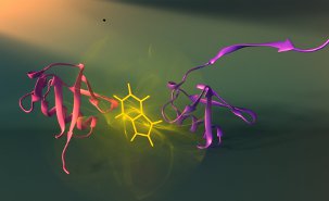
Here’s an image of protein self-assembly from the University of Montreal (Université de Montréal) website (Montréal, Québec, Canada),

Vallée-Bélisle and Michnick have developed a new approach to visualize how proteins assemble, which may also significantly aid our understanding of diseases such as Alzheimer's and Parkinson's, which are caused by errors in assembly. Here shown are two different assembly stages (purple and red) of the protein ubiquitin and the fluorescent probe used to visualize these stage (tryptophan: see yellow). Credit: Peter Allen

I would have liked a little more detail (e.g. how little time is there to capture the images?) but there isn’t always time either for the people who write these news releases or for me to follow up with questions. Given the huge political unrest amongst students over the proposed tuition fees and the Québec government’s attempts (sometimes described as draconian) to impose order, I’m impressed this news release was pulled together.