Tag Archives: Au nanoparticles

Measure the maturity of cask-aged whisky with gold nanoparticle test

it’s not altogether a surprise that this research on whisky has Scottish origins. From an October 11, 2022 news item on Nanowerk (Note: A link has been removed),

Researchers at a Scottish university have found a way to use gold nanoparticles to measure the maturity of whisky, which could help distillers with one of the key challenges in the production process (ACS Applied Nano Materials, “Growth of Plasmonic Nanoparticles for Aging Cask-Matured Whisky”).

Chemists and bioscientists from the University of Glasgow developed the test, which harnesses a unique property of cask-aged whisky to measure its maturity.

An October 11, 2022 University of Glasgow press release, which originated the news item, delves further into the work,

Each variety of whisky gains some of its colour flavour profile from being stored in wooden casks while it matures over a period of months or years. The flavour of the final product is created by a complex mix of factors known as ‘congeners’ – chemicals left in the spirit after it is distilled and other chemicals absorbed from the wood casks, which react with oxygen over time.

The unpredictable interactions of congeners, along with other factors like the size and shape of the cask and the number of times it has been used before, mean that each cask matures in its own way, and in its own time.

To ensure the consistency of their products, distillers employ highly experienced master blenders. They regularly sample the casks to check the whisky’s readiness for blending, bottling and sale as either a single malt or a mixed blend – a laborious and expensive task.

The researchers set out to develop a test which could do some of the work of the master blenders by using chemical reactions to determine the maturity or ‘age’ of whisky samples.

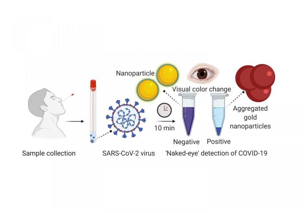
They built their test on a reaction which occurs when samples of whisky are mixed with a solution containing small quantities of a special type of gold. A chemical reaction in the whisky causes distinctively-coloured gold nanoparticles to form in the sample over a short period of time at room temperature.

The researchers mixed the gold solution with samples from 15 different whiskies distilled in Scotland, Japan and the US. They also tested multiple samples taken at regular intervals from a single cask over a period of six years, which were supplied by the Scotch Whisky Research Institute.

By measuring a property of each sample known as its localised surface plasmon resonance, they found that the unique chemical composition of the whiskies resulted in the creation of gold nanoparticles with distinctly different shapes, sizes and colours in each sample.

They also discovered that the speed of the production of the nanoparticles was connected with its maturity – the faster the nanoparticles formed, the more mature the whisky was.

The results suggest that the process could be used to develop a quick, reliable test for distillers to measure the maturity of their whiskies, reducing the need for master blenders to be involved in every step of the process.

Dr Will Peveler, of the University of Glasgow’s School of Chemistry, is the paper’s lead author. Dr Peveler said: “Age is more than just a number when it comes to whisky – the complex chemical reactions which occur in each cask make it impossible to estimate whisky’s maturity of flavour simply based on how long it’s been ageing.

“For as long as there’s been a whisky industry, distillers have been trying to find better ways to measure the maturity of individual casks to help them understand when they will be ready to use in a single malt or a mixed blend.

“What we’ve been able to do for the first time is show that the ageing-related chemistry of the whisky controls the formation of gold nanoparticles. That has allowed us to develop a unique ‘fingerprint’ not just for types of whisky we tested but also for how whiskies mature over time.

Co-author Dr Jenny Gracie, also of the School of Chemistry, added: “Currently, there are a number of other tests available to measure whisky maturity, which use specialist processes like chromatography and mass spectrometry. However, they are rarely available on the warehouse floor, and if samples have to be sent offsite for analysis, this slows everything down.

“We hope that in the future we can develop this initial finding into a quick, easy and portable kit that distillers can use to measure the maturity of their whiskies without having to send samples for time-consuming tests with specialist equipment.”

Here’s a link to and a citation for the paper,

Growth of Plasmonic Nanoparticles for Aging Cask-Matured Whisky by Jennifer Gracie, Francesco Zamberlan, Iain B. Andrews, Brian O. Smith, and William J. Peveler. ACS Appl. Nano Mater. 2022, XXXX, XXX, XXX-XXX DOI: https://doi.org/10.1021/acsanm.2c03406 Publication Date: October 6, 2022 © 2022 The Authors. Published by American Chemical Society

This paper appears to be open access.

Using natural proteins to grow gold nanoclusters for hybrid bionanomaterials

While there’s a January 10, 2022 news item on Nanowerk, the research being announced was made available online in the Fall of 2021 and is now available in print,

Gold nanoclusters are groups of a few gold atoms with interesting photoluminescent properties. The features of gold nanoclusters depend not only on their structure, but their size and also by the ligands coordinated to them. These inorganic nanomaterials have been used in sensing, biomedicine and optics and their coordination with biomolecules can endow multiple capabilities in biological media.

A research collaboration between the groups of Dr. Juan Cabanillas, Research Professor at IMDEA Nanociencia and Dr. Aitziber L. Cortajarena, Ikerbasque Professor and Principal Investigator at CIC biomaGUNE have explored the use of natural proteins to grow gold nanoclusters, resulting in hybrid bionanomaterials with tunable photoluminescent properties and with a plethora of potential applications.

A January 10, 2022 IMDEA Nanociencia press release, which originated the news item, provides more technical detail about the research,

The nanoclusters –with less than 2 nm in size- differentiate from larger nanoparticles (plasmonic) since they present discrete energy levels coupled optically. The groups of amino acids within the proteins coordinate the gold atoms and allow the groups to be arranged around the gold nanocluster, facilitating the stabilization and adding an extra level of tailoring. These nanoclusters have interesting energy harvesting features. Since the discrete energy levels are optically coupled, the absorption of a photon leads to promotion of an electron to higher levels, which can trigger a photophysical process or a photochemical reaction.  

The results by Cabanillas and Cortajarena groups, published in Advanced Optical Materials and Nano Letters, explore the origin of the photoluminescence in protein-designed gold nanoclusters and shed light into the strong influence of environmental conditions on the nature of luminescence. Nanocluster capping by two types of amino acids (histidine and cysteine) allow for changing the emission spectral range from blue to red, paving the way to tune the optical properties by an appropriate ligand choice. The nature of emission is also changed with capping, from fluorescence to phosphorescence, respectively. The synergistic protein-nanocluster effects on emission are still not clear, and the groups at IMDEA Nanociencia and CIC biomaGUNE are working to elucidate the mechanisms behind. There are potential applications for the aforementioned nanoclusters, in solid state as active medium in laser cavities. Optical gain properties from these nanoclusters are yet to be demonstrated, which could pave the way to a new generation of potentially interesting laser devices. As the combination of gold plus proteins is potentially biocompatible, many potential applications in biomedicine can also be envisaged.

A related publication of the groups in Nano Letters demonstrates that the insertion of tryptophans, amino acids with high electron density, in the vicinity of the nanocluster boosts its photoluminescence quantum efficiency up to 40% in some cases, values relevant for solid state light emission applications. Researchers also observed an antenna effect: the tryptophans can absorb light in a discrete manner and transfer the energy to the cluster. This effect has interest for energy harvesting and for sensing purposes as well.

The proteins through the biocapping enable the synthesis of the nanoclusters and largely improve their quantum efficiency. “The photoluminescence quantum efficiency is largely improved when using the biocapping” Dr. Cabanillas says. He believes this research work means “a new field opening for the tuning of optical properties of nanoclusters through protein engineering, and much work is ahead for the understanding of the amplification mechanism”. Dr. Cortajarena emphasizes “we have already demonstrated the great potential of engineered photoluminescent protein-nanocluster in biomedical and technological fields, and understanding the fundamental emission mechanisms is pivotal for future applications“. A variety of further applications include biosensors, as the protein admits functionalization with recognition molecules, energy harvesting, imaging and photodynamic therapies. Further work is ahead this opening avenue for photophysics research.

This research is a collaboration led by Dr. Juan Cabanillas and Dr. Aitziber L. Cortajarena research groups at IMDEA Nanociencia and CIC biomaGUNE, with contributions from researchers at the Diamond Light Source Ltd. [synchrotron] and DIPC. It has been cofounded by the projects AMAPOLA, NMAT2D, FULMATEN, Atracción de Talento from Comunidad de Madrid and the Severo Ochoa Centre of Excellence award to IMDEA Nanociencia. CIC biomaGUNE acknowledges support by the projects ERC-ProNANO, ERC-NIMM, ProTOOLs and the Maria de Maeztu Units of Excellence Programme.

Here are links to and citations for the papers,

Tuning the Optical Properties of Au Nanoclusters by Designed Proteins by Elena Lopez-Martinez, Diego Gianolio, Saül Garcia-Orrit, Victor Vega-Mayoral, Juan Cabanillas-Gonzalez, Carlos Sanchez-Cano, Aitziber L. Cortajarena. Advanced Optical Materials Volume 10, Issue 1 January 4, 2022 2101332 DOI: https://doi.org/10.1002/adom.202101332 First published: 31 October 2021

This paper is open access.

Boosting the Photoluminescent Properties of Protein-Stabilized Gold Nanoclusters through Protein Engineering by Antonio Aires, Ahmad Sousaraei, Marco Möller, Juan Cabanillas-Gonzalez, and Aitziber L. Cortajarena. Nano Lett. 2021, 21, 21, 9347–9353 DOI: https://doi.org/10.1021/acs.nanolett.1c03768 Publication Date: November 1, 2021 Copyright © 2021 American Chemical Society

This paper is behind a paywall.

Not being familiar with either of the two research institutions mentioned in the press release, I did a little digging.

Here’s a little information about IMDEA Nanociencia (IMDEA Nanoscience Institute), from its Wikipedia entry, Note: All links have been removed,

IMDEA Nanoscience Institute is a private non-profit foundation within the IMDEA Institutes network, created in 2006-2007 as a result of collaboration agreement between the Community of Madrid and Spanish Ministry of Education and Science. The foundation manages IMDEA-Nanoscience Institute,[1] a scientific centre dedicated to front-line research in nanoscience, nanotechnology and molecular design and aiming at transferable innovations and close contact with industries. IMDEA Nanoscience is a member of the Campus of International excellence, a consortium of research institutes promoted by the Autonomous University of Madrid and Spanish National Research Council (UAM/CSIC).[2]

As for CIC biomaGUNE, here’s more from its institutional profile on the science.eus website,

The Centre for Cooperative Research in Biomaterials-CIC biomaGUNE, located in San Sebastian (Spain), was officially opened in December 2006. CIC biomaGUNE is a non-profit research organization created to promote scientific research and technological innovation at the highest levels in the Basque Country following the BioBasque policy in order to create a new business sector based on biosciences. Established by the Department of Industry, Technology & Innovation of the Government of the Autonomous Community of the Basque Country, CIC biomaGUNE constitutes one of the Centres of the CIC network, the largest Basque Country research network on specific strategic areas, having the mission to contribute to the economical and social development of the country through the generation of knowledge and speeding up the process that leads to technological innovation.

‘Playing telephone’ with multivalent gold nanoparticles

A July 7, 2021 news item on phys.org describes what ‘playing telephone’ has to do with gold nanoparticles,

Cells play a precise game of telephone, sending messages to each other that trigger actions further on. With clear signaling, the cells achieve their goals. In disease, however, the signals break up and result in confused messaging and unintended consequences. To help parse out these signals and how they function in health—and go awry in disease—scientists tag proteins with labels they can follow as the proteins interact with the molecular world around them.

The challenge is figuring out which proteins to label in the first place. Now, a team led by researchers from Tokyo University of Agriculture and Technology (TUAT) has developed a new approach to identifying and tagging the specific proteins. They published their results on June 1 [2021] in Angewandte Chemie.

A July 8, 2021 TUAT press release on EurekAlert, which originated the news item, delves further into the research (I appreciate how clearly the work is explained),

“We are interested in exploring protein receptors of certain carbohydrate molecules that are involved in mediating cell signaling, particularly in cancer cells,” said paper author Kaori Sakurai, associate professor in the Department of Biotechnology and Life Science at TUAT.

The carbohydrate molecules, called ligands, are typically expressed on the surface of cells and are known to dynamically form complexes with protein receptors to coordinate complicated cellular functions. However, Sakurai said, the proteins responsible for binding the carbohydrates have been difficult to identify because they bond so weakly with the molecules.

The researchers designed a new type of carbohydrate probe that would not only link to the molecules, but tightly bind to them.

“We used gold nanoparticles as a scaffold to attach both carbohydrate ligands and electrophiles — a chemical that loves to react with other molecules — in a multivalent fashion,” Sakurai said. “This way, we were able to greatly increase binding affinity and reaction efficiency toward carbohydrate-binding proteins.”

The researchers applied the designed probes to cell lysate, a fluid containing the innards of broken-apart cells.

“The probes quickly found the target carbohydrate-binding proteins, triggering the electrophilic groups to react with electron-donating amino acid residues on nearby proteins,” Sakurai said. “This resulted in proteins firmly cross-linked to the gold nanoparticles’ surface, making it easy to subsequently analyze their identities.”

The team evaluated several electrophilic groups to identify the most efficient type for labeling their target proteins.

“We found that a particular electrophilic group called aryl sulfonyl fluoride is best suited for affinity labeling of carbohydrate-binding proteins,” said co-author Nanako Suto, a graduate student in the Department of Biotechnology and Life Science of TUAT. “However, they have rarely been used to identify target proteins, presumably because they would non-selectively react with various other, undesired proteins.”

However, the scale of aryl sulfonyl fluoride use appears to mitigate the issue.

“The non-selectivity isn’t a problem if aryl sulfonyl fluoride is used at very low concentrations, at the range of the nanoscale,” said co-author Shione Kamoshita, also a graduate student in the Department of Biotechnology and Life Science, TUAT.

The gold nanoparticle scaffolding displays many copies of the electrophilic group, which keeps aryl sulfonyl fluoride’s local concentration high on the nanoparticle surface but restrains them from the general cell system and reacting to undesired proteins. With the high concentration at the nano-level, some copies of electrophilic groups can efficiently react with target proteins.

“Through this process, we were able to achieve highly efficient and selective affinity labeling of carbohydrate-binding proteins in cell lysate,” Sakurai said. “We will apply the new method in target identification of several cancer-related carbohydrate ligands and investigate their function in cancer development. In parallel, we aim to explore the general utility of this new probe design for various other bioactive small molecules, so that we can accelerate the elucidation of their mechanisms.”

Here’s a link to and a citation for the paper,

Exploration of the Reactivity of Multivalent Electrophiles for Affinity Labeling: Sulfonyl Fluoride as a Highly Efficient and Selective Label by Nanako Suto, Shione Kamoshita, Dr. Shoichi Hosoya, Prof. Kaori Sakurai. Angewandte Chemie Volume 60, Issue 31 July 26, 2021 Pages 17080-17087 DOI: https://doi.org/10.1002/anie.202104347 First published: 01 June 2021

This paper is behind a paywall.

Put a ring on it: preventing clumps of gold nanoparticles

Caption: A comparison of how linear PEG (left) and cyclic PEG (right) attach to a gold nanoparticle Credit: Yubo Wang, Takuya Yamamoto

A January 20, 2021 news item on phys.org focuses on work designed to stop gold nanoparticles from clumping together (Note: A link has been removed),

Hokkaido University scientists have found a way to prevent gold nanoparticles from clumping, which could help towards their use as an anti-cancer therapy.

Attaching ring-shaped synthetic compounds to gold nanoparticles helps them retain their essential light-absorbing properties, Hokkaido University researchers report in the journal Nature Communications.

A January 20, 2021 Hokkaido University press release (also on EurekAlert but published Jan. 21, 2020), which originated the news item, elaborates on the work,

Metal nanoparticles have unique light-absorbing properties, making them interesting for a wide range of optical, electronic and biomedical applications. For example, if delivered to a tumour, they could react with applied light to kill cancerous tissue. A problem with this approach, though, is that they easily clump together in solution, losing their ability to absorb light. This clumping happens in response to a variety of factors, including temperature, salt concentration and acidity.

Scientists have been trying to find ways to ensure nanoparticles stay dispersed in their target environments. Covering them with polyethylene glycol, otherwise known as PEG, has been relatively successful at this in the case of gold nanoparticles. PEG is biocompatible and can prevent gold surfaces from clumping together in the laboratory and in living organisms, but improvements are still needed.

Applied chemist Takuya Yamamoto and colleagues at Hokkaido University, The University of Tokyo, and Tokyo Institute of Technology found that mixing gold nanoparticles with ring-shaped PEG, rather than the normally linear PEG, significantly improved dispersion. The ‘cyclic-PEG’ (c-PEG) attaches to the surfaces of the nanoparticles without forming chemical bonds with them, a process called physisorption. The coated nanoparticles remained dispersed when frozen, freeze-dried and heated.

The team tested the c-PEG-covered gold nanoparticles in mice and found that they cleared slowly from the blood and accumulated better in tumours compared to gold nanoparticles coated with linear PEG. However, accumulation was lower than desired levels, so the researchers recommend further investigations to fine-tune the nanoparticles for this purpose.

Associate Professor Takuya Yamamoto is part of the Laboratory of Chemistry of Molecular Assemblies at Hokkaido University, where he studies the properties and applications of various cyclic chemical compounds.

Here’s a link to and a citation for the paper,

Enhanced dispersion stability of gold nanoparticles by the physisorption of cyclic poly(ethylene glycol) by Yubo Wang, Jose Enrico Q. Quinsaat, Tomoko Ono, Masatoshi Maeki, Manabu Tokeshi, Takuya Isono, Kenji Tajima, Toshifumi Satoh, Shin-ichiro Sato, Yutaka Miura & Takuya Yamamoto. Nature Communications volume 11, Article number: 6089 (2020) DOI: https://doi.org/10.1038/s41467-020-19947-8 Published: 30 November 2020

This paper is open access.

Gold nanoparticles could help detect the presence of COVID-19 in ten minutes

If this works out, it would make testing for COVID-19 an infinitely easier task. From a May 29, 2020 news item on phys.org,

Scientists from the University of Maryland School of Medicine (UMSOM) developed an experimental diagnostic test for COVID-19 that can visually detect the presence of the virus in 10 minutes. It uses a simple assay containing plasmonic gold nanoparticles to detect a color change when the virus is present. The test does not require the use of any advanced laboratory techniques, such as those commonly used to amplify DNA, for analysis. The authors published their work last week [May 21, 2020] in the American Chemical Society’s nanotechnology journal ACS Nano.

“Based on our preliminary results, we believe this promising new test may detect RNA [ribonucleic acid] material from the virus as early as the first day of infection. Additional studies are needed, however, to confirm whether this is indeed the case,” said study leader Dipanjan Pan, PhD, Professor of Diagnostic Radiology and Nuclear Medicine and Pediatrics at the UMSOM.

Caption: A nasal swab containing a test sample is mixed with a simple lab test. It contains a liquid mixed with gold nanoparticles attached to a molecule that binds to the novel coronavirus. If the virus is present, the gold nanoparticles turns the solution a deep blue color (bottom of the tube) and a precipitation is noticed. If it is not present, the solution retains its original purple color. Credit: University of Maryland School of Medicine

A May 28, 2020 University of Maryland news release (also on EurekAlert), which originated the news item, provides more detail,

Once a nasal swab or saliva sample is obtained from a patient, the RNA is extracted from the sample via a simple process that takes about 10 minutes. The test uses a highly specific molecule attached to the gold nanoparticles to detect a particular protein. This protein is part of the genetic sequence that is unique to the novel coronavirus. When the biosensor binds to the virus’s gene sequence, the gold nanoparticles respond by turning the liquid reagent from purple to blue.

“The accuracy of any COVID-19 test is based on being able to reliably detect any virus. This means it does not give a false negative result if the virus actually is present, nor a false positive result if the virus is not present,” said Dr. Pan. “Many of the diagnostic tests currently on the market cannot detect the virus until several days after infection. For this reason, they have a significant rate of false negative results.”

Dr. Pan created a company called VitruVian Bio to develop the test for commercial application. He plans to have a pre-submission meeting with the U.S. Food and Drug Administration (FDA) within the next month to discuss requirements for getting an emergency use authorization for the test. New FDA policy allows for the marketing of COVID-19 tests without requiring them to go through the usual approval or clearance process. These tests do, however, need to meet certain validation testing requirements to ensure that they provide reliable results.

“This RNA-based test appears to be very promising in terms of detecting the virus. The innovative approach provides results without the need for a sophisticated laboratory facility,” said study co-author Matthew Frieman, PhD, Associate Professor of Microbiology and Immunology at UMSOM.

Although more clinical studies are warranted, this test could be far less expensive to produce and process than a standard COVID-19 lab test; it does not require laboratory equipment or trained personnel to run the test and analyze the results. If this new test meets FDA expectations, it could potentially be used in daycare centers, nursing homes, college campuses, and work places as a surveillance technique to monitor any resurgence of infections.

In Dr. Pan’s laboratory, research scientist Parikshit Moitra, PhD, and UMSOM research fellow Maha Alafeef conducted the studies along with research fellow Ketan Dighe from UMBC.

Dr. Pan holds a joint appointment with the College of Engineering at the University of Maryland Baltimore County and is also a faculty member of the Center for Blood Oxygen Transport and Hemostasis (CBOTH).

“This is another example of how our faculty is driving innovation to fulfill a vital need to expand the capacity of COVID-19 testing,” said Dean E. Albert Reece, MD, PhD, MBA, who is also Executive Vice President for Medical Affairs, UM Baltimore, and the John Z. and Akiko K. Bowers Distinguished Professor, University of Maryland School of Medicine. “Our nation will be relying on inexpensive, rapid tests that can be dispersed widely and used often until we have effective vaccines against this pandemic.”

Here’s a link to and a citation for the paper,

Selective Naked-Eye Detection of SARS-CoV-2 Mediated by N Gene Targeted Antisense Oligonucleotide Capped Plasmonic Nanoparticles by Parikshit Moitra, Maha Alafeef, Ketan Dighe, Matthew B. Frieman, and Dipanjan Pan. ACS Nano 2020, XXXX, XXX, XXX-XXX DOI: https://doi.org/10.1021/acsnano.0c03822 Publication Date:May 21, 2020 Copyright © 2020 American Chemical Society

This paper appears to be open access.

I tried to find Dr. Pan’s company, VitruVian Bio and found a business with an almost identical name, Vitruvian Biomedical, which does not include Dr. Pan on its management team list and this company’s focus is on Alzheimer’s Disease. Finally, there is no mention of the COVID-19 test anywhere on the Vitruvian Biomedical website.

Non-viral ocular gene therapy with gold nanoparticles and femtosecond lasers

I love the stylistic choice the writer made (pay special attention to the second paragraph) when producing this November 19, 2018 Polytechnique Montréal news release (also on EurekAlert),

A scientific breakthrough by Professor Michel Meunier of Polytechnique Montréal and his collaborators offers hope for people with glaucoma, retinitis or macular degeneration.

In January 2009, the life of engineer Michel Meunier, a professor at Polytechnique Montréal, changed dramatically. Like others, he had observed that the extremely short pulse of a femtosecond laser (0.000000000000001 second) could make nanometre-sized holes appear in silicon when it was covered by gold nanoparticles. But this researcher, recognized internationally for his skills in laser and nanotechnology, decided to go a step further with what was then just a laboratory curiosity. He wondered if it was possible to go from silicon to living matter, from inorganic to organic. Could the gold nanoparticles and the femtosecond laser, this “light scalpel,” reproduce the same phenomenon with living cells?

A very pretty image illustrating the work,

Caption: Gold nanoparticles, which act like “nanolenses,” concentrate the energy produced by the extremely short pulse of a femtosecond laser to create a nanoscale incision on the surface of the eye’s retina cells. This technology, which preserves cell integrity, can be used to effectively inject drugs or genes into specific areas of the eye, offering new hope to people with glaucoma, retinitis or macular degeneration. Credit and Copyright: Polytechnique Montréal

The news release goes on to describe the technology in more detail,

Professor Meunier started working on cells in vitro in his Polytechnique laboratory. The challenge was to make a nanometric incision in the cells’ extracellular membrane without damaging it. Using gold nanoparticles that acted as “nanolenses,” Professor Meunier realized that it was possible to concentrate the light energy coming from the laser at a wavelength of 800 nanometres. Since there is very little energy absorption by the cells at this wavelength, their integrity is preserved. Mission accomplished!

Based on this finding, Professor Meunier decided to work on cells in vivo, cells that are part of a complex living cell structure, such as the eye for example.

The eye and the light scalpel

In April 2012, Professor Meunier met Przemyslaw Sapieha, an internationally renowned eye specialist, particularly recognized for his work on the retina. “Mike”, as he goes by, is a professor in the Department of Ophthalmology at Université de Montréal and a researcher at Centre intégré universitaire de santé et de services sociaux (CIUSSS) de l’Est-de-l’Île-de-Montréal. He immediately saw the potential of this new technology and everything that could be done in the eye if you could block the ripple effect that occurs following a trigger that leads to glaucoma or macular degeneration, for example, by injecting drugs, proteins or even genes.

Using a femtosecond laser to treat the eye–a highly specialized and fragile organ–is very complex, however. The eye is part of the central nervous system, and therefore many of the cells or families of cells that compose it are neurons. And when a neuron dies, it does not regenerate like other cells do. Mike Sapieha’s first task was therefore to ensure that a femtosecond laser could be used on one or several neurons without affecting them. This is what is referred to as “proof of concept.”

Proof of concept

Mike and Michel called on biochemistry researcher Ariel Wilson, an expert in eye structures and vision mechanisms, as well as Professor Santiago Costantino and his team from the Department of Ophthalmology at Université de Montréal and the CIUSSS de l’Est-de-l’Île-de-Montréal for their expertise in biophotonics. The team first decided to work on healthy cells, because they are better understood than sick cells. They injected gold nanoparticles combined with antibodies to target specific neuronal cells in the eye, and then waited for the nanoparticles to settle around the various neurons or families of neurons, such as the retina. Following the bright flash generated by the femtosecond laser, the expected phenomenon occurred: small holes appeared in the cells of the eye’s retina, making it possible to effectively inject drugs or genes in specific areas of the eye. It was another victory for Michel Meunier and his collaborators, with these conclusive results now opening the path to new treatments.

The key feature of the technology developed by the researchers from Polytechnique and CIUSSS de l’Est-de-l’Île-de-Montréal is its extreme precision. With the use of functionalized gold nanoparticles, the light scalpel makes it possible to precisely locate the family of cells where the doctor will have to intervene.

Having successfully demonstrated proof of concept, Professor Meunier and his team filed a patent application in the United States. This tremendous work was also the subject of a paper reviewed by an impressive reading committee and published in the renowned journal Nano Letters in October 2018.

While there is still a lot of research to be done–at least 10 years’ worth, first on animals and then on humans–this technology could make all the difference in an aging population suffering from eye deterioration for which there are still no effective long-term treatments. It also has the advantage of avoiding the use of viruses commonly employed in gene therapy. These researchers are looking at applications of this technology in all eye diseases, but more particularly in glaucoma, retinitis and macular degeneration.

This light scalpel is unprecedented.

Here’s a link to and a citation for the paper,

In Vivo Laser-Mediated Retinal Ganglion Cell Optoporation Using KV1.1 Conjugated Gold Nanoparticles by Ariel M. Wilson, Javier Mazzaferri, Éric Bergeron, Sergiy Patskovsky, Paule Marcoux-Valiquette, Santiago Costantino, Przemyslaw Sapieha, Michel Meunier. Nano Lett.201818116981-6988 DOI: https://doi.org/10.1021/acs.nanolett.8b02896 Publication Date: October 4, 2018  Copyright © 2018 American Chemical Society

This paper is behind a paywall.