Tag Archives: bacteria

A 3D printed ‘living’ tattoo

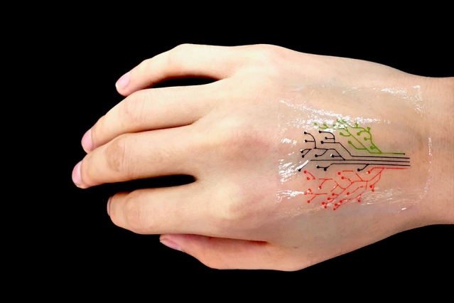
MIT engineers have devised a 3-D printing technique that uses a new kind of ink made from genetically programmed living cells. Courtesy of the researchers [and MIT]

If that image isn’t enough, there’s also a video abstract (I don’t think I’ve seen one of these before) for the paper,

For those who’d still like to read the text, here’s more from a December 5, 2017 MIT (Massachusetts Institute of Technology) news release (also on EurekAlert),

MIT engineers have devised a 3-D printing technique that uses a new kind of ink made from genetically programmed living cells.

The cells are engineered to light up in response to a variety of stimuli. When mixed with a slurry of hydrogel and nutrients, the cells can be printed, layer by layer, to form three-dimensional, interactive structures and devices.

The team has then demonstrated its technique by printing a “living tattoo” — a thin, transparent patch patterned with live bacteria cells in the shape of a tree. Each branch of the tree is lined with cells sensitive to a different chemical or molecular compound. When the patch is adhered to skin that has been exposed to the same compounds, corresponding regions of the tree light up in response.

The researchers, led by Xuanhe Zhao, the Noyce Career Development Professor in MIT’s Department of Mechanical Engineering, and Timothy Lu, associate professor of biological engineering and of electrical engineering and computer science, say that their technique can be used to fabricate “active” materials for wearable sensors and interactive displays. Such materials can be patterned with live cells engineered to sense environmental chemicals and pollutants as well as changes in pH and temperature.

What’s more, the team developed a model to predict the interactions between cells within a given 3-D-printed structure, under a variety of conditions. The team says researchers can use the model as a guide in designing responsive living materials.

Zhao, Lu, and their colleagues have published their results today [December 5, 2017] in the journal Advanced Materials. The paper’s co-authors are graduate students Xinyue Liu, Hyunwoo Yuk, Shaoting Lin, German Alberto Parada, Tzu-Chieh Tang, Eléonore Tham, and postdoc Cesar de la Fuente-Nunez.

A hardy alternative

In recent years, scientists have explored a variety of responsive materials as the basis for 3D-printed inks. For instance, scientists have used inks made from temperature-sensitive polymers to print heat-responsive shape-shifting objects. Others have printed photoactivated structures from polymers that shrink and stretch in response to light.

Zhao’s team, working with bioengineers in Lu’s lab, realized that live cells might also serve as responsive materials for 3D-printed inks, particularly as they can be genetically engineered to respond to a variety of stimuli. The researchers are not the first to consider 3-D printing genetically engineered cells; others have attempted to do so using live mammalian cells, but with little success.

“It turns out those cells were dying during the printing process, because mammalian cells are basically lipid bilayer balloons,” Yuk says. “They are too weak, and they easily rupture.”

Instead, the team identified a hardier cell type in bacteria. Bacterial cells have tough cell walls that are able to survive relatively harsh conditions, such as the forces applied to ink as it is pushed through a printer’s nozzle. Furthermore, bacteria, unlike mammalian cells, are compatible with most hydrogels — gel-like materials that are made from a mix of mostly water and a bit of polymer. The group found that hydrogels can provide an aqueous environment that can support living bacteria.

The researchers carried out a screening test to identify the type of hydrogel that would best host bacterial cells. After an extensive search, a hydrogel with pluronic acid was found to be the most compatible material. The hydrogel also exhibited an ideal consistency for 3-D printing.

“This hydrogel has ideal flow characteristics for printing through a nozzle,” Zhao says. “It’s like squeezing out toothpaste. You need [the ink] to flow out of a nozzle like toothpaste, and it can maintain its shape after it’s printed.”

From tattoos to living computers

Lu provided the team with bacterial cells engineered to light up in response to a variety of chemical stimuli. The researchers then came up with a recipe for their 3-D ink, using a combination of bacteria, hydrogel, and nutrients to sustain the cells and maintain their functionality.

“We found this new ink formula works very well and can print at a high resolution of about 30 micrometers per feature,” Zhao says. “That means each line we print contains only a few cells. We can also print relatively large-scale structures, measuring several centimeters.”

They printed the ink using a custom 3-D printer that they built using standard elements combined with fixtures they machined themselves. To demonstrate the technique, the team printed a pattern of hydrogel with cells in the shape of a tree on an elastomer layer. After printing, they solidified, or cured, the patch by exposing it to ultraviolet radiation. They then adhere the transparent elastomer layer with the living patterns on it, to skin.

To test the patch, the researchers smeared several chemical compounds onto the back of a test subject’s hand, then pressed the hydrogel patch over the exposed skin. Over several hours, branches of the patch’s tree lit up when bacteria sensed their corresponding chemical stimuli.

The researchers also engineered bacteria to communicate with each other; for instance they programmed some cells to light up only when they receive a certain signal from another cell. To test this type of communication in a 3-D structure, they printed a thin sheet of hydrogel filaments with “input,” or signal-producing bacteria and chemicals, overlaid with another layer of filaments of an “output,” or signal-receiving bacteria. They found the output filaments lit up only when they overlapped and received input signals from corresponding bacteria .

Yuk says in the future, researchers may use the team’s technique to print “living computers” — structures with multiple types of cells that communicate with each other, passing signals back and forth, much like transistors on a microchip.

“This is very future work, but we expect to be able to print living computational platforms that could be wearable,” Yuk says.

For more near-term applications, the researchers are aiming to fabricate customized sensors, in the form of flexible patches and stickers that could be engineered to detect a variety of chemical and molecular compounds. They also envision their technique may be used to manufacture drug capsules and surgical implants, containing cells engineered produce compounds such as glucose, to be released therapeutically over time.

“We can use bacterial cells like workers in a 3-D factory,” Liu says. “They can be engineered to produce drugs within a 3-D scaffold, and applications should not be confined to epidermal devices. As long as the fabrication method and approach are viable, applications such as implants and ingestibles should be possible.”

Here’s a link to and a citation for the paper,

3D Printing of Living Responsive Materials and Devices by Xinyue Liu, Hyunwoo Yuk, Shaoting Lin, German Alberto Parada, Tzu-Chieh Tang, Eléonore Tham, Cesar de la Fuente-Nunez, Timothy K. Lu, and Xuanhe Zhao. Advanced Materials DOI: 10.1002/adma.201704821 Version of Record online: 5 DEC 2017

© 2017 WILEY-VCH Verlag GmbH & Co. KGaA, Weinheim

This paper is behind a paywall.

Cotton that glows ‘naturally’

Interesting, non? This is causing a bit of excitement but before first, here’s more from the Sept. 14, 2017 American Association for the Advancement of Science (AAAS) news release on EurekAlert,

Cotton that’s grown with molecules that endow appealing properties – like fluorescence or magnetism – may one day eliminate the need for applying chemical treatments to fabrics to achieve such qualities, a new study suggests. Applying synthetic polymers to fabrics can result in a range of appealing properties, but anything added to a fabric can get washed or worn away. Furthermore, while many fibers used in fabrics are synthetic (e.g., polyester), some consumers prefer natural fibers to avoid issues related to sensation, skin irritation, smoothness, and weight. Here, Filipe Natalio and colleagues created cotton fibers that incorporate composites with fluorescent and magnetic properties. They synthesized glucose derivatives that deliver the desirable molecules into the growing ovules of the cotton plant, Gossypium hirsutum. Thus, the molecules are embedded into the cotton fibers themselves, rather than added in the form of a chemical treatment. The resulting fibers exhibited fluorescent or magnetic properties, respectively, although they were weaker than raw fibers lacking the embedded composites, the authors report. They propose that similar techniques could be expanded to other biological systems such as bacteria, bamboo, silk, and flax – essentially opening a new era of “material farming.”

Robert Service’s Sept. 14, 2017 article for Science explores the potential of growing cotton with new properties (Note: A link has been removed),

You may have heard about smartphones and smart homes. But scientists are also designing smart clothes, textiles that can harvest energy, light up, detect pollution, and even communicate with the internet. The problem? Even when they work, these often chemically treated fabrics wear out rapidly over time. Now, researchers have figured out a way to “grow” some of these functions directly into cotton fibers. If the work holds, it could lead to stronger, lighter, and brighter textiles that don’t wear out.

Yet, as the new paper went to press today in Science, editors at the journal were made aware of mistakes in a figure in the supplemental material that prompted them to issue an Editorial Expression of Concern, at least until they receive clarification from the authors. Filipe Natalio, lead author and chemist at the Weizmann Institute of Science in Rehovot, Israel, says the mistakes were errors in the names of pigments used in control experiments, which he is working with the editors to fix.

That hasn’t dampened enthusiasm for the work. “I like this paper a lot,” says Michael Strano, a chemical engineer at the Massachusetts Institute of Technology in Cambridge. The study, he says, lays out a new way to add new functions into plants without changing their genes through genetic engineering. Those approaches face steep regulatory hurdles for widespread use. “Assuming the methods claimed are correct, that’s a big advantage,” Strano says.

Sam Lemonick’s Sept. 14, 2017 article for forbes.com describes how the researchers introduced new properties (in this case, glowing colours) into the cotton plants,

His [Filipe Natalio] team of researchers in Israel, Germany, and Austria used sugar molecules to sneak new properties into cotton. Like a Trojan horse, Natalio says. They tested the method by tagging glucose with a fluorescent dye molecule that glows green when hit with the right kind of light.

They bathed cotton ovules—the part of the plant that makes the fibers—in the glucose. And just like flowers suck up dyed water in grade school experiments, the ovules absorbed the sugar solution and piped the tagged glucose molecules to their cells. As the fibers grew, they took on a yellowish tinge—and glowed bright green under ultraviolet light.

Glowing cotton wasn’t enough for Natalio. It took his group about six months to be sure they were actually delivering the fluorescent protein into the cotton cells and not just coating the fibers in it. Once they were certain, they decided to push the envelope with something very unnatural: magnets.

This time, Natalio’s team modified glucose with the rare earth metal dysprosium, making a molecule that acts like a magnet. And just like they did with the dye, the researchers fed it to cotton ovules and ended up with fibers with magnetic properties.

Both Service and Lemonwick note that the editor of the journal Science (where the research paper was published) Jeremy Berg has written an expression of editorial concern as of Sept. 14, 2017,

In the 15 September [2017] issue, Science published the Report “Biological fabrication of cellulose fibers with tailored properties” by F. Natalio et al. (1). After the issue went to press, we became aware of errors in the labeling and/or identification of the pigments used for the control experiments detailed in figs. S1 and S2 of the supplementary materials. Science is publishing this Editorial Expression of Concern to alert our readers to this information as we await full explanation and clarification from the authors.

The problem seems to be one of terminology (from the Lemonwick article),

… Filipe Natalio, lead author and chemist at the Weizmann Institute of Science in Rehovot, Israel, says the mistakes were errors in the names of pigments used in control experiments, which he is working with the editors to fix.

These things happen. Terminology and spelling aren’t always the same from one country to the next and it can result in confusion. I’m glad to see the discussion is being held openly.

Here’s a link to and a citation for the paper,

Biological fabrication of cellulose fibers with tailored properties by Filipe Natalio, Regina Fuchs, Sidney R. Cohen, Gregory Leitus, Gerhard Fritz-Popovski, Oskar Paris, Michael Kappl, Hans-Jürgen Butt. Science 15 Sep 2017: Vol. 357, Issue 6356, pp. 1118-1122 DOI: 10.1126/science.aan5830

This paper is behind a paywall.

Historic and other buildings get protection from pollution?

This Sept. 15, 2017 news item on Nanowerk announces a new product for protecting buildings from pollution,

The organic pollution decomposing properties of titanium dioxide (TiO2 ) have been known for about half a century. However, practical applications have been few and hard to develop, but now a Greek paint producer claims to have found a solution

A Sept. 11, 2017 Youris (European Research Media Center) press release by Koen Mortelmans which originated the news item expands on the theme,

The photocatalytic properties of anatase, one of the three naturally occurring forms of titanium dioxide, were discovered in Japan in the late 1960s. Under the influence of the UV-radiation in sunlight, it can decompose organic pollutants such as bacteria, fungi and nicotine, and some inorganic materials into carbon dioxide. The catalytic effect is caused by the nanostructure of its crystals.

Applied outdoors, this affordable and widely available material could represent an efficient self-cleaning solution for buildings. This is due to the chemical reaction, which leaves a residue on building façades, a residue then washed away when it rains. Applying it to monuments in urban areas may save our cultural heritage, which is threatened by pollutants.

However, “photocatalytic paints and additives have long been a challenge for the coating industry, because the catalytic action affects the durability of resin binders and oxidizes the paint components,” explains Ioannis Arabatzis, founder and managing director of NanoPhos, based in the Greek town of Lavrio, in one of the countries home to some of the most important monuments of human history. The Greek company is testing a paint called Kirei, inspired by a Japanese word meaning both clean and beautiful.

According to Arabatzis, it’s an innovative product because it combines the self-cleaning action of photocatalytic nanoparticles and the reflective properties of cool wall paints. “When applied on exterior surfaces this paint can reflect more than 94% of the incident InfraRed radiation (IR), saving energy and reducing costs for heating and cooling”, he says. “The reflection values are enhanced by the self-cleaning ability. Compared to conventional paints, they remain unchanged for longer.”

The development of Kirei has been included in the European project BRESAER (BREakthrough Solutions for Adaptable Envelopes in building Refurbishment) which is studying a sustainable and adaptable “envelope system” to renovate buildings. The new paint was tested and subjected to quality controls following ISO standard procedures at the company’s own facilities and in other independent laboratories. “The lab results from testing in artificial, accelerated weathering conditions are reliable,” Arabatzis claims. “There was no sign of discolouration, chalking, cracking or any other paint defect during 2,000 hours of exposure to the simulated environmental conditions. We expect the coating’s service lifetime to be at least ten years.”

Many studies are being conducted to exploit the properties of titanium dioxide. Jan Duyzer, researcher at the Netherlands Organisation for Applied Scientific Research (TNO) in Utrecht, focused on depollution: “There is no doubt about the ability of anatase to decrease the levels of nitrogen oxides in the air. But in real situations, there are many differences in pollution, wind, light, and temperature. We were commissioned by the Dutch government specifically to find a way to take nitrogen oxides out of the air on roads and in traffic tunnels. We used anatase coated panels. Our results were disappointing, so the government decided to discontinue the research. Furthermore, we still don’t know what caused the difference between lab and life. Our best current hypothesis is that the total surface of the coated panels is very small compared to the large volumes of polluted air passing over them,” he tells youris.com.

Experimental deployment of titanium dioxide panels on an acoustic wall along a Dutch highway – Courtesy of Netherlands Organisation for Applied Scientific Research (TNO)

“In laboratory conditions the air is blown over the photocatalytic surface with a certain degree of turbulence. This results in the NOx-particles and the photocatalytic material coming into full contact with one another,” says engineer Anne Beeldens, visiting professor at KU Leuven, Belgium. Her experience with photocatalytic TiO2 is also limited to nitrogen dioxide (NOx) pollution.

In real applications, the air stream at the contact surface becomes laminar. This results in a lower velocity of the air at the surface and a lower depollution rate. Additionally, not all the air will be in contact with the photocatalytic surfaces. To ensure a good working application, the photocatalytic material needs to be positioned so that all the air is in contact with the surface and flows over it in a turbulent manner. This would allow as much of the NOx as possible to be in contact with photocatalytic material. In view of this, a good working application could lead to a reduction of 5 to 10 percent of NOx in the air, which is significant compared to other measures to reduce pollutants.”

The depollution capacity of TiO2 is undisputed, but most applications and tests have only involved specific kinds of substances. More research and measurements are required if we are to benefit more from the precious features of this material.

I think the most recent piece here on protecting buildings, i.e., the historic type, from pollution is an Oct. 21, 2014 posting: Heart of stone.

Cyborg bacteria to reduce carbon dioxide

This video is a bit technical but then it is about work being presented to chemists at the American Chemical Society’s (ACS) at the 254th National Meeting & Exposition Aug. 20 -24, 2017,

For a more plain language explanation, there’s an August 22, 2017 ACS news release (also on EurekAlert),

Photosynthesis provides energy for the vast majority of life on Earth. But chlorophyll, the green pigment that plants use to harvest sunlight, is relatively inefficient. To enable humans to capture more of the sun’s energy than natural photosynthesis can, scientists have taught bacteria to cover themselves in tiny, highly efficient solar panels to produce useful compounds.

“Rather than rely on inefficient chlorophyll to harvest sunlight, I’ve taught bacteria how to grow and cover their bodies with tiny semiconductor nanocrystals,” says Kelsey K. Sakimoto, Ph.D., who carried out the research in the lab of Peidong Yang, Ph.D. “These nanocrystals are much more efficient than chlorophyll and can be grown at a fraction of the cost of manufactured solar panels.”

Humans increasingly are looking to find alternatives to fossil fuels as sources of energy and feedstocks for chemical production. Many scientists have worked to create artificial photosynthetic systems to generate renewable energy and simple organic chemicals using sunlight. Progress has been made, but the systems are not efficient enough for commercial production of fuels and feedstocks.

Research in Yang’s lab at the University of California, Berkeley, where Sakimoto earned his Ph.D., focuses on harnessing inorganic semiconductors that can capture sunlight to organisms such as bacteria that can then use the energy to produce useful chemicals from carbon dioxide and water. “The thrust of research in my lab is to essentially ‘supercharge’ nonphotosynthetic bacteria by providing them energy in the form of electrons from inorganic semiconductors, like cadmium sulfide, that are efficient light absorbers,” Yang says. “We are now looking for more benign light absorbers than cadmium sulfide to provide bacteria with energy from light.”

Sakimoto worked with a naturally occurring, nonphotosynthetic bacterium, Moorella thermoacetica, which, as part of its normal respiration, produces acetic acid from carbon dioxide (CO2). Acetic acid is a versatile chemical that can be readily upgraded to a number of fuels, polymers, pharmaceuticals and commodity chemicals through complementary, genetically engineered bacteria.

When Sakimoto fed cadmium and the amino acid cysteine, which contains a sulfur atom, to the bacteria, they synthesized cadmium sulfide (CdS) nanoparticles, which function as solar panels on their surfaces. The hybrid organism, M. thermoacetica-CdS, produces acetic acid from CO2, water and light. “Once covered with these tiny solar panels, the bacteria can synthesize food, fuels and plastics, all using solar energy,” Sakimoto says. “These bacteria outperform natural photosynthesis.”

The bacteria operate at an efficiency of more than 80 percent, and the process is self-replicating and self-regenerating, making this a zero-waste technology. “Synthetic biology and the ability to expand the product scope of CO2 reduction will be crucial to poising this technology as a replacement, or one of many replacements, for the petrochemical industry,” Sakimoto says.

So, do the inorganic-biological hybrids have commercial potential? “I sure hope so!” he says. “Many current systems in artificial photosynthesis require solid electrodes, which is a huge cost. Our algal biofuels are much more attractive, as the whole CO2-to-chemical apparatus is self-contained and only requires a big vat out in the sun.” But he points out that the system still requires some tweaking to tune both the semiconductor and the bacteria. He also suggests that it is possible that the hybrid bacteria he created may have some naturally occurring analog. “A future direction, if this phenomenon exists in nature, would be to bioprospect for these organisms and put them to use,” he says.

For more insight into the work, check out Dexter Johnson’s Aug. 22, 2017 posting on his Nanoclast blog (on the IEEE [Institute of Electrical and Electronics Engineers] website),

“It’s actually a natural, overlooked feature of their biology,” explains Sakimoto in an e-mail interview with IEEE Spectrum. “This bacterium has a detoxification pathway, meaning if it encounters a toxic metal, like cadmium, it will try to precipitate it out, thereby detoxifying it. So when we introduce cadmium ions into the growth medium in which M. thermoacetica is hanging out, it will convert the amino acid cysteine into sulfide, which precipitates out cadmium as cadmium sulfide. The crystals then assemble and stick onto the bacterium through normal electrostatic interactions.”

I’ve just excerpted one bit, there’s more in Dexter’s posting.

Monitoring the life of bacteria in microdroplets

Trying to establish better ways to test the effect of drugs on bacteria has led the Institute of Physical Chemistry of the Polish Academy of Sciences to develop a new monitoring technique. From a Jan.  11, 2017 news item on Nanowerk,

So far, however, there has been no quick or accurate method of assessing the oxygen conditions in individual microdroplets. This key obstacle has been overcome at the Institute of Physical Chemistry of the Polish Academy of Sciences.

Not in rows of large industrial tanks, nor on shelves laden with test tubes and beakers. The future of chemistry and biology is barely visible to the eye: it’s hundreds and thousands of microdroplets, whizzing through thin tubules of microfluidic devices. The race is on to find technologies that will make it possible to carry out controlled chemical and biological experiments in microdroplets. At the Institute of Physical Chemistry of the Polish Academy of Sciences (IPC PAS) in Warsaw a method of remote, yet rapid and accurate assessment of oxygen consumption by micro-organisms living in individual microdroplets has been demonstrated for the first time.

“Devices for the cultivation of bacteria in microdroplets have the chance to revolutionize work on the development of new antibiotics and the study of mechanisms responsible for the acquisition of drug resistance by bacteria. In one small microfluidic system it is possible to accommodate several hundred or even several thousand microdroplets – and to carry out a different experiment in each of them, for example with different types of microorganisms and at different concentrations of antibiotic in each drop,” describes Prof. Piotr Garstecki (IPC PAS), then explains: “For such studies to be possible, one has to provide the bacteria with conditions for development for even a few weeks. Thus, knowledge about the flow of oxygen to the droplets and the rate of its consumption by the microorganisms becomes extremely important. In our latest system we demonstrate how to read this key information.”

A Jan. 11, 2017 IPC PAS press release on EurekAlert, which originated the  news item, describes the work in more detail,

The bioreactors of the future are water droplets with culture medium suspended in a carrier liquid with which they are immiscible (usually this is oil). In the channel of the microfluidic device each droplet is longer than it is wide and it almost completely fills its lumen; sizes matched in this manner ensure that the drops do not swop places in the channel and throughout the duration of the experiment they can be identified without any problems. At the same time, there has to be a thin layer of oil maintained continuously between each microdroplet and the wall of the channel. Without this, the bacteria would be in direct contact with the walls of the channel so they would be able to settle on them and move from drop to drop. Unfortunately, when the microdroplet is stationary, with time it pushes out the oil separating it from the walls, laying it open to contamination. For this reason the drops must be kept in constant motion – even for weeks.

Growing bacteria need culture medium, and waste products need to be removed from their environment at an appropriate rate. Information about the bacterial oxygen consumption in individual droplets is therefore crucial to the operation of microbioreactors.

“It is immediately obvious where the problem lies. In each of the hundreds of moving droplets measurements need to be carried out at a frequency corresponding to the frequency of division of the bacteria or more, in practice at least once every 15 minutes. In addition, the measurement cannot cause any interference in the microdroplets,” says PhD student Michal Horka (IPC PAS), a co-author of the publication in the journal Analytical Chemistry.

Help was at hand for the Warsaw researchers from chemists from the Austrian Institute of Analytical Chemistry and Food Chemistry at the Graz University of Technology. They provided polymer nanoparticles with a phosphorescent dye, which after excitation emit light for longer the higher the concentration of oxygen in the surrounding solution (the nanoparticles underwent tests at the IPC PAS on bacteria in order to determine their possible toxicity – none was found).

Research on monitoring oxygen consumption in the droplets commenced with the preparation of an aqueous solution with the bacteria, the culture medium and a suitable quantity of nanoparticles. The mixture was injected into the microfluidic system constructed of tubing with Teflon connectors with correspondingly shaped channels. The first module formed droplets with a volume of approx. 4 microlitres, which were directed to the incubation tube wound on a spool. In the middle of its length there was another module, with detectors for measuring oxygen and absorbance.

“In the incubation part in one phase of the cycle the droplets flowed in one direction, in the second – in another, electronically controlled by means of suitable solenoid valves. All this looks seemingly simple enough, but in practice one of the biggest challenges was to ensure a smooth transition between the detection module and the tubing, so that bacterial contamination did not occur at the connections,” explains PhD student Horka.

During their passage through the detection module the droplets flowed under an optical sensor which measured the so-called optical density, which is the standard parameter used to evaluate the number of cells (the more bacteria in the droplets, the less light passes through them). In turn, the measurement of the duration of the phosphorescence of the nanoparticles, evaluating the concentration of oxygen in the microdroplets, was carried out using the Piccolo2 optical detector, provided by the Austrian group. This detector, which looks like a big pen drive, was connected directly to the USB port on the control computer. Comparing information from both sensors, IPC PAS researchers showed that the microfluidic device they had constructed made it possible to regularly and quickly monitor the metabolic activity of bacteria in the individual microdroplets.

“We carried out our tests both with bacteria floating in water singly – this is how the common Escherichia coli bacteria behave – as well as with those having a tendency to stick together in clumps – as is the case for tuberculosis bacilli or others belonging to the same family including Mycobacterium smegmatis which we studied. Evaluation of the rate of oxygen consumption by both species of microorganisms proved to be not only possible, but also reliable,” stresses PhD student Artur Ruszczak (IPC PAS).

The results of the research, funded by the European ERC Starting Grant (Polish side) and the Maria Sklodowska-Curie grant (Austrian side) are an important step in the process of building fully functional microfluidic devices for conducting biological experiments lasting many weeks. A system for culturing bacteria in microdroplets was developed at the IPC PAS a few years ago, however it was constructed on a polycarbonate plate. The maximum dimensions of the plate did not exceed 10 cm, which greatly limited the number of droplets; in addition, as a result of interaction with the polycarbonate, after four days the channels were contaminated with bacteria. Devices of Teflon modules and tubing would not have these disadvantages, and would be suitable for practical applications.

Here’s a link to and a citation for the paper,

Lifetime of Phosphorescence from Nanoparticles Yields Accurate Measurement of Concentration of Oxygen in Microdroplets, Allowing One To Monitor the Metabolism of Bacteria by Michał Horka, Shiwen Sun, Artur Ruszczak, Piotr Garstecki, and Torsten Mayr. Anal. Chem., 2016, 88 (24), pp 12006–12012 DOI: 10.1021/acs.analchem.6b03758 Publication Date (Web): November 23, 2016

Copyright © 2016 American Chemical Society

This paper is behind a paywall.

Testing ‘smart’ antibacterial surfaces and eating haute cuisine in space

Housekeeping in space, eh? This seems to be a French initiative. From a Nov. 15, 2016 news item on Nanowerk,

Leti [Laboratoire d’électronique des technologies de l’information (LETI)], an institute of CEA [French Alternative Energies and Atomic Energy Commission or Commissariat a l’Energie Atomique (CEA)] Tech, and three French partners are collaborating in a “house-cleaning” project aboard the International Space Station that will investigate antibacterial properties of new materials in a zero-gravity environment to see if they can improve and simplify cleaning inside spacecraft.

The Matiss experiment, as part of the Proxima Mission sponsored by France’s CNES space agency [Centre national d’études spatiales (CNES); National Centre for Space Studies (CNES)], is based on four identical plaques that European Space Agency (ESA) astronaut Thomas Pesquet, the 10th French citizen to go into space, will take with him and install when he joins the space station in November for a six-month mission. The plaques will be in the European Columbus laboratory in the space station for at least three months, and Pesquet will bring them back to earth for analysis at the conclusion of his mission.

A November 15, 2016 CEA-LETI press release on Business Wire (you may also download it from here), which originated the news item, describes the proposed experiments in more detail,

Leti, in collaboration with the ENS de Lyon, CNRS, the French company Saint Gobain and CNES, selected five advanced materials that could stop bacteria from settling and growing on “smart” surfaces. A sixth material, made of glass, will be used as control material.

The experiment will test the new smart surfaces in a gravity-free, enclosed environment. These surfaces are called “smart” because of their ability to provide an appropriate response to a given stimulus. For example, they may repel bacteria, prevent them from growing on the surface, or create their own biofilms that protect them from the bacteria.

The materials are a mix of advanced technology – from self-assembly monolayers and green polymers to ceramic polymers and water-repellent hybrid silica. By responding protectively to air-borne bacteria they become easier to clean and more hygienic. The experiment will determine which one is most effective and could lead to antibacterial surfaces on elevator buttons and bars in mass-transit cars, for example.

“Leveraging its unique chemistry platform, Leti has been developing gas, liquid and supercritical-phase-collective processes of surface functionalization for more than 10 years,” said Guillaume Nonglaton, Leti’s project manager for surface chemistry for biology and health-care applications. “Three Leti-developed surfaces will be part of the space-station experiment: a fluorinated thin layer, an organic silica and a biocompatible polymer. They were chosen for their hydrophobicity, or lack of attraction properties, their level of reproducibility and their rapid integration within Pesquet’s six-month mission.”

Now, for Haute Cusine

Pesquet is bringing meals from top French chefs Alain Ducasse and Thierry Marx for delectation. The menu includes beef tongue with truffled foie gras and duck breast confit. Here’s more from a Nov. 17, 2016 article by Thibault Marchand (Agence France Presse) ong phys.org,

“We will have food prepared by a Michelin-starred chef at the station. We have food for the big feasts: for Christmas, New Year’s and birthdays. We’ll have two birthdays, mine and Peggy’s,” said the Frenchman, who is also taking a saxophone up with him.

French space rookie Thomas Pesquet, 38, will lift off from the Baikonur cosmodrome in Kazakhstan with veteran US and Russian colleagues Peggy Whitson and Oleg Novitsky, for a six-month mission to the ISS.

Bon appétit! By the way, this is not the first time astronauts have been treated to haute cuisine (see a Dec. 2, 2006 article on the BBC [British Broadcasting Corporation] website.)

The launch

Mark Garcia’s Nov. 17, 2016 posting on one of the NASA (US National Aeronautics and Space Administration) blogs describes this latest launch into space,

The Soyuz MS-03 launched from the Baikonur Cosmodrome in Kazakhstan to the International Space Station at 3:20 p.m. EST Thursday, Nov. 17 (2:20 a.m. Baikonur time, Nov. 18). At the time of launch, the space station was flying about 250 miles over the south Atlantic east of Argentina. NASA astronaut Peggy Whitson, Oleg Novitskiy of Roscosmos and Thomas Pesquet of ESA (European Space Agency) are now safely in orbit.

Over the next two days, the trio will orbit the Earth for approximately two days before docking to the space station’s Rassvet module, at 5:01 p.m. on Saturday, Nov. 19. NASA TV coverage of the docking will begin at 4:15 p.m. Saturday.

Garcia’s post gives you details about how to access more information about the mission. The European Space Agency also offers more information as does Thomas Pesquet on his website.

Nanoparticles for breaking up plaque and preventing cavities

There may be iron in your tooth care future if a team of researchers at the University of Pennsylvania have their way. From a July 26, 2016 news item on ScienceDaily,

The bacteria that live in dental plaque and contribute to tooth decay often resist traditional antimicrobial treatment, as they can “hide” within a sticky biofilm matrix, a glue-like polymer scaffold.

A new strategy conceived by University of Pennsylvania researchers took a more sophisticated approach. Instead of simply applying an antibiotic to the teeth, they took advantage of the pH-sensitive and enzyme-like properties of iron-containing nanoparticles to catalyze the activity of hydrogen peroxide, a commonly used natural antiseptic. The activated hydrogen peroxide produced free radicals that were able to simultaneously degrade the biofilm matrix and kill the bacteria within, significantly reducing plaque and preventing the tooth decay, or cavities, in an animal model.

“Even using a very low concentration of hydrogen peroxide, the process was incredibly effective at disrupting the biofilm,” said Hyun (Michel) Koo, a professor in the Penn School of Dental Medicine’s Department of Orthodontics and divisions of Pediatric Dentistry and Community and Oral Health and the senior author of the study, which was published in the journal Biomaterials. “Adding nanoparticles increased the efficiency of bacterial killing more than 5,000-fold.”

A July 25, 2016 University of Pennsylvania news release, which originated the news item, describes the genesis of the work and provides more details about the current research (Note: A link has been removed),

The work built off a seminal finding by Gao [Lizeng Gao, a postdoctoral researcher in Koo’s lab] and colleagues, published in 2007 in Nature Nanotechnology, showing that nanoparticles, long believed to be biologically and chemically inert, could in fact possess enzyme-like properties. In that study, Gao showed that an iron oxide nanoparticle behaved similarly to a peroxidase, an enzyme found naturally that catalyzes oxidative reactions, often using hydrogen peroxide.

When Gao joined Koo’s lab in 2013, he proposed using these nanoparticles in an oral setting, as the oxidation of hydrogen peroxide produces free radicals that can kill bacteria.

“When he first presented it to me, I was very skeptical,” Koo said, “because these free radicals can also damage healthy tissue. But then he refuted that and told me this is different because the nanoparticles’ activity is dependent on pH.”

Gao had found that the nanoparticles had no catalytic activity at neutral or near-neutral pH of 6.5 or 7, physiological values typically found in blood or in a healthy mouth. But when pH was acidic, closer to 5, they become highly active and can rapidly produce free radicals.

The scenario was ideal for targeting plaque, which can produce an acidic microenvironment when exposed to sugars.

Gao and Koo reached out to Cormode [David Cormode, an assistant professor of radiology and bioengineering], who had experience working with iron oxide nanoparticles in a radiological imaging context, to help them synthesize, characterize and test the effectiveness of the nanoparticles, several forms of which are already FDA-approved for imaging in humans.

Beginning with in vitro studies, which involved growing a biofilm containing the cavity-causing bacteria Streptococcus mutans on a tooth-enamel-like surface and then exposing it to sugar, the researchers confirmed that the nanoparticles adhered to the biofilm, were retained even after treatment stopped and could effectively catalyze hydrogen peroxide in acidic conditions.

They also showed that the nanoparticles’ reaction with a 1 percent or less hydrogen peroxide solution was remarkably effective at killing bacteria, wiping out more than 99.9 percent of the S. mutans in the biofilm within five minutes, an efficacy more than 5,000 times greater than using hydrogen peroxide alone. Even more promising, they demonstrated that the treatment regimen, involving a 30-second topical treatment of the nanoparticles followed by a 30-second treatment with hydrogen peroxide, could break down the biofilm matrix components, essentially removing the protective sticky scaffold.

Moving to an animal model, they applied the nanoparticles and hydrogen peroxide topically to the teeth of rats, which can develop tooth decay when infected with S. mutans just as humans do. Twice-a-day, one-minute treatments for three weeks significantly reduced the onset and severity of carious lesions, the clinical term for tooth decay, compared to the control or treatment with hydrogen peroxide alone. The researchers observed no adverse effects on the gum or oral soft tissues from the treatment.

“It’s very promising,” said Koo. “The efficacy and toxicity need to be validated in clinical studies, but I think the potential is there.”

Among the attractive features of the platform is the fact that the components are relatively inexpensive.

“If you look at the amount you would need for a dose, you’re looking at something like 5 milligrams,” Cormode said. “It’s a tiny amount of material, and the nanoparticles are fairly easily synthesize, so we’re talking about a cost of cents per dose.”

In addition, the platform uses a concentration of hydrogen peroxide, 1 percent, which is lower than many currently available tooth-whitening systems that use 3 to 10 percent concentrations, minimizing the chance of negative side effects.

Looking ahead, Gao, Koo, Cormode and colleagues hope to continue refining and improving upon the effectiveness of the nanoparticle platform to fight biofilms.

“We’re studying the role of nanoparticle coatings, composition, size and so forth so we can engineer the particles for even better performance,” Cormode said.

The funding agencies provide a note of interest (Note: Links have been removed),

The study was funded by the International Association for Dental Research/GlaxoSmithKline Innovation in Oral Health Award, National Science Foundation and University of Pennsylvania Research Foundation.

Presumably the industry as represented by the GlaxoSmithKline Innovation in Oral Health Award is keeping a close eye on this work.

Here’s a link to and a citation for the paper,

Nanocatalysts promote Streptococcus mutans biofilm matrix degradation and enhance bacterial killing to suppress dental caries in vivo by Lizeng Gao, Yuan Liu, Dongyeop Kim, Yong Li, Geelsu Hwang, Pratap C. Naha, David P. Cormode, & Hyun Koo. Biomaterials Volume 101, September 2016, Pages 272–284 doi:10.1016/j.biomaterials.2016.05.051

This paper is behind a paywall.

Nano and food discussion for beginners

I try to make sure there are a range of posts here for various levels of ‘nanotechnology sophistication’ but over time I’ve given less attention to ‘beginner’ posts, i.e., pieces where nanotechnology basics are explained as best as possible. This is largely due to concerns about repetition; I mean, how many times do you want to read that nano means one billionth?

In that spirit, this June 22, 2016 news item on Nanowerk about food and nanotechnology provides a good entry piece that is not terribly repetitive,

Every mouthful of food we eat is teeming with chemical reactions. Adding ingredients and cooking helps us control these reactions and makes the food taste better and last longer. So what if we could target food at the molecular level, sending in specially designed particles to control reactions even more tightly? Well, this is exactly what scientists are trying to do and it has already produced some impressive results – from food that tastes salty without the health risks of adding salt, to bread that contains healthy fish oil but without any fishy aftertaste.

But while this nanotechnology could significantly enhance our food, it also raises big questions about safety. We only have to look at the strong reaction against genetically modified foods to see how important this issue is. How can we ensure that nanotechnology in food will be different? Will our food be safe? And will people accept these new foods?

Nanotechnology is an emerging technology that creates and uses materials and particles at the scale of a nanometre, one billionth of a metre. To get an understanding of just how small this is, if you imagine a nanoparticle was the size of a football then an animal like a sheep would be as big as our planet.

Working with such small particles allows us to create materials and products with improved properties, from lighter bicycles and more durable beer bottles to cosmetic creams with better absorption and toothpastes that stop bacteria from growing. Being able to change a material’s properties means nanotechnology can help create many innovative food products and applications that change the way we process, preserve and package foods.

For example, nanotechnology can be used for “smart” packaging that can monitor the condition of foods while they are stored and transported. When foods are contaminated or going off, the sensors on the packaging pick up gases produced by bacteria and change colour to alert anyone who wants to eat the food.

A June 22, 2016 essay by Seda Erdem (University of Stirling; UK) on The Conversation, which originated the news item, provides more information in this excerpt,

Silver is already used in healthcare products such as dental equipment for its antibacterial properties. Nano-sizing silver particles improves their ability to kill bacteria because it increases the surface area of silver the bacteria are exposed to. Israeli scientists found that also coating packaging paper with nano-sized silver particles [also known as silver nanoparticles] combats bacteria such as E. coli and extends product shelf life.

Another example of nanotechnology’s use in food manufacturing is nano-encapsulation. This technology has been used to mask the taste and odour of tuna fish oil so that it could be used to enrich bread with heart healthy Omega-3 fatty acids. Fish oil particles are packed into a film coating that prevents the fish oil from reacting with oxygen and releasing its smell. The nanocapsules break open only when they reach the stomach so you can receive the health benefits of eating them without experiencing the odour.

Meanwhile, researchers at Nottingham University are looking into nanoscale salt particles than can increase the saltiness of food without increasing the amount of salt.

As with silver, breaking salt into smaller nanosize increases its surface area. This means its flavour can be spread more efficiently. The researchers claim this can reduce the salt content of standard crisps by 90% while keeping the same flavour.

Despite all the opportunities nanotechnology offers the food industry, most developments remain at the research and development stage. This slow uptake is due to the lack of information about the health and environmental impacts of the technology. For example, there is a concern whether ingested nanomaterials migrate to different parts of the body and accumulate in certain organs, such as liver and kidneys. This may then affect the functionality of these organs in the medium to long term.

Unknown risks

However, our knowledge of the risks associated with the use of nanomaterials is incomplete. These issues need to be better understood and addressed for the public to accept nanotechnology in food. This will also depend on the public’s understanding of the technology and how much they trust the food industry and the regulatory process watching over it.

Research has shown, for example, that consumers are more likely to accept nanotechnology when it is used in food packaging rather than in food processing. But nanotechnology in food production was seen as more acceptable if it increased the food’s health benefits, although consumers weren’t necessarily willing to pay more for this.

In our recent research, we found no strong attitudes towards or resistance to nanotechnology in food packaging in the UK. But there was still concern among a small group of consumers about the safety of foods. This shows how important it will be for food producers and regulators to provide consumers with the best available information about nanotechnology, including any uncertainties about the technology.

There you have it.

Bacteria and an anti-superbug coating from Ireland’s Sligo Institute of Technology

Unlike today’s (April 28, 2016) earlier piece about dealing with bacteria, the focus for this research is on superbugs and not the bacteria which form biofilm on medical implants and such. An April 21, 2016 news item on RTE News makes the announcement about a new means of dealing with superbugs,

A discovery by a team of scientists in Ireland could stem the spread of deadly superbugs predicted to kill millions of people worldwide over the coming decades.

The research has found an agent that can be baked into everyday items like smart-phones and door handles to combat the likes of MRSA and E. coli.

The nanotechnology has a 99.9 % kill rate of potentially lethal and drug-resistant bacteria, they say.

Lead scientist Professor Suresh C. Pillai, of Sligo Institute of Technology’s Nanotechnology Research Group, says the discovery is the culmination of 12 years work.

“This is a game changer,” he said.

“This breakthrough will change the whole fight against superbugs. It can effectively control the spread of bacteria.”

An April 21, 2016 Sligo Institute of Technology press release provides some context for the work and a few details about the coating,

News of the discovery comes just days after UK Chancellor of the Exchequer George Osborne warned that superbugs could become deadlier than cancer and are on course to kill 10 million people globally by 2050.

Speaking at the International Monetary Fund (IMF) in Washington, Mr Osborne warned that the problem would slash global GDP by around €100 trillion if it was not tackled.

Using nanotechnology, the discovery is an effective and practical antimicrobial solution — an agent that kills microorganisms or inhibits their growth — that can be used to protect a range of everyday items.

Items include anything made from glass, metallics and ceramics including computer or tablet screens, smartphones, ATMs, door handles, TVs, handrails, lifts, urinals, toilet seats, fridges, microwaves and ceramic floor or wall tiles.

It will be of particular use in hospitals and medical facilities which are losing the battle against the spread of killer superbugs.

Other common uses would include in swimming pools and public buildings, on glass in public buses and trains, sneeze guards protecting food in delis and restaurants as well as in clean rooms in the medical sector.

“It’s absolutely wonderful to finally be at this stage. This breakthrough will change the whole fight against superbugs. It can effectvely control the spread of bacteria,” said Prof. Pillai.

He continued: “Every single person has a sea of bacteria on their hands. The mobile phone is the most contaminated personal item that we can have. Bacteria grows on the phone and can live there for up to five months. As it is contaminated with proteins from saliva and from the hand, It’s fertile land for bacteria and has been shown to carry 30 times more bacteria than a toilet seat.”

The research started at Dublin Institute of Technology (DIT)’s CREST and involves scientists now based at IT Sligo, Dublin City University (DCU) and the University of Surrey. Major researchers included Dr Joanna Carroll and Dr Nigel S. Leyland.

It has been funded for the past eight years by John Browne, founder and CEO of Kastus Technologies Ltd, who is bringing the product to a global market. He was also supported by significant investment from Enterprise Ireland.

As there is nothing that will effectively kill antibiotic-resistant superbugs completely from the surface of items, scientists have been searching for a way to prevent the spread.

This has been in the form of building or ‘baking’ antimicrobial surfaces into products during the manufacturing process.

However, until now, all these materials were toxic or needed UV light in order to make them work. This meant they were not practical for indoor use and had limited commercial application.

“The challenge was the preparation of a solution that was activated by indoor light rather than UV light and we have now done that,” said Prof Pillai.

The new water-based solution can be sprayed onto any glass, ceramic or metallic surface during the production process, rendering the surface 99.9 per cent resistant to superbugs like MRSA, E. coli and other fungi. [emphasis mine]

The solution is sprayed on the product — such as a smartphone glass surface — and then ‘baked’ into it, forming a super-hard surface. The coating is transparent, permanent and scratch resistant and actually forms a harder surface than the original glass or ceramic material.

The team first developed the revolutionary material to work on ceramics and has spent the last five years adapting the formula – which is non-toxic and has no harmful bi-products ‑- to make it work on glass and metallic surfaces.

Research is now underway by the group on how to adapt the solution for use in plastics and paint, allowing even wider use of the protective material.

Prof Pillai, Kastus and the team have obtained a US and a UK patent on the unique process with a number of global patent applications pending. It is rare for such an academic scientific discovery to have such commercial viability.

“I was sold on this from the first moment I heard about it. It’s been a long road to here but it was such a compelling story that it was hard to walk away from so I had to see it through to the end,” said John Browne, Kastus CEO.

He continued: “This is a game changer. The uniqueness of antimicrobia surface treatment means that the applications for it in the real world are endless. The multinational glass manufacturers we are in negotiations with to sell the product to have been searching for years to come up with such a solution but have failed.”

If the coating kills 99.9%, doesn’t that mean 0.1% are immune? If that’s the case, won’t they reproduce and eventually establish themselves as a new kind of superbug?

Here’s a link to and a citation for the paper,

Highly Efficient F, Cu doped TiO2 anti-bacterial visible light active photocatalytic coatings to combat hospital-acquired infections by Nigel S. Leyland, Joanna Podporska-Carroll, John Browne, Steven J. Hinder, Brid Quilty, & Suresh C. Pillai. Scientific Reports 6, Article number: 24770 (2016) doi:10.1038/srep24770 Published online: 21 April 2016

This paper is open access.

Nanotechnology-enabled cleansers in Turkish baths

This item about Turkish baths came to me via Chinese news agency Xinhua. In a March 10, 2016 news item on ShanghaiDaily.com,

It is very common to take a bath, yet it is quite a different experience to bathe in Istanbul’s famed hamams, or bath houses.

Bathing in a hamam is similar to that of a sauna, but is more closely related to ancient Greek and ancient Roman bathing practices, and it involves services like washing, aromatherapy oil massage, reflexology, Indian head massage and facial clay mask.

Both tourists and local Turks alike are fans of Turkish baths, said Banu Cagdas, the owner of Cagaloglu.

As customers are flocking and their number growing, hygiene appears to be the most important issue for Turkish baths.

“Visually there is nothing,” said Cagdas. “It looks like every corner is clean and no one can see the germs and viruses with the naked eye.”

Generally, Turkish baths have been using the traditional ways to maintain the state of hygiene, like bleach.

“The sterilization with bleach, especially a long-lasting sterilization, is very difficult to achieve,” Cagdas said, noting that after two hours of the cleaning, micro-organisms and bacteria start to reproduce again due to the warm and humid environment.

Fungal infections are among the most common diseases in Turkish baths. “Then comes all kind of genital diseases,” said Cagdas.

The team is turning to a cleaning agent developed by Turkish engineers from Sabanci University in Istanbul. The product, the result of five-year efforts based on nanotechnology, is called Antimics.

Antimics can stunt the production of germs, viruses, bacteria and fungi.

“We have been applying the solution to Cagaloglu bath once a month and we observe the rate of bacterium has been dropping each time even further,” Menceoglu told Xinhua.

She explained that Antimics enables the bath’s surface to be covered with a tiny antimicrobial coating and “no single microbe, virus or bacterium can hold on to after the application.”

“Every time we do the cleaning we witness that the bacteria level has been dropping drastically,” she said.

In addition, the eco-friendly new product is not harmful to humans, as opposed to the traditional disinfectant detergents that contain chemicals.

It is possible to get more information about the product (Antimic Nanotego Facebook page and on antimic.com) but you do need Turkish language reading skills.