Tag Archives: bandage

Bandage with nanogenerator promotes healing

This bandage not only heals wounds (on rats) much faster; it’s cheap, according to a November 29, 2018 news item on Nanowerk,

A new, low-cost wound dressing developed by University of Wisconsin-Madison engineers could dramatically speed up healing in a surprising way.

The method leverages energy generated from a patient’s own body motions to apply gentle electrical pulses at the site of an injury.

In rodent tests, the dressings reduced healing times to a mere three days compared to nearly two weeks for the normal healing process.

“We were surprised to see such a fast recovery rate,” says Xudong Wang, a professor of materials science and engineering at UW-Madison. “We suspected that the devices would produce some effect, but the magnitude was much more than we expected.”

A November 29, 2018 University of Wisconsin-Madison news release (also on EurekAlert) by Sam Million-Weaver, which originated the news item, expands on the theme,

Researchers have known for several decades that electricity can be beneficial for skin healing, but most electrotherapy units in use today require bulky electrical equipment and complicated wiring to deliver powerful jolts of electricity.

“Acute and chronic wounds represent a substantial burden in healthcare worldwide,” says collaborator Angela Gibson, professor of surgery at UW-Madison and a burn surgeon and director of wound healing services at UW Health. “The use of electrical stimulation in wound healing is uncommon.”

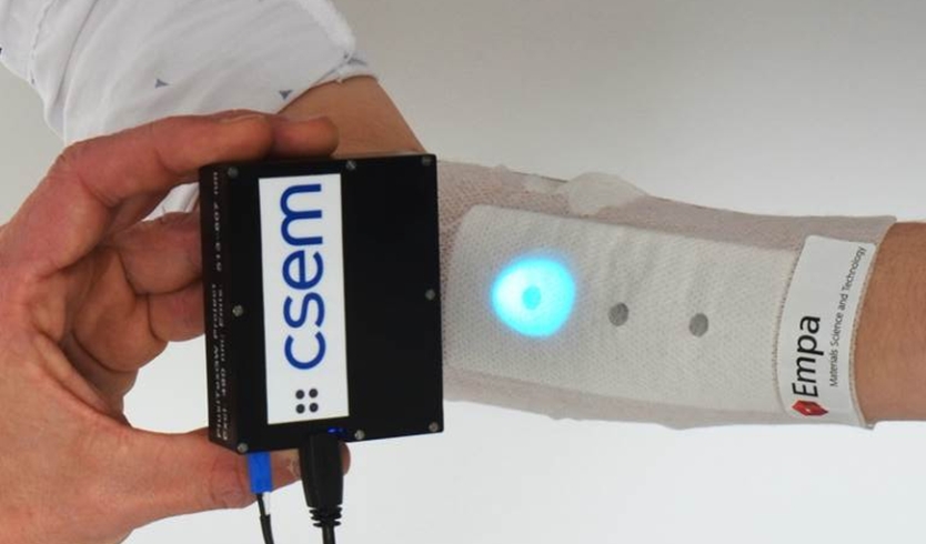
In contrast with existing methods, the new dressing is much more straightforward.

“Our device is as convenient as a bandage you put on your skin,” says Wang.

The new dressings consist of small electrodes for the injury site that are linked to a band holding energy-harvesting units called nanogenerators, which are looped around a wearer’s torso. The natural expansion and contraction of the wearer’s ribcage during breathing powers the nanogenerators, which deliver low-intensity electric pulses.

“The nature of these electrical pulses is similar to the way the body generates an internal electric field,” says Wang.

And, those low-power pulses won’t harm healthy tissue like traditional, high-power electrotherapy devices might.

In fact, the researchers showed that exposing cells to high-energy electrical pulses caused them to produce almost five times more reactive oxygen species — major risk factors for cancer and cellular aging — than did cells that were exposed to the nanogenerators.

Also a boon to healing: They determined that the low-power pulses boosted viability for a type of skin cell called fibroblasts, and exposure to the nanogenerator’s pulses encouraged fibroblasts to line up (a crucial step in wound healing) and produce more biochemical substances that promote tissue growth.

“These findings are very exciting,” says collaborator Weibo Cai, a professor of radiology at UW-Madison. “The detailed mechanisms will still need to be elucidated in future work.”

In that vein, the researchers aim to tease out precisely how the gentle pulses aid in healing. The scientists also plan to test the devices on pig skin, which closely mimics human tissue.

And, they are working to give the nanogenerators additional capabilities–tweaking their structure to allow for energy harvesting from small imperceptible twitches in the skin or the thrumming pulse of a heartbeat.

“The impressive results in this study represent an exciting new spin on electrical stimulation for many different wound types, given the simplicity of the design,” says Gibson, who will collaborate with the team to confirm the reproducibility of these results in human skin models.

If the team is successful, the devices could help solve a major challenge for modern medicine.

“We think our nanogenerator could be the most effective electrical stimulation approach for many therapeutic purposes,” says Wang.

And because the nanogenerators consist of relatively common materials, price won’t be an issue.

“I don’t think the cost will be much more than a regular bandage,” says Wang. “The device in itself is very simple and convenient to fabricate.”

Here’s a link to and a citation for the paper,

Effective Wound Healing Enabled by Discrete Alternative Electric Fields from Wearable Nanogenerators by Yin Long, Hao Wei, Jun Li, Guang Yao, Bo Yu, Dalong Ni, Angela LF Gibson, Xiaoli Lan, Yadong Jiang, Weibo Cai, and Xudong Wang. ACS Nano, Article ASAP DOI: 10.1021/acsnano.8b07038 Publication Date (Web): November 29, 2018

Copyright © 2018 American Chemical Society

This paper is open access.

I assume it will be a while before there are human clinical trials.

Colo(u)r-changing bandage for better compression

This is a structural colo(u)r story, from a May 29, 2018 news item on Nanowerk,

Compression therapy is a standard form of treatment for patients who suffer from venous ulcers and other conditions in which veins struggle to return blood from the lower extremities. Compression stockings and bandages, wrapped tightly around the affected limb, can help to stimulate blood flow. But there is currently no clear way to gauge whether a bandage is applying an optimal pressure for a given condition.

Now engineers at MIT {Massachusetts Institute of Technology] have developed pressure-sensing photonic fibers that they have woven into a typical compression bandage. As the bandage is stretched, the fibers change color. Using a color chart, a caregiver can stretch a bandage until it matches the color for a desired pressure, before, say, wrapping it around a patient’s leg.

The photonic fibers can then serve as a continuous pressure sensor — if their color changes, caregivers or patients can use the color chart to determine whether and to what degree the bandage needs loosening or tightening.

A May 29, 2018 MIT news release (also on EurekAlert), which originated the news item, provides more detail,

“Getting the pressure right is critical in treating many medical conditions including venous ulcers, which affect several hundred thousand patients in the U.S. each year,” says Mathias Kolle, assistant professor of mechanical engineering at MIT. “These fibers can provide information about the pressure that the bandage exerts. We can design them so that for a specific desired pressure, the fibers reflect an easily distinguished color.”

Kolle and his colleagues have published their results in the journal Advanced Healthcare Materials. Co-authors from MIT include first author Joseph Sandt, Marie Moudio, and Christian Argenti, along with J. Kenji Clark of the Univeristy of Tokyo, James Hardin of the United States Air Force Research Laboratory, Matthew Carty of Brigham and Women’s Hospital-Harvard Medical School, and Jennifer Lewis of Harvard University.

Natural inspiration

The color of the photonic fibers arises not from any intrinsic pigmentation, but from their carefully designed structural configuration. Each fiber is about 10 times the diameter of a human hair. The researchers fabricated the fiber from ultrathin layers of transparent rubber materials, which they rolled up to create a jelly-roll-type structure. Each layer within the roll is only a few hundred nanometers thick.

In this rolled-up configuration, light reflects off each interface between individual layers. With enough layers of consistent thickness, these reflections interact to strengthen some colors in the visible spectrum, for instance red, while diminishing the brightness of other colors. This makes the fiber appear a certain color, depending on the thickness of the layers within the fiber.

“Structural color is really neat, because you can get brighter, stronger colors than with inks or dyes just by using particular arrangements of transparent materials,” Sandt says. “These colors persist as long as the structure is maintained.”

The fibers’ design relies upon an optical phenomenon known as “interference,” in which light, reflected from a periodic stack of thin, transparent layers, can produce vibrant colors that depend on the stack’s geometric parameters and material composition. Optical interference is what produces colorful swirls in oily puddles and soap bubbles. It’s also what gives peacocks and butterflies their dazzling, shifting shades, as their feathers and wings are made from similarly periodic structures.

“My interest has always been in taking interesting structural elements that lie at the origin of nature’s most dazzling light manipulation strategies, to try recreating and employing them in useful applications,” Kolle says.

A multilayered approach

The team’s approach combines known optical design concepts with soft materials, to create dynamic photonic materials.

While a postdoc at Harvard in the group of Professor Joanna Aizenberg, Kolle was inspired by the work of Pete Vukusic, professor of biophotonics at the University of Exeter in the U.K., on Margaritaria nobilis, a tropical plant that produces extremely shiny blue berries. The fruits’ skin is made up of cells with a periodic cellulose structure, through which light can reflect to give the fruit its signature metallic blue color.

Together, Kolle and Vukusic sought ways to translate the fruit’s photonic architecture into a useful synthetic material. Ultimately, they fashioned multilayered fibers from stretchable materials, and assumed that stretching the fibers would change the individual layers’ thicknesses, enabling them to tune the fibers’ color. The results of these first efforts were published in Advanced Materials in 2013.

When Kolle joined the MIT faculty in the same year, he and his group, including Sandt, improved on the photonic fiber’s design and fabrication. In their current form, the fibers are made from layers of commonly used and widely available transparent rubbers, wrapped around highly stretchable fiber cores. Sandt fabricated each layer using spin-coating, a technique in which a rubber, dissolved into solution, is poured onto a spinning wheel. Excess material is flung off the wheel, leaving a thin, uniform coating, the thickness of which can be determined by the wheel’s speed.

For fiber fabrication, Sandt formed these two layers on top of a water-soluble film on a silicon wafer. He then submerged the wafer, with all three layers, in water to dissolve the water-soluble layer, leaving the two rubbery layers floating on the water’s surface. Finally, he carefully rolled the two transparent layers around a black rubber fiber, to produce the final colorful photonic fiber.

Reflecting pressure

The team can tune the thickness of the fibers’ layers to produce any desired color tuning, using standard optical modeling approaches customized for their fiber design.

“If you want a fiber to go from yellow to green, or blue, we can say, ‘This is how we have to lay out the fiber to give us this kind of [color] trajectory,'” Kolle says. “This is powerful because you might want to have something that reflects red to show a dangerously high strain, or green for ‘ok.’ We have that capacity.”

The team fabricated color-changing fibers with a tailored, strain-dependent color variation using the theoretical model, and then stitched them along the length of a conventional compression bandage, which they previously characterized to determine the pressure that the bandage generates when it’s stretched by a certain amount.

The team used the relationship between bandage stretch and pressure, and the correlation between fiber color and strain, to draw up a color chart, matching a fiber’s color (produced by a certain amount of stretching) to the pressure that is generated by the bandage.

To test the bandage’s effectiveness, Sandt and Moudio enlisted over a dozen student volunteers, who worked in pairs to apply three different compression bandages to each other’s legs: a plain bandage, a bandage threaded with photonic fibers, and a commercially-available bandage printed with rectangular patterns. This bandage is designed so that when it is applying an optimal pressure, users should see that the rectangles become squares.

Overall, the bandage woven with photonic fibers gave the clearest pressure feedback. Students were able to interpret the color of the fibers, and based on the color chart, apply a corresponding optimal pressure more accurately than either of the other bandages.

The researchers are now looking for ways to scale up the fiber fabrication process. Currently, they are able to make fibers that are several inches long. Ideally, they would like to produce meters or even kilometers of such fibers at a time.

“Currently, the fibers are costly, mostly because of the labor that goes into making them,” Kolle says. “The materials themselves are not worth much. If we could reel out kilometers of these fibers with relatively little work, then they would be dirt cheap.”

Then, such fibers could be threaded into bandages, along with textiles such as athletic apparel and shoes as color indicators for, say, muscle strain during workouts. Kolle envisions that they may also be used as remotely readable strain gauges for infrastructure and machinery.

“Of course, they could also be a scientific tool that could be used in a broader context, which we want to explore,” Kolle says.

Here’s what the bandage looks like,

Caption: Engineers at MIT have developed pressure-sensing photonic fibers that they have woven into a typical compression bandage. Credit Courtesy of the researchers

Here’s a link to and a citation for the paper,

Stretchable Optomechanical Fiber Sensors for Pressure Determination in Compressive Medical Textiles by Joseph D. Sandt, Marie Moudio, J. Kenji Clark, James Hardin, Christian Argenti, Matthew Carty, Jennifer A. Lewis, Mathias Kolle. Advanced Healthcare Materials https://doi.org/10.1002/adhm.201800293 First published: 29 May 2018

This paper is behind a paywall.

Bandage with a voice (sort of)

Researchers at Empa (Swiss Federal Laboratories for Materials Testing and Research) have not developed a talking bandage despite the title (Bandage with a Voice) for a July 4, 2017 Empa press release  (also a July 4, 2017 news item on Nanowerk),

A novel bandage alerts the nursing staff as soon as a wound starts healing badly. Sensors incorporated into the base material glow with a different intensity if the wound’s pH level changes. This way even chronic wounds could be monitored at home.

/documents/56164/1274117/bild1-CSEM-Bild-quer.jpg/a6a827b3-c7e7-49a4-86f2-73c2c7b15ce1?t=1499165473227

Using a UV lamp, the pH level in the wound can be verified without removing the bandage and the healing process can continue unimpeded. Image: Empa / CSEM

All too often, changing bandages is extremely unpleasant, even for smaller, everyday injuries. It stings and pulls, and sometimes a scab will even start bleeding again. And so we prefer to wait until the bandage drops off by itself.

It’s a different story with chronic wounds, though: normally, the nursing staff has to change the dressing regularly – not just for reasons of hygiene, but also to examine the wound, take swabs and clean it. Not only does this irritate the skin unnecessarily; bacteria can also get in, the risk of infection soars. It would be much better to leave the bandage on for longer and have the nursing staff “read” the condition of the wound from outside.

The idea of being able to see through a wound dressing gave rise to the project Flusitex (Fluorescence sensing integrated into medical textiles), which is being funded by the Swiss initiative Nano-Tera. Researchers from Empa teamed up with ETH Zurich, Centre Suisse d’Electronique et de Microtechnique (CSEM) and University Hospital Zurich to develop a high-tech system that is supposed to supply the nursing staff with relevant data about the condition of a wound. As Luciano Boesel from Empa’s Laboratory for Biomimetic Membranes and Textiles, who is coordinating the project at Empa, explains: “The idea of a smart wound dressing with integrated sensors is to provide continuous information on the state of the healing process without the bandages having to be changed any more frequently than necessary.” This would mean a gentler treatment for patients, less work for the nursing staff and, therefore, lower costs: globally, around 17 billion $ were spent on treating wounds last year.

When wounds heal, the body produces specific substances in a complex sequence of biochemical processes, which leads to a significant variation in a number of metabolic parameters. For instance, the amount of glucose and oxygen rises and falls depending on the phase of the healing process; likewise does the pH level change. All these variations can be detected with specialized sensors. With this in mind, Empa teamed up with project partner CSEM to develop a portable, cheap and easy-to-use device for measuring fluorescence that is capable of monitoring several parameters at once. It should enable nursing staff to keep tabs on the pH as well as on glucose and oxygen levels while the wound heals. If these change, conclusions about other key biochemical processes involved in wound healing can be drawn.

/documents/56164/1274117/bild2-Guido%26Alina_0788.jpg/6024b368-b367-42e1-a6d9-d376c27e179a?t=1499165473430
The bandage reveals ist measurings in UV light.
A high pH signals chronic wounds

The pH level is particularly useful for chronic wounds. If the wound heals normally, the pH rises to 8 before falling to 5 or 6. If a wound fails to close and becomes chronic, however, the pH level fluctuates between 7 and 8. Therefore, it would be helpful if a signal on the bandage could inform the nursing staff that the wound pH is permanently high. If the bandage does not need changing for reasons of hygiene and pH levels are low, on the other hand, they could afford to wait.

But how do the sensors work? The idea: if certain substances appear in the wound fluid, “customized” fluorescent sensor molecules respond with a physical signal. They start glowing and some even change color in the visible or ultra-violet (UV) range. Thanks to a color scale, weaker and stronger changes in color can be detected and the quantity of the emitted substance be deduced.

Empa chemist Guido Panzarasa from the Laboratory for Biomimetic Membranes and Textiles vividly demonstrates how a sample containing sensor molecules begins to fluoresce in the lab. He carefully drips a solution with a pH level of 7.5 into a dish. Under a UV light, the change is plain to see. He adds another solution and the luminescence fades. A glance at the little bottle confirms it: the pH level of the second solution is lower.

Luminous molecules under UV

The Empa team designed a molecule composed of benzalkonium chloride and pyranine. While benzalkonium chloride is a substance also used for conventional medical soap to combat bacteria, fungi and other microorganisms, pyranine is a dye found in highlighters that glows under UV light. “This biomarker works really well,” says Panzarasa; “especially at pH levels between 5.5 and 7.5. The colors can be visualized with simple UV lamps available in electronics stores.” The Empa team recently published their results in the journal “Sensors and Actuators”.

The designer molecule has another advantage: thanks to the benzalkonium chloride, it has an antimicrobial effect, as researchers from Empa’s Laboratory for Biointerfaces confirmed for the bacteria strain Staphylococcus aureus. Unwelcome bacteria might potentially also be combatted by selecting the right bandage material in future. As further investigations, such as on the chemical’s compatibility with cells and tissues, are currently lacking, however, the researchers do not yet know how their sensor works in a complex wound.

Keen interest from industry

In order to illustrate what a smart wound dressing might actually look like in future, Boesel places a prototype on the lab bench. “You don’t have to cover the entire surface of wound dressings with sensors,” he explains. “It’s enough for a few small areas to be impregnated with the pyranine benzalkonium molecules and integrated into the base material. This means the industrial wound dressings won’t be much pricier than they are now – only up to 20% more expensive.” Empa scientists are currently working on this in the follow-up project FlusiTex-Gateway in cooperation with industrial partners Flawa, Schöller, Kenzen and Theranoptics.
Panzarasa now drips various liquids with different pH levels onto all the little cylinders on the wound pad prototype. Sure enough, the lighter and darker dots are also clearly discernible as soon as the UV lamp is switched on. They are even visible to the naked eye and glow in bright yellow if liquids with a high pH come into contact with the sensor. The scientists are convinced: since the pH level is so easy to read and provides precise information about the acidic or alkaline state of the sample, this kind of wound dressing is just the ticket as a diagnostic tool. Using the fluorescence meter developed by CSEM, more accurate, quantitative measure-ments of the pH level can be accomplished for medical purposes.

According to Boesel, it might one day even be possible to read the signals with the aid of a smartphone camera. Combined with a simple app, nursing staff and doctors would have a tool that enables them to easily and conveniently read the wound status “from outside”, even without a UV lamp. And patients would then also have the possibility of detecting the early onset of a chronic wound at home.

I wonder how long or even if this innovation will ever make its way into medical practice. I’m guessing this stage would be described as ‘proof of concept’ and that clinical testing is still many years away.

The metaphor in the press release’s title helped to wake me up. Thank you to whoever wrote it.