Tag Archives: BESSY II

Artificial graphene with buckyballs

A July 21, 2022 news item on Nanowerk describes graphene in its ‘natural’ state and explains what ‘artificial’ graphene is although there is no mention of why variants are a hot topic,

Graphene consists of carbon atoms that crosslink in a plane to form a flat honeycomb structure. In addition to surprisingly high mechanical stability, the material has exciting electronic properties: The electrons behave like massless particles, which can be clearly demonstrated in spectrometric experiments.

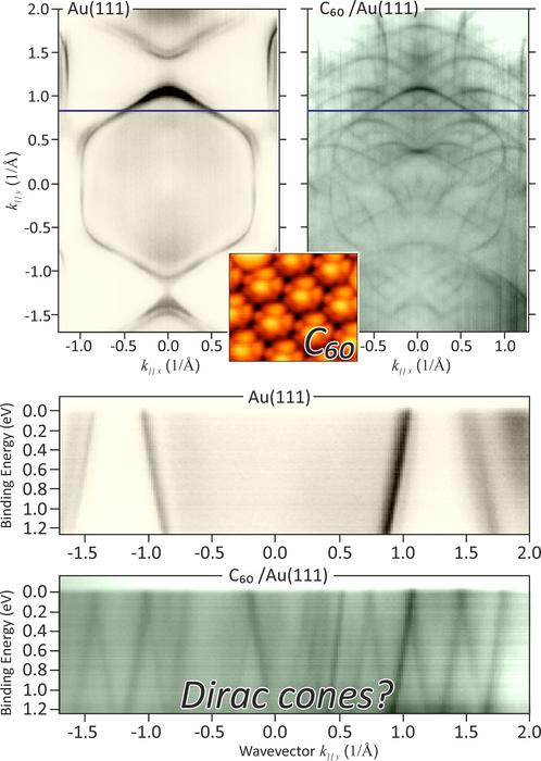
Measurements reveal a linear dependence of energy on momentum, namely the so-called Dirac cones – two lines that cross without a band gap – i.e. an energy difference between electrons in the conduction band and those in the valence bands.

Variants in graphene architecture

Artificial variants of graphene architecture are a hot topic in materials research right now. Instead of carbon atoms, quantum dots of silicon have been placed, ultracold atoms have been trapped in the honeycomb lattice with strong laser fields, or carbon monoxide molecules have been pushed into place on a copper surface piece by piece with a scanning tunneling microscope, where they could impart the characteristic graphene properties to the electrons of the copper.

A July 21, 2022 Helmholtz-Zentrum Berlin (HZB) press release (also on EurekAlert), which originated the news item, describes research into whether or not layering buckyballs onto gold would result in artificial graphene,

Artificial graphene with buckyballs?

A recent study suggested that it is infinitely easier to make artificial graphene using C60 molecules called buckyballs [or buckminsterfullerenes or, more generically, fullerenes]. Only a uniform layer of these needs to be vapor-deposited onto gold for the gold electrons to take on the special graphene properties. Measurements of photoemission spectra appeared to show a kind of Dirac cone.

Analysis of band structures at BESSY II

“That would be really quite amazing,” says Dr. Andrei Varykhalov, of HZB, who heads a photoemission and scanning tunneling microscopy group. “Because the C60 molecule is absolutely nonpolar, it was hard for us to imagine how such molecules would exert a strong influence on the electrons in the gold.” So Varykhalov and his team launched a series of measurements to test this hypothesis.

In tricky and detailed analyses, the Berlin team was able to study C60 layers on gold over a much larger energy range and for different measurement parameters. They used angle-resolved ARPES spectroscopy at BESSY II [third-generation synchrotron radiation source], which enables particularly precise measurements, and also analysed electron spin for some measurements.

Normal behavior

“We see a parabolic relationship between momentum and energy in our measured data, so it’s a very normal behavior. These signals come from the electrons deep in the substrate (gold or copper) and not the layer, which could be affected by the buckyballs,” explains Dr. Maxim Krivenkov, lead author of the study. The team was also able to explain the linear measurement curves from the previous study. “These measurement curves merely mimic the Dirac cones; they are an artifact, so to speak, of a deflection of the photoelectrons as they leave the gold and pass through the C60 layer,” Varykhalov explains. Therefore, the buckyball layer on gold cannot be considered an artificial graphene.

Caption: Measurement data from BESSY II before and after deposition of C60 molecules demonstrate the replication of the band structure and the emergence of cone-like band crossings. A scanning electron microscopy of the buckyballs on gold is superimposed in the centre. Credit: HZB

Here’s a link to and a citation for the paper,

On the problem of Dirac cones in fullerenes on gold by M. Krivenkov, D. Marchenko, M. Sajedi, A. Fedorov, O. J. Clark, J. Sánchez-Barriga, E. D. L. Rienks, O. Rader and A. Varykhalov. Nanoscale, 2022,14, 9124-9133 First published: 23 May 2022

This paper is open access.

Japanese researchers note the emergence of the ‘Devil’s staircase’

I wanted to know why it’s called the ‘Devil’s staircase’ and this is what I found. According to Wikipedia there are several of them,

I gather the scientists are referring to the Cantor function (mathematics), Note: Links have been removed,

In mathematics, the Cantor function is an example of a function that is continuous, but not absolutely continuous. It is also referred to as the Cantor ternary function, the Lebesgue function, Lebesgue’s singular function, the Cantor-Vitali function, the Devil’s staircase,[1] the Cantor staircase function,[2] and the Cantor-Lebesgue function.[3]

Here’s a diagram illustrating the Cantor function (from the Wikipedia entry),

CC BY-SA 3.0 File:CantorEscalier.svg Uploaded by Theon Created: January 24, 2009

CC BY-SA 3.0
File:CantorEscalier.svg
Uploaded by Theon
Created: January 24, 2009

As for this latest ‘Devil’s staircase’, a June 17, 2015 news item on Nanowerk announces the research (Note: A link has been removed),

Researchers at the University of Tokyo have revealed a novel magnetic structure named the “Devil’s staircase” in cobalt oxides using soft X-rays (“Observation of a Devil’s Staircase in the Novel Spin-Valve System SrCo6O11“). This is an important result since the researchers succeeded in determining the detailed magnetic structure of a very small single crystal invisible to the human eye.

A June 17, 2015 University of Tokyo press release, which originated the news item on Nanowerk, describes why this research is now possible and the impact it could have,

Recent remarkable progress in resonant soft x-ray diffraction performed in synchrotron facilities has made it possible to determine spin ordering (magnetic structure) in small-volume samples including thin films and nanostructures, and thus is expected to lead not only to advances in materials science but also application to spintronics, a technology which is expected to form the basis of future electronic devices. Cobalt oxide is known as one material that is suitable for spintronics applications, but its magnetic structure was not fully understood.

The research group of Associate Professor Hiroki Wada at the University of Tokyo Institute for Solid State Physics, together with the researchers at Kyoto University and in Germany, performed a resonant soft X-ray diffraction study of cobalt (Co) oxides in the synchrotron facility BESSY II in Germany. They observed all the spin orderings which are theoretically possible and determined how these orderings change with the application of magnetic fields. The plateau-like behavior of magnetic structure as a function of magnetic field is called the “Devil’s staircase,” and is the first such discovery in spin systems in 3D transition metal oxides including cobalt, iron, manganese.

By further resonant soft X-ray diffraction studies, one can expect to find similar “Devil’s staircase” behavior in other materials. By increasing the spatial resolution of microscopic observation of the “Devil’s staircase” may lead to the development of novel types of spintronics materials.

Here’s an example of the ‘cobalt’ Devil’s staircase,

The magnetic structure that gives rise to the Devil's Staircase Magnetization (vertical axis) of cobalt oxide shows plateau like behaviors as a function of the externally-applied magnetic field (horizontal axis). The researchers succeeded in determining the magnetic structures which create such plateaus. Red and blue arrows indicate spin direction. © 2015 Hiroki Wadati.

The magnetic structure that gives rise to the Devil’s Staircase
Magnetization (vertical axis) of cobalt oxide shows plateau like behaviors as a function of the externally-applied magnetic field (horizontal axis). The researchers succeeded in determining the magnetic structures which create such plateaus. Red and blue arrows indicate spin direction.
© 2015 Hiroki Wadati.

Here’s a link to and a citation for the paper,

Observation of a Devil’s Staircase in the Novel Spin-Valve System SrCo6O11 by T. Matsuda, S. Partzsch, T. Tsuyama, E. Schierle, E. Weschke, J. Geck, T. Saito, S. Ishiwata, Y. Tokura, and H. Wadati. Phys. Rev. Lett. 114, 236403 – Published 11 June 2015 (paper: Vol. 114, Iss. 23 — 12 June 2015)  DOI: 10.1103/PhysRevLett.114.236403

This paper is behind a paywall.

Discovering why your teeth aren’t perfectly crack-resistant

This helps make your teeth crack-resistant?

Caption: Illustration shows complex biostructure of dentin: the dental tubuli (yellow hollow cylinders, diameters appr. 1 micrometer) are surrounded by layers of mineralized collagen fibers (brown rods). The tiny mineral nanoparticles are embedded in the mesh of collagen fibers and not visible here. Credit: JB Forien @Charité

Caption: Illustration shows complex biostructure of dentin: the dental tubuli (yellow hollow cylinders, diameters appr. 1 micrometer) are surrounded by layers of mineralized collagen fibers (brown rods). The tiny mineral nanoparticles are embedded in the mesh of collagen fibers and not visible here. Credit: JB Forien @Charité

A June 10, 2015 Helmholtz Zentrum Berlin (HZB) press release (also on EurekAlert) explains how the illustration above relates to the research,

Human teeth have to serve for a lifetime, despite being subjected to huge forces. But the high failure resistance of dentin in teeth is not fully understood. An interdisciplinary team led by scientists of Charite Universitaetsmedizin Berlin has now analyzed the complex structure of dentin. At the synchrotron sources BESSY II at HZB, Berlin, Germany, and the European Synchrotron Radiation Facility ESRF, Grenoble, France, they could reveal that the mineral particles are precompressed.

The internal stress works against crack propagation and increases resistance of the biostructure.

Engineers use internal stresses to strengthen materials for specific technical purposes. Now it seems that evolution has long ‘known’ about this trick, and has put it to use in our natural teeth. Unlike bones, which are made partly of living cells, human teeth are not able to repair damage. Their bulk is made of dentin, a bonelike material consisting of mineral nanoparticles. These mineral nanoparticles are embedded in collagen protein fibres, with which they are tightly connected. In every tooth, such fibers can be found, and they lie in layers, making teeth tough and damage resistant. Still, it was not well understood, how crack propagation in teeth can be stopped.

The press release goes on to describe the new research and the teams which investigated the role of the mineral nanoparticles with regard to compression and cracking,

Now researchers from Charite Julius-Wolff-Institute, Berlin have been working with partners from Materials Engineering Department of Technische Universitaets Berlin, MPI of Colloids and Interfaces, Potsdam and Technion – Israel Institute of Technology, Haifa, to examine these biostructures more closely. They performed Micro-beam in-situ stress experiments in the mySpot BESSY facility of HZB, Berlin, Germany and analyzed the local orientation of the mineral nanoparticles using the nano-imaging facility of the European Synchrotron Radiation Facility (ESRF) in Grenoble, France.

When the tiny collagen fibers shrink, the attached mineral particles become increasingly compressed, the science team found out. “Our group was able to use changes in humidity to demonstrate how stress appears in the mineral in the collagen fibers, Dr. Paul Zaslansky from Julius Wolff-Institute of Charite Berlin explains. “The compressed state helps to prevents cracks from developing and we found that compression takes place in such a way that cracks cannot easily reach the tooth inner parts, which could damage the sensitive pulp. In this manner, compression stress helps to prevent cracks from rushing through the tooth.

The scientists also examined what happens if the tight mineral-protein link is destroyed by heating: In that case, dentin in teeth becomes much weaker. We therefore believe that the balance of stresses between the particles and the protein is important for the extended survival of teeth in the mouth, Charite scientist Jean-Baptiste Forien says. Their results may explain why artificial tooth replacements usually do not work as well as healthy teeth do: they are simply too passive, lacking the mechanisms found in the natural tooth structures, and consequently fillings cannot sustain the stresses in the mouth as well as teeth do. “Our results might inspire the development of tougher ceramic structures for tooth repair or replacement, Zaslansky hopes.

Experiments took place as part of the DFG project “Biomimetic Materials Research: Functionality by Hierarchical Structuring of Materials (SPP1420).

Here’s a link to and a citation for the paper,

Compressive Residual Strains in Mineral Nanoparticles as a Possible Origin of Enhanced Crack Resistance in Human Tooth Dentin by Jean-Baptiste Forien, Claudia Fleck, Peter Cloetens, Georg Duda, Peter Fratzl, Emil Zolotoyabko, and Paul Zaslansky. Nano Lett., 2015, 15 (6), pp 3729–3734 DOI: 10.1021/acs.nanolett.5b00143 Publication Date (Web): May 26, 2015

Copyright © 2015 American Chemical Society

This paper is behind a paywall.