Tag Archives: bionanotechnology

Researchers at University of Montéal decode how molecules “talk” to each

An August 15, 2023 news item on ScienceDaily breaks news from the University of Montréal,

Two molecular languages at the origin of life have been successfully recreated and mathematically validated, thanks to pioneering work by Canadian scientists at Université de Montréal.

Fascinating, non? An August 15, 2023 Université de Montréal news release (also on EurekAlert), which originated the news item, explaining how this leads to nanotechnology-enabled applications, Note: A link has been removed,

Published this week in the Journal of American Chemical Society, the breakthrough opens new doors for the development of nanotechnologies with applications ranging from biosensing, drug delivery and molecular imaging.

Living organisms are made up of billions of nanomachines and nanostructures that communicate to create higher-order entities able to do many essential things, such as moving, thinking, surviving and reproducing.

“The key to life’s emergence relies on the development of molecular languages – also called signalling mechanisms – which ensure that all molecules in living organisms are working together to achieve specific tasks,” said the study’s principal investigator, UdeM bioengineering professor Alexis Vallée-Bélisle.

In yeasts, for example, upon detecting and binding a mating pheromone, billions of molecules will communicate and coordinate their activities to initiate union, said Vallée-Bélisle, holder of a Canada Research Chair in Bioengineering and Bionanotechnology.

“As we enter the era of nanotechnology, many scientists believe that the key to designing and programming more complex and useful artificial nanosystems relies on our ability to understand and better employ molecular languages developed by living organisms,” he said.

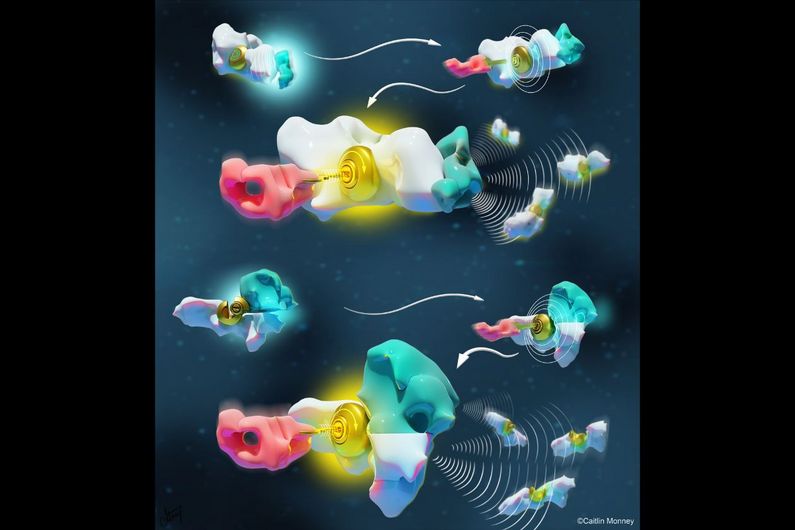
Two types of languages

One well-known molecular language is allostery. The mechanism of this language is “lock-and-key”: a molecule binds and modifies the structure of another molecule, directing it to trigger or inhibit an activity.

Another, lesser-known molecular language is multivalency, also known as the chelate effect. It works like a puzzle: as one molecule binds to another, it facilitates (or not) the binding of a third molecule by simply increasing its binding interface.

Although these two languages are observed in all molecular systems of all living organisms, it is only recently that scientists have started to understand their rules and principles – and so use these languages to design and program novel artificial nanotechnologies.

“Given the complexity of natural nanosystems, before now nobody was able to compare the basic rules, advantage or limitations of these two languages on the same system,” said Vallée-Bélisle.

To do so, his doctoral student Dominic Lauzon, first author of the study, had the idea of creating a DNA-based molecular system that could function using both languages. “DNA is like Lego bricks for nanoengineers,” said Lauzon. “It’s a remarkable molecule that offers simple, programmable and easy-to-use chemistry.”

Simple mathematical equations to detect antibodies

The researchers found that simple mathematical equations could well describe both languages, which unravelled the parameters and design rules to program the communication between molecules within a nanosystem.

For example, while the multivalent language enabled control of both the sensitivity and cooperativity of the activation or deactivation of the molecules, the corresponding allosteric translation only enabled control of the sensitivity of the response.

With this new understanding at hand, the researchers used the language of multivalency to design and engineer a programmable antibody sensor that allows the detection of antibodies over different ranges of concentration.

“As shown with the recent pandemic, our ability to precisely monitor the concentration of antibodies in the general population is a powerful tool to determine the people’s individual and collective immunity,” said Vallée-Bélisle.

In addition to expanding the synthetic toolbox to create the next generation of nanotechnology, the scientist’s discovery also shines a light on why some natural nanosystems may have selected one language over another to communicate chemical information.

Caption; The illustration depicts two chemical languages at the basis of molecular communication. The same white molecule, represented as a lock, is activated either via allostery (top) or multivalency (bottom). The allosteric activator (cyan) induces a conformational change of the lock while the multivalent activator provides the missing part of the lock, both enabling the activation by the key (pink). Credit: Monney Medical Media / Caitlin Monney

Here’s a link to and a citation for the paper,

Programing Chemical Communication: Allostery vs Multivalent Mechanism by Dominic Lauzon and Alexis Vallée-Bélisle. J. Am. Chem. Soc. 2023, XXXX, XXX, XXX-XXX DOI: https://doi.org/10.1021/jacs.3c04045 Online Publication Date: August 15, 2023 © 2023 American Chemical Society

This paper is behind a paywall.

‘Origami organs’ for tissue engineering

This is a different approach to tissue engineering and its the consequence of a serendipitous accident.  From an Aug. 7, 2017 Northwestern University news release (also on EurekAlert),

Northwestern Medicine scientists and engineers have invented a range of bioactive “tissue papers” made of materials derived from organs that are thin and flexible enough to even fold into an origami bird. The new biomaterials can potentially be used to support natural hormone production in young cancer patients and aid wound healing.

The tissue papers are made from structural proteins excreted by cells that give organs their form and structure. The proteins are combined with a polymer to make the material pliable.

In the study, individual types of tissue papers were made from ovarian, uterine, kidney, liver, muscle or heart proteins obtained by processing pig and cow organs. Each tissue paper had specific cellular properties of the organ from which it was made.

The article describing the tissue paper and its function will be published Aug. 7 in the journal Advanced Functional Materials.

“This new class of biomaterials has potential for tissue engineering and regenerative medicine as well as drug discovery and therapeutics,” corresponding author Ramille Shah said. “It’s versatile and surgically friendly.”

Shah is an assistant professor of surgery at the Feinberg School of Medicine and an assistant professor of materials science and engineering at McCormick School of Engineering. She also is a member of the Simpson Querrey Institute for BioNanotechnology.

For wound healing, Shah thinks the tissue paper could provide support and the cell signaling needed to help regenerate tissue to prevent scarring and accelerate healing.

The tissue papers are made from natural organs or tissues. The cells are removed, leaving the natural structural proteins – known as the extracellular matrix – that then are dried into a powder and processed into the tissue papers. Each type of paper contains residual biochemicals and protein architecture from its original organ that can stimulate cells to behave in a certain way.

In the lab of reproductive scientist Teresa Woodruff, the tissue paper made from a bovine ovary was used to grow ovarian follicles when they were cultured in vitro. The follicles (eggs and hormone-producing cells) grown on the tissue paper produced hormones necessary for proper function and maturation.

“This could provide another option to restore normal hormone function to young cancer patients who often lose their hormone function as a result of chemotherapy and radiation,” Woodruff, a study coauthor, said.

A strip of the ovarian paper with the follicles could be implanted under the arm to restore hormone production for cancer patients or even women in menopause.

Woodruff is the director of the Oncofertility Consortium and the Thomas J. Watkins Memorial Professor of Obstetrics and Gynecology at Feinberg.

In addition, the tissue paper made from various organs separately supported the growth of adult human stem cells. Scientists placed human bone marrow stem cells on the tissue paper, and all the stem cells attached and multiplied over four weeks.

“That’s a good sign that the paper supports human stem cell growth,” said first author Adam Jakus, who developed the tissue papers. “It’s an indicator that once we start using tissue paper in animal models it will be biocompatible.”

The tissue papers feel and behave much like standard office paper when they are dry, Jakus said. Jakus simply stacks them in a refrigerator or a freezer. He even playfully folded them into an origami bird.

“Even when wet, the tissue papers maintain their mechanical properties and can be rolled, folded, cut and sutured to tissue,” he said.

Jakus was a Hartwell postdoctoral fellow in Shah’s lab for the study and is now chief technology officer and cofounder of the startup company Dimension Inx, LLC, which was also cofounded by Shah. The company will develop, produce and sell 3-D printable materials primarily for medical applications. The Intellectual Property is owned by Northwestern University and will be licensed to Dimension Inx.

An Accidental Spill Sparked Invention

An accidental spill of 3-D printing ink in Shah’s lab by Jakus sparked the invention of the tissue paper. Jakus was attempting to make a 3-D printable ovary ink similar to the other 3-D printable materials he previously developed to repair and regenerate bone, muscle and nerve tissue. When he went to wipe up the spill, the ovary ink had already formed a dry sheet.

“When I tried to pick it up, it felt strong,” Jakus said. “I knew right then I could make large amounts of bioactive materials from other organs. The light bulb went on in my head. I could do this with other organs.”

“It is really amazing that meat and animal by-products like a kidney, liver, heart and uterus can be transformed into paper-like biomaterials that can potentially regenerate and restore function to tissues and organs,” Jakus said. “I’ll never look at a steak or pork tenderloin the same way again.”

For those who like their news in a video,

As someone who once made baklava, that does not look like filo pastry, where an individual sheet is quite thin and rips easily. Enough said.

Here’s a link to and a citation for the paper,

“Tissue Papers” from Organ-Specific Decellularized Extracellular Matrices by Adam E. Jakus, Monica M. Laronda, Alexandra S. Rashedi, Christina M. Robinson, Chris Lee, Sumanas W. Jordan, Kyle E. Orwig, Teresa K. Woodruff, and Ramille N. Shah. Advnaced Functional Materials DOI: 10.1002/adfm.201700992 Version of Record online: 7 AUG 2017

© 2017 WILEY-VCH Verlag GmbH & Co. KGaA, Weinheim

This paper is behind a paywall.

‘Smart dress’ for oil-degrading bacteria (marine oil spill remediation)

This July 22, 2016 news item (on Nanowerk) about bacteria and marine oil spill remediation was a little challenging (for me) to read (Note: A link has been removed),

Bionanotechnology research is targeted on functional structures synergistically combining macromolecules, cells, or multicellular assemblies with a wide range of nanomaterials. Providing micrometer-sized cells with tiny nanodevices expands the uses of the cultured microorganisms and requires nanoassembly on individual live cells (“Nanoshell Assembly for Magnet-Responsive Oil-Degrading Bacteria”).

Surface engineering functionalizes the cell walls with polymer layers and/or nanosized particles and has been widely employed to modify the intrinsic properties of microbial cells. Cell encapsulation allows fabricating live microbial cells with magnetic nanoparticles onto cell walls, which mimics natural magnetotactic bacteria.

For this study researchers from Kazan Federal University and Louisiana Tech University chose Alcanivorax borkumensis marine bacteria as a target microorganism for cell surface engineering with magnetic nanoparticles for the following reasons: (1) these hydrocarbon-degrading bacteria are regarded as an important tool in marine oil spill remediation and potentially can be used in industrial oil-processing bioreactors, therefore the external magnetic manipulations with these cells seems to be practically relevant; (2) A. borkumensis are marine Gram-negative species having relatively fragile and thin cell walls, which makes cell wall engineering of these bacteria particularly challenging.

Rendering oil-degrading bacteria with artificially added magnetic functionality is important to attenuate their properties and to expand their practical use.

[downloaded from http://pubs.acs.org/doi/abs/10.1021/acs.langmuir.6b01743]

[downloaded from http://pubs.acs.org/doi/abs/10.1021/acs.langmuir.6b01743]

A July 22, 2016 Kazan Federal University (Russia) press release (also on EurekAlert), which originated the news item, has more detail about the research,

Cell surface engineering was performed using polycation-coated magnetic nanoparticles, which is a fast and straightforward process utilizing the direct deposition of positively charged iron oxide nanoparticles onto microbial cells during a brief incubation in excessive concentrations of nanoparticles. Gram-negative bacteria cell walls are built from the thin peptidoglycan layer sandwiched between the outer membrane and inner plasma membrane, with lipopolysaccharides rendering the overall negative cell charge, therefore cationic particles will attach to the cell walls due to electrostatic interactions.

Rod-like 0.5-μm diameter Gram-negative bacteria A. borkumensis were coated with 70?100 nm [sic] magnetite shells. The deposition of nanoparticles was performed with extreme care to ensure the survival of magnetized cells.

The development of biofilms on hydrophobic surface is a very important feature of A. borkumensis cells because this is how these cells attach to the oil droplets in natural environments. Consequently, any cell surface modification should not reduce their ability to attach and proliferate as biofilms. Here, at all concentrations of PAH- magnetite nanoparticles investigated, authors of the study detected the similar biofilm growth patterns. Overall, the magnetized cells were able to proliferate and exhibited normal physiological activity.

The next generations of the bacteria have a tendency to remove the artificial shell returning to the native form. Such magnetic nanoencapsulation may be used for the A. borkumensis transportation in the bioreactors to enhance the spill oil decomposition at certain locations.

If I read this rightly, the idea, in future iterations of this research, is to destroy the oil once it’s been gathered by the biofilm. This seems a different approach where other oil spill remediation techniques have hydrophobic/oleophilic sponges absorbing the oil, which could potentially be used in the future. There are carbon nanotube sponges (my April 17, 2012 posting) and boron nitride sponges (my Dec. 7, 2015 posting).

Here’s a link to and a citation for the paper,

Nanoshell Assembly for Magnet-Responsive Oil-Degrading Bacteria by Svetlana A. Konnova, Yuri M. Lvov, and Rawil F. Fakhrullin. Langmuir, Article ASAP DOI: 10.1021/acs.langmuir.6b01743 Publication Date (Web): June 09, 2016

Copyright © 2016 American Chemical Society

This paper is behind a paywall.

Using mung bean extract to synthesize silver nanoparticles

Not everyone is quite as enthusiastic as whoever wrote this press release about silver nanoparticles (due to potential environmental issues as more manufactured silver nanoparticles enter the ecosystem). Still, it’s good news that there may be a greener way to synthesize them. A July 20, 2016 news item on Azonano ‘spills the beans’,

… researchers from the Guru Nanak National College, the Institute of Microbial Technology, Mata Gujri College and Punjab University, have found a simple, non-toxic and environmentally friendly way to synthesize AgNPs [silver nanoparticles] using seed extract of Vigna radiata, commonly known as the mung bean or green gram.

Manoj Kumar Choudhary and colleagues used aqueous seed extract of mung beans to break up aqueous silver nitrate solution into NPs, as well as to reduce and stabilize the particles. The NPs were characterized by UV–visible spectroscopy, Fourier transform infrared spectroscopy, transmission electron microscopy, atomic absorption spectroscopy and X-ray diffraction. They were then tested for antimicrobial effectiveness.

As reported in Applied Nanoscience, the researchers found that phytochemicals present in the seed extract were effective at reducing and stabilizing the Ag metal ions. They found they could synthesize crystalline, spherically-shaped NPs, with a size range of 5 to 30 nm. The particles remained highly stable for months at room temperature, even after five months.

Antibacterial activity was assayed by the standard well-diffusion method, which showed that the biogenic silver NPs had broad-spectrum antibacterial activity against the Gram-negative bacteria Escherichia coli and the Gram-positive bacteria Staphylococcus aureus.

“In the present paper, we report a simple, eco-friendly and cost-effective synthesis method of AgNPs at ambient conditions using seed extract of Vigna radiata as a reducing and stabilizing agent,” say Choudhary and team. “The AgNPs synthesized by this method have efficient antimicrobial activity against pathogenic bacteria.”

The researchers say the next step would be further investigation of the potential applications of the synthesized AgNPs, as the outcome of the study could be useful for applications in nanotechnology-based applications in pharmacology and medicine.

Fig. 1: A Vigna radiata seeds. b Reddish brown solution of silver nanoparticles formed after 3 h due to reduction of silver ions [downloaded from http://link.springer.com/article/10.1007/s13204-015-0418-6]

Fig. 1: A Vigna radiata seeds. b Reddish brown solution of silver nanoparticles formed after 3 h due to reduction of silver ions [downloaded from http://link.springer.com/article/10.1007/s13204-015-0418-6]

Here’s a link to and a citation for the paper,

A facile biomimetic preparation of highly stabilized silver nanoparticles derived from seed extract of Vigna radiata and evaluation of their antibacterial activity by Manoj Kumar Choudhary, Jyoti Kataria, Swaranjit Singh Cameotra, Jagdish Singh. Appl Nanosci (2016) 6: 105. doi:10.1007/s13204-015-0418-6 First Online: 19 February 2015

This paper is open access.

A bioelectronic future made possible with DNA-based electromechanical switch

DNA-based electronics are discussed in the context of a Dec. 14, 2015 news item by Beth Ellison for Azonano about research into electromechanical switches at the University of California at Davis,

Researchers from the University of California, Davis (UC Davis) and the University of Washington have shown the possibility of using DNA-based electromechanical switches for nanoscale computing.

DNA is considered to be the molecule of life, and researchers have shown considerable interest in utilizing DNA as a nanoscale material in various applications.

A Dec. 14, 2015 UC Davis news release on EurekAlert, which originated the news item, provides more detail,

In their paper published in Nature Communications, the team demonstrated that changing the structure of the DNA double helix by modifying its environment allows the conductance (the ease with which an electric current passes) to be reversibly controlled. This ability to structurally modulate the charge transport properties may enable the design of unique nanodevices based on DNA. These devices would operate using a completely different paradigm than today’s conventional electronics.

“As electronics get smaller they are becoming more difficult and expensive to manufacture, but DNA-based devices could be designed from the bottom-up using directed self-assembly techniques such as ‘DNA origami’,” said Josh Hihath, assistant professor of electrical and computer engineering at UC Davis and senior author on the paper. DNA origami is the folding of DNA to create two- and three-dimensional shapes at the nanoscale level.

“Considerable progress has been made in understanding DNA’s mechanical, structural, and self-assembly properties and the use of these properties to design structures at the nanoscale. The electrical properties, however, have generally been difficult to control,” said Hihath.

New Twist on DNA? Possible Paradigms for Computing

In addition to potential advantages in fabrication at the nanoscale level, such DNA-based devices may also improve the energy efficiency of electronic circuits. The size of devices has been significantly reduced over the last 40 years, but as the size has decreased, the power density on-chip has increased. Scientists and engineers have been exploring novel solutions to improve the efficiency.

“There’s no reason that computation must be done with traditional transistors. Early computers were fully mechanical and later worked on relays and vacuum tubes,” said Hihath. “Moving to an electromechanical platform may eventually allow us to improve the energy efficiency of electronic devices at the nanoscale.”

This work demonstrates that DNA is capable of operating as an electromechanical switch and could lead to new paradigms for computing.

To develop DNA into a reversible switch, the scientists focused on switching between two stable conformations of DNA, known as the A-form and the B-form. In DNA, the B-form is the conventional DNA duplex that is commonly associated with these molecules. The A-form is a more compact version with different spacing and tilting between the base pairs. Exposure to ethanol forces the DNA into the A-form conformation resulting in an increased conductance. Similarly, by removing the ethanol, the DNA can switch back to the B-form and return to its original reduced conductance value.

One Step Toward Molecular Computing

In order to develop this finding into a technologically viable platform for electronics, the authors also noted that there is still a great deal of work to be done. Although this discovery provides a proof-of-principle demonstration of electromechanical switching in DNA, there are generally two major hurdles yet to be overcome in the field of molecular electronics. First, billions of active molecular devices must be integrated into the same circuit as is done currently in conventional electronics. Next, scientists must be able to gate specific devices individually in such a large system.

“Eventually, the environmental gating aspect of this work will have to be replaced with a mechanical or electrical signal in order to locally address a single device,” noted Hihath.

Here’s a link to and a citation for the paper,

Conformational gating of DNA conductance by Juan Manuel Artés, Yuanhui Li, Jianqing Qi, M. P. Anantram, & Joshua Hihath. Nature Communications 6, Article number: 8870 doi:10.1038/ncomms9870 Published 09 December 2015

This paper is open access.

Cyborgs (a presentation) at the American Chemical Society’s 248th meeting

There will be a plethora of chemistry news online over the next few days as the American Society’s (ACS) 248th meeting in San Francisco, CA from Aug. 10 -14, 2014 takes place. Unexpectedly, an Aug. 11, 2014 news item on Azonano highlights a meeting presentation focused on cyborgs,

No longer just fantastical fodder for sci-fi buffs, cyborg technology is bringing us tangible progress toward real-life electronic skin, prosthetics and ultraflexible circuits. Now taking this human-machine concept to an unprecedented level, pioneering scientists are working on the seamless marriage between electronics and brain signaling with the potential to transform our understanding of how the brain works — and how to treat its most devastating diseases.

An Aug. 10, 2014 ACS news release on EurekAlert provides more detail about the presentation (Note: Links have been removed),

“By focusing on the nanoelectronic connections between cells, we can do things no one has done before,” says Charles M. Lieber, Ph.D. “We’re really going into a new size regime for not only the device that records or stimulates cellular activity, but also for the whole circuit. We can make it really look and behave like smart, soft biological material, and integrate it with cells and cellular networks at the whole-tissue level. This could get around a lot of serious health problems in neurodegenerative diseases in the future.”

These disorders, such as Parkinson’s, that involve malfunctioning nerve cells can lead to difficulty with the most mundane and essential movements that most of us take for granted: walking, talking, eating and swallowing.

Scientists are working furiously to get to the bottom of neurological disorders. But they involve the body’s most complex organ — the brain — which is largely inaccessible to detailed, real-time scrutiny. This inability to see what’s happening in the body’s command center hinders the development of effective treatments for diseases that stem from it.

By using nanoelectronics, it could become possible for scientists to peer for the first time inside cells, see what’s going wrong in real time and ideally set them on a functional path again.

For the past several years, Lieber has been working to dramatically shrink cyborg science to a level that’s thousands of times smaller and more flexible than other bioelectronic research efforts. His team has made ultrathin nanowires that can monitor and influence what goes on inside cells. Using these wires, they have built ultraflexible, 3-D mesh scaffolding with hundreds of addressable electronic units, and they have grown living tissue on it. They have also developed the tiniest electronic probe ever that can record even the fastest signaling between cells.

Rapid-fire cell signaling controls all of the body’s movements, including breathing and swallowing, which are affected in some neurodegenerative diseases. And it’s at this level where the promise of Lieber’s most recent work enters the picture.

In one of the lab’s latest directions, Lieber’s team is figuring out how to inject their tiny, ultraflexible electronics into the brain and allow them to become fully integrated with the existing biological web of neurons. They’re currently in the early stages of the project and are working with rat models.

“It’s hard to say where this work will take us,” he says. “But in the end, I believe our unique approach will take us on a path to do something really revolutionary.”

Lieber acknowledges funding from the U.S. Department of Defense, the National Institutes of Health and the U.S. Air Force.

I first covered Lieber’s work in an Aug. 27, 2012 posting  highlighting some good descriptions from Lieber and his colleagues of their work. There’s also this Aug. 26, 2012 article by Peter Reuell in the Harvard Gazette (featuring a very good technical description for someone not terribly familiar with the field but able to grasp some technical information while managing their own [mine] ignorance). The posting and the article provide details about the foundational work for Lieber’s 2014 presentation at the ACS meeting.

Lieber will be speaking next at the IEEE (Institute for Electrical and Electronics Engineers) 14th International Conference on Nanotechnology sometime between August 18 – 21, 2014 in Toronto, Ontario, Canada.

As for some of Lieber’s latest published work, there’s more information in my Feb. 20, 2014 posting which features a link to a citation for the paper (behind a paywall) in question.

Cloud and molecular aesthetics; an art/science conference features a bionanotechnology speaker

Here’s a notice from a June 19, 2014 from OCR (Operational and Curatorial Research in Art Design Science and Technology) organization newsletter highlighting an upcoming conference in Istanbul, Turkey, which includes a nanotechnology speaker,

Lanfranco Aceti, the founder of OCR; Edward Colless Head of Critical and Theoretical Studies and Paul Thomas, Program Director of Fine Art at COFA, are the lead chairs and organizers of the conference Cloud & Molecular Aesthetics from June 26 to 28, 2014, at the Pera Museum.

We invite you to three stimulating days that explores new perspectives and evolutions in contemporary art were acclaimed professionals including curators,historians, creative arts practitioners, critics and theorists consider transdisciplinary imaging relating to the theme of cloud, dispersal, infinitesimally small and molecular aesthetics. The conference is free and open to all. The program is available here.

The conference keynotes are Professor Anne Balsamo, Dean of the School of Media Studies at The New School, Dr. Ljiljana Fruk co-author of Molecular Aesthetics, Dr. Jussi Parikka who authored Insect Media: An Archaeology of Animals and Technology; and Prof. Darren Tofts author of Alephbet: Essays on Ghost-writing, Nutshells & Infinite Space.

The notice doesn’t mention the most interesting aspect (for me, anyway) of Dr. Ljiljana Fruk’s work. Here’s more from her OCR Cloud and Molecular Aesthetics Keynote bio page,

Dr. Fruk is a scientist and lecturer at Karlsruhe Institute of Technology, Germany working on the development of photosensitive bio nano hybrid systems to be used in the design of new catalysts, artificial enzymes and biosensors for nanomedicinal applications. [emphases mine] She studied chemistry at University of Zagreb and continued to pursue her PhD at the University of Strathclyde in Glasgow, where she worked on the development of advanced tools for DNA detection. After award of Humboldt Fellowship and Marie Curie International Incoming Fellowship she conducted a postdoctoral research on artificial enzyme catalysts at the University of Dortmund in Germany. Since 2009 she leads her own research group and is also active in exploring the interface of art and science, in particular the cultural and societal impact of new technologies such as nanotechnology and synthetic biology. Besides number of scientific activities, she was also a co-organizer of the first symposium on Molecular Aesthetics (2011), 3D interactive exhibition on Molecules that Changed the World, and together with artist Peter Weibel, a co-editor of Molecular Aesthetic book (2013).

The official title for the conference is this: ‘The Third International Conference on Transdisciplinary Imaging at the Intersections of Art, Science and Culture’ although the organizers seem to be using the theme, Cloud and Molecular Aesthetics, as an easy way to refer to it. You can still register for the conference here: http://ocradst.org/cloudandmolecularaesthetics/registration/

I last mentioned the OCR in a March 24, 2014 posting about a call for papers for a conference on sound curation.

Oxford’s 2014 Nanotechnology Summer School

Here’s some information about Oxford’s sixth annual nanotechnology summer programme from a March 25, 2014 news item on Nanowerk (Note: A link has been removed),

The theme of the sixth annual Oxford Nanotechnology Summer School in 2014 will be ‘An Introduction to Bionanotechnology’.

Each year Oxford’s Nanotechnology Summer School focuses on applications of nanotechnologies in a different field. Comprising presentations from leading researchers and practitioners from the University of Oxford and beyond, the Nanotechnology Summer School is essential for anyone with an interest in these topics.

There’s more about the summer school on the University of Oxford’s Nanotechnology Summer School 2014’s course page,

This five-day intensive course provides a thorough introduction to the exciting and emerging field of bionanotechnology. Each of the five days of the Nanotechnology Summer School has a dedicated theme and is led by key researchers in the field. The course will be valuable to those seeking an introduction to current research and applications in the subject.

The first day of the Summer School gives an introduction to cell biology and bionanotechnology. The following four days focus on bioanalytical techniques; applied genomics and proteomics; nanoparticles, nanostructures and biomimetics; and the interaction of nanomaterials with biological systems, respectively.

The full Summer School programme will be as follows:

For those who like to know about the costs and attendance options (from the course page),

Payment

Summer School fees include electronic course materials, tuition, refreshments and three-course lunches. The price does not include accommodation. All courses are VAT exempt. There may also be some social events on certain days of the Summer School.

Student discounts

We offer a discounted fee to students in higher education. The student fee rate for five days of the Nanotechnology Summer School is £680.00. It is not possible to enrol online if you wish to take the course at a discounted rate. To apply at the discounted rate, please contact us for details: email nano@conted.ox.ac.uk.

Alumni Card-holders discount

Alumni Card-holders benefit from a 10% discount* on the Nanotechnology Summer School. If you wish to enrol, please remember to quote the code given in e-Pidge to ensure you receive your discount.

* This offer is subject to availability, cannot be used retrospectively or in conjunction with any other offers or concessions available from either the University of Oxford or the Department for Continuing Education.

Fee options

Programme Fee
Five Days – Standard Fee: £1340.00
Five Days – Student Fee: £680.00
One Day – Standard Rate: £295.00
One Day – Student Rate: £150.00

Here’s how you can apply,

Please note that we cannot accept applications from those who are under 18 years of age.

You can apply for this course in the following ways:

Apply online
enrol onlineto secure your place on this course now
Apply by post, email or fax
PDF application form PDF document.

Terms and Conditions (important: please read before applying) .
Guidance Notes (important: please read before applying) PDF document.

Good luck1

A twist in my DNA

Professor Hao Yan’s team at Arizona State University (ASU) has created some new 2D and 3D DNA objects according to a Mar. 21, 2013 news release on EurekAlert,

In their latest twist to the technology, Yan’s team made new 2-D and 3-D objects that look like wire-frame art of spheres as well as molecular tweezers, scissors, a screw, hand fan, and even a spider web.

The Yan lab, which includes ASU Biodesign Institute colleagues Dongran Han, Suchetan Pal, Shuoxing Jiang, Jeanette Nangreave and assistant professor Yan Liu, published their results in the March 22 issue of Science.

Here’s where the twist comes in,

The twist in their ‘bottom up,’ molecular Lego design strategy focuses on a DNA structure called a Holliday junction. In nature, this cross-shaped, double-stacked DNA structure is like the 4-way traffic stop of genetics — where 2 separate DNA helices temporality meet to exchange genetic information. The Holliday junction is the crossroads responsible for the diversity of life on Earth, and ensures that children are given a unique shuffling of traits from a mother and father’s DNA.

In nature, the Holliday junction twists the double-stacked strands of DNA at an angle of about 60-degrees, which is perfect for swapping genes but sometimes frustrating for DNA nanotechnology scientists, because it limits the design rules of their structures.

“In principal, you can use the scaffold to connect multiple layers horizontally,” [which many research teams have utilized since the development of DNA origami by Cal Tech’s Paul Rothemund in 2006]. However, when you go in the vertical direction, the polarity of DNA prevents you from making multiple layers,” said Yan. “What we needed to do is rotate the angle and force it to connect.”

Making the new structures that Yan envisioned required re-engineering the Holliday junction by flipping and rotating around the junction point about half a clock face, or 150 degrees. Such a feat has not been considered in existing designs.

“The initial idea was the hardest part,” said Yan. “Your mind doesn’t always see the possibilities so you forget about it. We had to break the conceptual barrier that this could happen.”

In the new study, by varying the length of the DNA between each Holliday junction, they could force the geometry at the Holliday junctions into an unconventional rearrangement, making the junctions more flexible to build for the first time in the vertical dimension. Yan calls the backyard barbeque grill-shaped structure a DNA Gridiron.

“We were amazed that it worked!” said Yan. “Once we saw that it actually worked, it was relatively easy to implement new designs. Now it seems easy in hindsight. If your mindset is limited by the conventional rules, it’s really hard to take the next step. Once you take that step, it becomes so obvious.”

The DNA Gridiron designs are programmed into a viral DNA, where a spaghetti-shaped single strand of DNA is spit out and folded together with the help of small ‘staple’ strands of DNA that help mold the final DNA structure. In a test tube, the mixture is heated, then rapidly cooled, and everything self-assembles and molds into the final shape once cooled. Next, using sophisticated AFM and TEM imaging technology, they are able to examine the shapes and sizes of the final products and determine that they had formed correctly.

This approach has allowed them to build multilayered, 3-D structures and curved objects for new applications.

In addition to the EurekAlert version, you can find the full text, images, and video about the team’s paper in the Mar. 21, 2013 news item on ScienceDaily (a citation and link to the team’s paper is also included) or you can read the original Mar. 21, 2013 ASU news release. (Hao Yan’s work was last mentioned here in an Aug. 7, 2012 post.)

All of this talk of twists reminded me of a song by Tanita Tikaram, Twist in My Sobriety. I found this video of an acoustic performance (two guitars and a bass [the musical instrument not the fish]) which is even more sultry than original hit version,

Happy weekend!

Protein cages, viruses, and nanoparticles

The Dec. 19, 2012 news release on EurekAlert about a study published by researchers at Aalto University (Finland) describes a project where virus particles are combined with nanoparticles to create new metamaterials,

Scientists from Aalto University, Finland, have succeeded in organising virus particles, protein cages and nanoparticles into crystalline materials. These nanomaterials studied by the Finnish research group are important for applications in sensing, optics, electronics and drug delivery.

… biohybrid superlattices of nanoparticles and proteins would allow the best features of both particle types to be combined. They would comprise the versatility of synthetic nanoparticles and the highly controlled assembly properties of biomolecules.

The gold nanoparticles and viruses adopt a special kind of crystal structure. It does not correspond to any known atomic or molecular crystal structure and it has previously not been observed with nano-sized particles.

Virus particles – the old foes of mankind – can do much more than infect living organisms. Evolution has rendered them with the capability of highly controlled self-assembly properties. Ultimately, by utilising their building blocks we can bring multiple functions to hybrid materials that consist of both living and synthetic matter, Kostiainen [Mauri A. Kostiainen, postdoctoral researcher] trusts.

The article which has been published in Nature Nanotechnology is free,

Electrostatic assembly of binary nanoparticle superlattices using protein cages by Mauri A. Kostiainen, Panu Hiekkataipale, Ari Laiho, Vincent Lemieux, Jani Seitsonen, Janne Ruokolainen & Pierpaolo Ceci in Nature Nanotechnology (2012) doi:10.1038/nnano.2012.220  Published online 16 December 2012

There’s a video demonstrating the assembly,

From the YouTube page, here’s a description of what the video is demonstrating,

Aalto University-led research group shows that CCMV virus or ferritin protein cages can be used to guide the assembly of RNA molecules or iron oxide nanoparticles into three-dimensional binary superlattices. The lattices are formed through tuneable electrostatic interactions with charged gold nanoparticles.

Bravo and thank  you to  Kostiainen who seems to have written the news release and prepared all of the additional materials (image and video). There are university press offices that could take lessons from Kostiainen’s efforts to communicate about the work.