Tag Archives: blood

Sponging up the toxins in your blood

It doesn’t sound like these nanosponges are going to help you with your hangover but should you have a snakebite, an E. coli infection or other such pore-forming toxin in your blood, engineers at the University of California at San Diego are working on a solution. From the University of California at San Diego Apr. 14, 2103 news release,

Engineers at the University of California, San Diego have invented a “nanosponge” capable of safely removing a broad class of dangerous toxins from the bloodstream – including toxins produced by MRSA, E. coli, poisonous snakes and bees. These nanosponges, which thus far have been studied in mice, can neutralize “pore-forming toxins,” which destroy cells by poking holes in their cell membranes. Unlike other anti-toxin platforms that need to be custom synthesized for individual toxin type, the nanosponges can absorb different pore-forming toxins regardless of their molecular structures. In a study against alpha-haemolysin toxin from MRSA, pre-innoculation with nanosponges enabled 89 percent of mice to survive lethal doses. Administering nanosponges after the lethal dose led to 44 percent survival.

They’ve produced a video about their work,

I like the fact that this therapy isn’t specific but can be used for different toxins (from the news release),

“This is a new way to remove toxins from the bloodstream,” said Liangfang Zhang, a nanoengineering professor at the UC San Diego Jacobs School of Engineering and the senior author on the study. “Instead of creating specific treatments for individual toxins, we are developing a platform that can neutralize toxins caused by a wide range of pathogens, including MRSA and other antibiotic resistant bacteria,” said Zhang. The work could also lead to non-species-specific therapies for venomous snake bites and bee stings, which would make it more likely that health care providers or at-risk individuals will have life-saving treatments available when they need them most.

Here’s how the nanosponges work (from the news release),

In order to evade the immune system and remain in circulation in the bloodstream, the nanosponges are wrapped in red blood cell membranes. This red blood cell cloaking technology was developed in Liangfang Zhang’s lab at UC San Diego. The researchers previously demonstrated that nanoparticles disguised as red blood cells could be used to deliver cancer-fighting drugs directly to a tumor. …

Red blood cells are one of the primary targets of pore-forming toxins. When a group of toxins all puncture the same cell, forming a pore, uncontrolled ions rush in and the cell dies.

The nanosponges look like red blood cells, and therefore serve as red blood cell decoys that collect the toxins. The nanosponges absorb damaging toxins and divert them away from their cellular targets. The nanosponges had a half-life of 40 hours in the researchers’ experiments in mice. Eventually the liver safely metabolized both the nanosponges and the sequestered toxins, with the liver incurring no discernible damage. [emphasis mine]

It’s reassuring to see that this therapy doesn’t damage as it heals.

For those interested, here’s some technical information about how the nanosponges are created in the laboratory (from the news release),

Each nanosponge has a diameter of approximately 85 nanometers and is made of a biocompatible polymer core wrapped in segments of red blood cells membranes.

Zhang’s team separates the red blood cells from a small sample of blood using a centrifuge and then puts the cells into a solution that causes them to swell and burst, releasing hemoglobin and leaving RBC [red blood cell] skins behind. The skins are then mixed with the ball-shaped nanoparticles until they are coated with a red blood cell membrane.

Just one red blood cell membrane can make thousands of nanosponges, which are 3,000 times smaller than a red blood cell. With a single dose, this army of nanosponges floods the bloodstream, outnumbering red blood cells and intercepting toxins. Based on test-tube experiments, the number of toxins each nanosponge could absorb depended on the toxin. For example, approximately 85 alpha-haemolysin toxin produced by MRSA, 30 stretpolysin-O toxins and 850 melittin monomoers, which are part of bee venom.

In mice, administering nanosponges and alpha-haemolysin toxin simultaneously at a toxin-to-nanosponge ratio of 70:1 neutralized the toxins and caused no discernible damage.

This seems like promising work and, hopefully, they will be testing these nanosponges in human clinical trials soon.

Here’s a link to and a citation for the researchers’ paper,

A biomimetic nanosponge that absorbs pore-forming toxins by Che-Ming J. Hu, Ronnie H. Fang, Jonathan Copp, Brian T. Luk,& Liangfang Zhang. Nature Nanotechnology (2013) doi:10.1038/nnano.2013.54 Published online 14 April 2013

This paper is behind a paywall. (H/T to EurekAlert [Apr. 14, 2013 news release].)

The last time I wrote about nanosponges it was in the context of oil spills in my Apr. 17, 2012 posting.
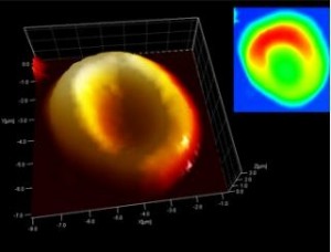
5,000 year old blood analyzed with an AFM (atomic force microscope)

They’ve been able to recover blood from the 5000 year old Iceman Mummy and answer a question regarding his (Ötzi’s) death according to the May 8, 2012 news item on Nanowerk,

His DNA has been decoded; samples from his stomach and intestines have allowed us to reconstruct his very last meal. The circumstances of his violent death appear to have been explained. However, what had, at least thus far, eluded the scientists, was identifying any traces of blood in Ötzi, the 5,000 year old glacier mummy. Examination of his aorta had yielded no results. Yet recently, a team of scientists from Italy and Germany, using nanotechnology, succeeded in locating red blood cells in Ötzi’s wounds, thereby discovering the oldest traces of blood to have been found anywhere in the world …

The research paper “Preservation of 5300 year old red blood cells in the Iceman” ([free access] Interface: Journal of the Royal Society) written by Marek Janko, Robert W. Stark, and Albert Zink helps outsiders like me better understand why there is excitement about finding blood, from the Introduction to the paper (footnotes have been edited out),

Examining mummies with sensitive analytic tools enables the reconstruction of their ancestry and genetic relationships, diet, diseases, living conditions, state of preservation and the mummification processes. While many studies provided molecular evidence for the presence of infectious diseases in ancient populations, leading to deep insights into the evolution of such diseases, only a few reports on the recovery of blood from mummified bodies are available. Previous investigations, based on optical or electron microscopy data, postulated that blood remains or fragments could be preserved in mummies as old as 2000 years. [emphases mine] Although molecular verification of blood findings was not performed, detection of blood components was of major interest because it could give new perspectives on the lives and fates of our ancestors. Blood can indicate the general health status of an individual and it can be analysed to detect pathological conditions or to provide valuable information in forensic crime scene investigations.

So finding blood in the 5000 year old Iceman Mummy means this sample is more than double the age of previous samples. It also answers a question about his death, from the May 8, 2012 news item on Nanowerk,

Whilst examining the wound at the point where the arrow entered the body, the team of scientists also identified fibrin, a protein involved in the clotting of blood. “Because fibrin is present in fresh wounds and then degrades, the theory that Ötzi died some days after he had been injured by the arrow, as had once been mooted, can no longer be upheld,” explains Albert Zink.

They used an atomic force microscope for part of this project,

The team of scientists used an atomic force microscope to investigate thin tissue sections from the wound where the arrow entered Ötzi’s back and from the laceration on his right hand. This nanotechnology instrument scans the surface of the tissue sections using a very fine probe. As the probe moves over the surface, sensors measure every tiny deflection of the probe, line by line and point by point, building up a three-dimensional image of the surface. What emerged was an image of red blood cells with the classic “doughnut shape”, exactly as we find them in healthy people today.

Here’s an image of the blood,

5,000 year old red blood cells discovered. Oldest blood known to modern science. Credit: EURAC

This finding, as exciting as it is from an historical perspective, also hints at possible future applications for modern forensic science, from the May 8, 2012 news item on Nanowerk,

“Up to now there had been uncertainty about how long blood could survive – let alone what human blood cells from the Chalcolithic period, the Copper Stone Age, might look like.” This is how Albert Zink, Head of the Institute for Mummies and the Iceman at the European Academy, Bozen-Bolzano (EURAC) explains the starting point for the investigations which he undertook with Marek Janko and Robert Stark, materials scientists at the Center of Smart Interfaces at Darmstadt Technical University.

Even in modern forensic medicine it has so far been almost impossible to determine how long a trace of blood had been present at a crime scene. Scientists Zink, Janko and Stark are convinced that the nanotechnological methods which they tested out on Ötzi’s blood to analyse the microstructure of blood cells and minute blood clots might possibly lead to a break-through in this area.

EURAC’s Institute for Mummies and the Iceman is rather interesting, from the Institute for Mummies and the Iceman webpage,

The EURAC-Institute for Mummies and the Iceman gathers and coordinates all currently available scientific data on the Iceman and various other mummies.
Founded in 2007, it also supplies new impulses for anthropological, palaeopathological, genetic and medical research. In addition, it promotes innovative techniques for mummy conservation.

The EURAC-Institute for Mummies and the Iceman strongly supports and promotes the use of non-and minimal invasive investigation methods, such as computer tomography, nanotechnology, molecular and biological approaches, as well as ancient DNA research.  It collaborates with several renowned universities and museums worldwide.

The creation of a mummy research centre in Bolzano was generally welcomed, in particular by those who were more or less directly involved with studies on the Iceman, not least because the EURAC-Institute for Mummies and the Iceman assures optimal conservation conditions for the mummy.

One of the EURAC-Institute for Mummies and the Iceman’s tasks is gathering all available scientific data on the Iceman. This includes archaeological site material, as well as papers, notes, documentation material from research groups from all over the world.

The Iceman was found in 1991 in the Alps and is the oldest wet mummy ever discovered (dated 3.300-3.150 BC). He spent seven years in Innsbruck (A) thereafter, where he was extensively studied at the Research Institute for Alpine Prehistory. Meanwhile, the South Tyrolean Museum of Archaeology was established in Bolzano and a specially designed refrigerating chamber was created in order to preserve this unique mummy. In 1998, once the mummy had been moved to Bolzano, the Research Institute for Alpine Prehistory in Innsbruck was closed and all studies concerning the Iceman were carried out at different Universities.

That excerpt from the Institute’s webpage was a bit off tangent but I do find the “specially designed refrigerating chamber” a rather intriguing detail.

Blood-, milk-, and mucus-powered electronics

Researchers at Tel Aviv University ([TAU] Israel) have already begun to develop biodegradable display screens in their quest to create electronic devices powered by blood, milk, and mucus proteins found in our bodies. From the March 7, 2012 news item on Nanowerk,

… a team including Ph.D. students Elad Mentovich and Netta Hendler of TAU’s Department of Chemistry and The Center for Nanoscience and Nanotechnology, with supervisor Dr. Shachar Richter and in collaboration with Prof. Michael Gozin and his Ph.D. student Bogdan Belgorodsky, has brought together cutting-edge techniques from multiple fields of science to create protein-based transistors — semi-conductors used to power electronic devices — from organic materials found in the human body. They could become the basis of a new generation of nano-sized technologies that are both flexible and biodegradable.

The March 7, 2012 news release on the American Friend of TAU website notes some of the issues with silicon-based electronics,

One of the challenges of using silicon as a semi-conductor is that a transistor must be created with a “top down” approach. Manufacturers start with a sheet of silicon and carve it into the shape that is needed, like carving a sculpture out of a rock. This method limits the capabilities of transistors when it comes to factors such as size and flexibility.

The TAU researchers turned to biology and chemistry for a different approach to building the ideal transistor. When they applied various combinations of blood, milk, and mucus proteins to any base material, the molecules self-assembled to create a semi-conducting film on a nano-scale. In the case of blood protein, for example, the film is approximately four nanometers high. The current technology in use now is 18 nanometers, says Mentovich.

Together, the three different kinds of proteins create a complete circuit with electronic and optical capabilities, each bringing something unique to the table. Blood protein has the ability to absorb oxygen, Mentovich says, which permits the “doping” of semi-conductors with specific chemicals in order to create specific technological properties. Milk proteins, known for their strength in difficult environments, form the fibers which are the building blocks of the transistors, while the mucosal proteins have the ability to keep red, green and, blue fluorescent dyes separate, together creating the white light emission that is necessary for advanced optics.

Overall, the natural abilities of each protein give the researchers “unique control” over the resulting organic transistor, allowing adjustments for conductivity, memory storage, and fluorescence among other characteristics.

I have previously featured work on vampire (blood-powered) fuel cells and batteries  in my July 18, 2012 posting and my April 3, 2009 posting so the notion of using blood (and presumably other bodily fluids) as a source for electrical power is generating (pun intended, weak though it is) interest in many research labs.

While the researchers don’t speculate about integrating these new carbon-based devices, which are smaller and more flexible than current devices, in bodies (from the American Friends of TAU news release),

Technology is now shifting from a silicon era to a carbon era, notes Mentovich, and this new type of transistor could play a big role. Transistors built from these proteins will be ideal for smaller, flexible devices that are made out of plastic rather than silicon, which exists in wafer form that would shatter like glass if bent. The breakthrough could lead to a new range of flexible technologies, such as screens, cell phones and tablets, biosensors, and microprocessor chips.

Just as significant, because the researchers are using natural proteins to build their transistor, the products they create will be biodegradable. It’s a far more environmentally friendly technology that addresses the growing problem of electronic waste, which is overflowing landfills worldwide.

The biodegradability of these proposed devices may be a problem if they are integrated into our bodies but it is certain that this will be attempted as we continue to explore machine/flesh possibilities.

Blood, memristors, cyborgs plus brain-controlled computers, prosthetics, and art

The memristor, a circuit element that quite interests me [April 7, 2010 posting], seems to be moving from being a purely electrical engineering term to one that’s used metaphorically to describe biological processes in a way that is transforming my understanding of machine/human (and other animal) interfaces from a science fiction concept to reality.

March 2, 2011 Kate McAlpine wrote an article for the New Scientist which suggested that skin has memristive properties while noting that the same has been said of the brain. From Sweat ducts make skin a memristor,

Synapses, junctions between neurons in the brain, display electrical behaviour that depends on past activity and are said to behave like memristors. This has raised the prospect of using memristors as the basis of an artificial brain.

Now, by re-examining data from the early 1980s on the electrical conductivity of human skin in response to various voltages, Gorm Johnsen and his colleagues at the University of Oslo in Norway have uncovered a more prosaic example of memristive behaviour in nature.

They found that when a negative electrical potential is applied to skin on various parts of the arm, creating a current, that stretch of skin exhibits a low resistance to a subsequent current flowing through the skin. But if the first potential is positive relative to the skin, then a subsequent potential produces a current that meets with a much higher resistance. In other words, the skin has a memory of previous currents. The finding is due to be published in Physical Review E.
The researchers attribute skin’s memristor behaviour to sweat pores.

More recently, there’s been some excitement about a research team in India that’s working with blood so they can eventually create a ‘liquid memristor’. Rachel Courtland wrote a brief item on the ‘blood memristor’ on April 1, 2011 for the IEEE Tech Talk blog,

S.P. Kosta of the Education Campus Changa in Gujarat, India and colleagues have published a paper in the International Journal of Medical Engineering and Informatics showing that human blood changes its electrical resistance depending on how much voltage is applied. It also seems to retain memory of this resistance for at least five minutes.

The team says that makes human blood a memristor: the fourth in the family of fundamental circuit elements that includes the resistor, the capacitor, and the inductor. Proposed in 1971, the memristor’s existence wasn’t proven until 2008, when HP senior fellow Stanley Williams and colleagues demonstrated a memristor device made of doped titanium dioxide.

There was also a March 30, 2011 news item about the Indian research titled, Blood simple circuitry for cyborgs, on Nanowerk, which provided this information,

They [the research team] constructed the laboratory-based biological memristor using a 10 ml test tube filled with human blood held at 37 Celsius into which two electrodes are inserted; appropriate measuring instrumentation was attached. The experimental memristor shows that resistance varies with applied voltage polarity and magnitude and this memory effect is sustained for at least five minutes in the device.

Having demonstrated memristor behavior in blood, the next step was to test that the same behavior would be observed in a device through which blood is flowing. This step was also successful. The next stage will be to develop a micro-channel version of the flow memristor device and to integrate several to carry out particular logic functions. This research is still a long way from an electronic to biological interface, but bodes well for the development of such devices in the future.

Kit Eaton in an April 4, 2011 article (Electronics Made from Human Blood Cells Suggest Cyborg Interfaces, Spark Nightmares) on the Fast Company website gives more details about possible future applications,

Ultimately, the fact that a biological system could be used to interact with a hard semiconductor system could revolutionize biomechanics. That’s because wiring devices like cochlear implants, nerve-triggered artificial limbs and artificial eyeballs into the body at the moment involves a terribly difficult integration of metal wiring–with all the associated risk of infection and rejection. Plus it’s really a very promising first step toward making a cyborg. Countdown to military interest in this tech in 5…4…3…

It should be noted that the team in India is working towards applications in neuroprosthetics. As for the Norwegian team with their ‘sweat duct/skin memristor’, the article did not specify what types of applications, if any, their work might lead to.

As evidenced by the research covered in these news items, the memristor seems to be drifting or, more accurately, developing a second identity/ghost identity as the term is applied to biological processes.

The body as a machine is a notion that’s been around for a while as has the notion of combining the two. The first notion is a metaphor while the second is a staple in science fiction which, in a minor way, has found a home in the real life practice of body hacking where someone implants a magnetic or computer chip into their body (my May 27, 2010 posting). So the memristor becoming a metaphor for certain biological processes doesn’t seem something new but rather the next step in a process that’s well on its way.

Two students at Ryerson University (Toronto, Canada) recently announced that they had developed a brain-controlled prosthetic. From the March 30, 2011 news item on Nanowerk,

Two Ryerson University undergraduate biomedical engineering students are changing the world of medical prosthetics with a newly developed prosthetic arm that is controlled by brain signals. The Artificial Muscle-Operated (AMO) Arm not only enables amputees more range of movement as compared to other prosthetic arms but it allows amputees to avoid invasive surgeries and could potentially save hundreds of thousands of dollars. The AMO Arm is controlled by the user’s brain signals and is powered by ‘artificial muscles’ – simple pneumatic pumps and valves – to create movements. In contrast, traditional prosthetic limbs – which typically offer more limited movements – rely on intricate and expensive electrical and mechanical components.

Developed by third-year student Thiago Caires and second-year student Michal Prywata, the AMO Arm is controlled by the brain and uses compressed air as the main source of power. The digital device makes use of signals in the brain that continue to fire even after a limb is amputated. Users wear a head-set that senses a signal – for example, the thought “up” – and sends it wirelessly to a miniature computer in the arm. The computer then compares the signal to others in a database. The resulting information is sent to the pneumatic system, which in turn, activates the arm to create the correct movement. Simulating the expansion and contraction of real muscles, the system makes use of compressed air from a small, refillable tank in the user’s pocket.

I think what they mean is that the components are not traditionally electrical and mechanical but in fact informed by emerging technologies and the science that supports them. After all, the computer must run on some kind of electricity and brain activity (wireless signals from the brain will be controlling the prosthetic) is often described as electrical. The result is that the human and the machine are effectively made one since the prosthetic arm is controlled as if it were ‘biological’ arm.

On another part of the spectrum, Iraqui artist Wafaa Bilal made headlines recently when he had a camera implanted into the back of his head creating a third eye. Designed to be a one year project, the artist had to remove the camera when he developed an infection at the site of one of the metal posts used to anchor the camera to his head. From the Feb. 11, 2011 BBC news item,

An artist who had a camera implanted into the back of his head has been forced to remove it after his body rejected part of the device.

Iraqi-born Wafaa Bilal had surgery last week to remove one of three posts holding the camera in place as it posed a risk of infection.

The camera had been taking a photo every minute as part of a year-long project.

Wafaa Bilal and camera (image downloaded from BBC website)

(The artist would like to try it again but, in the meantime, has slung the camera around his neck as a substitute.)

In Bilal’s case, the body is being significantly altered as the machine (camera) is implanted in a place (back of head) where no animal has them located.

What I’m getting at with all of this is that at the same time we seem to be expanding the memristor’s meaning from a term used to describe a concept in electrical engineering to include biological processes, we are exploring new ways of integrating machinery into our bodies. In effect our relationships to our bodies and machines are changing and that change can be traced in the language we use to describe ourselves.