Tag Archives: blood vessels

First 3D heart printed using patient’s biological materials

This is very exciting news and it’s likely be at least 10 years before this technology could be made available to the public.
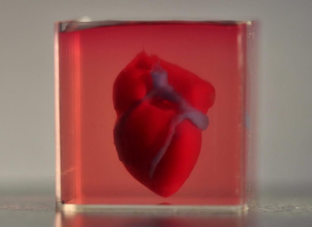
Caption: A 3D-printed, small-scaled human heart engineered from the patient’s own materials and cells. Credit: Advanced Science. © 2019 The Authors.

An April 15, 2019 news item on ScienceDaily makes a remarkable announcement,

In a major medical breakthrough, Tel Aviv University researchers have “printed” the world’s first 3D vascularised engineered heart using a patient’s own cells and biological materials. Their findings were published on April 15 [2019] in a study in Advanced Science.

Until now, scientists in regenerative medicine — a field positioned at the crossroads of biology and technology — have been successful in printing only simple tissues without blood vessels.

“This is the first time anyone anywhere has successfully engineered and printed an entire heart replete with cells, blood vessels, ventricles and chambers,” says Prof. Tal Dvir of TAU’s School of Molecular Cell Biology and Biotechnology, Department of Materials Science and Engineering, Center for Nanoscience and Nanotechnology and Sagol Center for Regenerative Biotechnology, who led the research for the study.

An April 15, 2019 Amricna Friends of Tel Aviv University (TAU) news release (also on EurekAlert), which originated the news item, provides more detail,

Heart disease is the leading cause of death among both men and women in the United States. Heart transplantation is currently the only treatment available to patients with end-stage heart failure. Given the dire shortage of heart donors, the need to develop new approaches to regenerate the diseased heart is urgent.

“This heart is made from human cells and patient-specific biological materials. In our process these materials serve as the bioinks, substances made of sugars and proteins that can be used for 3D printing of complex tissue models,” Prof. Dvir says. “People have managed to 3D-print the structure of a heart in the past, but not with cells or with blood vessels. Our results demonstrate the potential of our approach for engineering personalized tissue and organ replacement in the future.

Research for the study was conducted jointly by Prof. Dvir, Dr. Assaf Shapira of TAU’s Faculty of Life Sciences and Nadav Moor, a doctoral student in Prof. Dvir’s lab.

“At this stage, our 3D heart is small, the size of a rabbit’s heart, [emphasis mine] ” explains Prof. Dvir. “But larger human hearts require the same technology.”

For the research, a biopsy of fatty tissue was taken from patients. The cellular and a-cellular materials of the tissue were then separated. While the cells were reprogrammed to become pluripotent stem cells, the extracellular matrix (ECM), a three-dimensional network of extracellular macromolecules such as collagen and glycoproteins, were processed into a personalized hydrogel that served as the printing “ink.”

After being mixed with the hydrogel, the cells were efficiently differentiated to cardiac or endothelial cells to create patient-specific, immune-compatible cardiac patches with blood vessels and, subsequently, an entire heart.

According to Prof. Dvir, the use of “native” patient-specific materials is crucial to successfully engineering tissues and organs.

“The biocompatibility of engineered materials is crucial to eliminating the risk of implant rejection, which jeopardizes the success of such treatments,” Prof. Dvir says. “Ideally, the biomaterial should possess the same biochemical, mechanical and topographical properties of the patient’s own tissues. Here, we can report a simple approach to 3D-printed thick, vascularized and perfusable cardiac tissues that completely match the immunological, cellular, biochemical and anatomical properties of the patient.”

The researchers are now planning on culturing the printed hearts in the lab and “teaching them to behave” like hearts, Prof. Dvir says. They then plan to transplant the 3D-printed heart in animal models.

“We need to develop the printed heart further,” he concludes. “The cells need to form a pumping ability; they can currently contract, but we need them to work together. Our hope is that we will succeed and prove our method’s efficacy and usefulness.

“Maybe, in ten years, there will be organ printers in the finest hospitals around the world, and these procedures will be conducted routinely.”

Growing the heart to human size and getting the cells to work together so the heart will pump makes it seem like the 10 years Dvir imagines as the future date when there will be organ printers in hospitals routinely printing up hearts seems a bit optimistic. Regardless, I hope he’s right. Bravo to these Israeli researchers!

Here’s a link to and a citation for the paper,

3D Printing of Personalized Thick and Perfusable Cardiac Patches and Hearts by Nadav Noor, Assaf Shapira, Reuven Edri, Idan Gal, Lior Wertheim, Tal Dvir. Advanced Science DOI: https://doi.org/10.1002/advs.201900344 First published: 15 April 2019

This paper is open access.

Growing perfect human blood vessels in a Petri dish

I had not realized that blood vessels are considered organs (Live and learn.) The big news about blood vessel organoids was announced in a January 16, 2019 news item on ScienceDaily,

Scientists have managed to grow perfect human blood vessels as organoids in a petri dish for the first time

The breakthrough engineering technology, outlined in a new study published today [January 16, 2019] in Nature, dramatically advances research of vascular diseases like diabetes, identifying a key pathway to potentially prevent changes to blood vessels — a major cause of death and morbidity among those with diabetes.

A January 16, 2019 University of British Columbia (UBC; Canada) news release (also on EurekAlert), which originated the news item, explains organoids and describes the work in more detail,

An organoid is a three-dimensional structure grown from stem cells that mimics an organ and can be used to study aspects of that organ in a petri dish.

“Being able to build human blood vessels as organoids from stem cells is a game changer,” said the study’s senior author Josef Penninger, the Canada 150 Research Chair in Functional Genetics, director of the Life Sciences Institute at UBC and founding director of the Institute for Molecular Biotechnology of the Austrian Academy of Sciences (IMBA).

“Every single organ in our body is linked with the circulatory system. This could potentially allow researchers to unravel the causes and treatments for a variety of vascular diseases, from Alzheimer’s disease, cardiovascular diseases, wound healing problems, stroke, cancer and, of course, diabetes.”

Diabetes affects an estimated 420 million people worldwide. Many diabetic symptoms are the result of changes in blood vessels that result in impaired blood circulation and oxygen supply of tissues. Despite its prevalence, very little is known about the vascular changes arising from diabetes. This limitation has slowed the development of much-needed treatment.

To tackle this problem, Penninger and his colleagues developed a groundbreaking model: three-dimensional human blood vessel organoids grown in a petri dish. These so-called “vascular organoids” can be cultivated using stem cells in the lab, strikingly mimicking the structure and function of real human blood vessels.

When researchers transplanted the blood vessel organoids into mice, they found that they developed into perfectly functional human blood vessels including arteries and capillaries. The discovery illustrates that it is possible to not only engineer blood vessel organoids from human stem cells in a dish, but also to grow a functional human vascular system in another species.

“What is so exciting about our work is that we were successful in making real human blood vessels out of stem cells,” said Reiner Wimmer, the study’s first author and a postdoctoral research fellow at IMBA. “Our organoids resemble human capillaries to a great extent, even on a molecular level, and we can now use them to study blood vessel diseases directly on human tissue.”

One feature of diabetes is that blood vessels show an abnormal thickening of the basement membrane. As a result, the delivery of oxygen and nutrients to cells and tissues is strongly impaired, causing a multitude of health problems, such as kidney failure, heart attacks, strokes, blindness and peripheral artery disease, leading to amputations.

The researchers then exposed the blood vessel organoids to a “diabetic” environment in a petri dish.

“Surprisingly, we could observe a massive expansion of the basement membrane in the vascular organoids,” said Wimmer. “This typical thickening of the basement membrane is strikingly similar to the vascular damage seen in diabetic patients.”

The researchers then searched for chemical compounds that could block thickening of the blood vessel walls. They found none of the current anti-diabetic medications had any positive effects on these blood vessel defects. However, they discovered that an inhibitor of γ-secretase, a type of enzyme in the body, prevented the thickening of the blood vessel walls, suggesting, at least in animal models, that blocking γ-secretase could be helpful in treating diabetes.

The researchers say the findings could allow them to identify underlying causes of vascular disease, and to potentially develop and test new treatments for patients with diabetes.

Here’s a link to and a citation for the paper,

Human blood vessel organoids as a model of diabetic vasculopathy by Reiner A. Wimmer, Alexandra Leopoldi, Martin Aichinger, Nikolaus Wick, Brigitte Hantusch, Maria Novatchkova, Jasmin Taubenschmid, Monika Hämmerle, Christopher Esk, Joshua A. Bagley, Dominik Lindenhofer, Guibin Chen, Manfred Boehm, Chukwuma A. Agu, Fengtang Yang, Beiyuan Fu, Johannes Zuber, Juergen A. Knoblich, Dontscho Kerjaschki & Josef M. Penninger. Nature volume 565, pages505–510 (2019) DOI: https://doi.org/10.1038/s41586-018-0858-8 Issue Date: 24 January 2019

This paper is behind a paywall. One other thing, a patent application has been filed according to the Author information section (subsection: Competing interests) of the abstract.

3D bioprinting: a conference about the latest trends (May 3 – 5, 2017 at the University of British Columbia, Vancouver)

The University of British Columbia’s (UBC) Peter Wall Institute for Advanced Studies (PWIAS) is hosting along with local biotech firm, Aspect Biosystems, a May 3 -5, 2017 international research roundtable known as ‘Printing the Future of Therapeutics in 3D‘.

A May 1, 2017 UBC news release (received via email) offers some insight into the field of bioprinting from one of the roundtable organizers,

This week, global experts will gather [4] at the University of British
Columbia to discuss the latest trends in 3D bioprinting—a technology
used to create living tissues and organs.

In this Q&A, UBC chemical and biological engineering professor
Vikramaditya Yadav [5], who is also with the Regenerative Medicine
Cluster Initiative in B.C., explains how bioprinting could potentially
transform healthcare and drug development, and highlights Canadian
innovations in this field.

WHY IS 3D BIOPRINTING SIGNIFICANT?

Bioprinted tissues or organs could allow scientists to predict
beforehand how a drug will interact within the body. For every
life-saving therapeutic drug that makes its way into our medicine
cabinets, Health Canada blocks the entry of nine drugs because they are
proven unsafe or ineffective. Eliminating poor-quality drug candidates
to reduce development costs—and therefore the cost to consumers—has
never been more urgent.

In Canada alone, nearly 4,500 individuals are waiting to be matched with
organ donors. If and when bioprinters evolve to the point where they can
manufacture implantable organs, the concept of an organ transplant
waiting list would cease to exist. And bioprinted tissues and organs
from a patient’s own healthy cells could potentially reduce the risk
of transplant rejection and related challenges.

HOW IS THIS TECHNOLOGY CURRENTLY BEING USED?

Skin, cartilage and bone, and blood vessels are some of the tissue types
that have been successfully constructed using bioprinting. Two of the
most active players are the Wake Forest Institute for Regenerative
Medicine in North Carolina, which reports that its bioprinters can make
enough replacement skin to cover a burn with 10 times less healthy
tissue than is usually needed, and California-based Organovo, which
makes its kidney and liver tissue commercially available to
pharmaceutical companies for drug testing.

Beyond medicine, bioprinting has already been commercialized to print
meat and artificial leather. It’s been estimated that the global
bioprinting market will hit $2 billion by 2021.

HOW IS CANADA INVOLVED IN THIS FIELD?

Canada is home to some of the most innovative research clusters and
start-up companies in the field. The UBC spin-off Aspect Biosystems [6]
has pioneered a bioprinting paradigm that rapidly prints on-demand
tissues. It has successfully generated tissues found in human lungs.

Many initiatives at Canadian universities are laying strong foundations
for the translation of bioprinting and tissue engineering into
mainstream medical technologies. These include the Regenerative Medicine
Cluster Initiative in B.C., which is headed by UBC, and the University
of Toronto’s Institute of Biomaterials and Biomedical Engineering.

WHAT ETHICAL ISSUES DOES BIOPRINTING CREATE?

There are concerns about the quality of the printed tissues. It’s
important to note that the U.S. Food and Drug Administration and Health
Canada are dedicating entire divisions to regulation of biomanufactured
products and biomedical devices, and the FDA also has a special division
that focuses on regulation of additive manufacturing – another name
for 3D printing.

These regulatory bodies have an impressive track record that should
assuage concerns about the marketing of substandard tissue. But cost and
pricing are arguably much more complex issues.

Some ethicists have also raised questions about whether society is not
too far away from creating Replicants, à la _Blade Runner_. The idea is
fascinating, scary and ethically grey. In theory, if one could replace
the extracellular matrix of bones and muscles with a stronger substitute
and use cells that are viable for longer, it is not too far-fetched to
create bones or muscles that are stronger and more durable than their
natural counterparts.

WILL DOCTORS BE PRINTING REPLACEMENT BODY PARTS IN 20 YEARS’ TIME?

This is still some way off. Optimistically, patients could see the
technology in certain clinical environments within the next decade.
However, some technical challenges must be addressed in order for this
to occur, beginning with faithful replication of the correct 3D
architecture and vascularity of tissues and organs. The bioprinters
themselves need to be improved in order to increase cell viability after
printing.

These developments are happening as we speak. Regulation, though, will
be the biggest challenge for the field in the coming years.

There are some events open to the public (from the international research roundtable homepage),

OPEN EVENTS

You’re invited to attend the open events associated with Printing the Future of Therapeutics in 3D.

Café Scientifique

Thursday, May 4, 2017
Telus World of Science
5:30 – 8:00pm [all tickets have been claimed as of May 2, 2017 at 3:15 pm PT]

3D Bioprinting: Shaping the Future of Health

Imagine a world where drugs are developed without the use of animals, where doctors know how a patient will react to a drug before prescribing it and where patients can have a replacement organ 3D-printed using their own cells, without dealing with long donor waiting lists or organ rejection. 3D bioprinting could enable this world. Join us for lively discussion and dessert as experts in the field discuss the exciting potential of 3D bioprinting and the ethical issues raised when you can print human tissues on demand. This is also a rare opportunity to see a bioprinter live in action!

Open Session

Friday, May 5, 2017
Peter Wall Institute for Advanced Studies
2:00 – 7:00pm

A Scientific Discussion on the Promise of 3D Bioprinting

The medical industry is struggling to keep our ageing population healthy. Developing effective and safe drugs is too expensive and time-consuming to continue unchanged. We cannot meet the current demand for transplant organs, and people are dying on the donor waiting list every day.  We invite you to join an open session where four of the most influential academic and industry professionals in the field discuss how 3D bioprinting is being used to shape the future of health and what ethical challenges may be involved if you are able to print your own organs.

ROUNDTABLE INFORMATION

The University of British Columbia and the award-winning bioprinting company Aspect Biosystems, are proud to be co-organizing the first “Printing the Future of Therapeutics in 3D” International Research Roundtable. This event will congregate global leaders in tissue engineering research and pharmaceutical industry experts to discuss the rapidly emerging and potentially game-changing technology of 3D-printing living human tissues (bioprinting). The goals are to:

Highlight the state-of-the-art in 3D bioprinting research
Ideate on disruptive innovations that will transform bioprinting from a novel research tool to a broadly adopted systematic practice
Formulate an actionable strategy for industry engagement, clinical translation and societal impact
Present in a public forum, key messages to educate and stimulate discussion on the promises of bioprinting technology

The Roundtable will bring together a unique collection of industry experts and academic leaders to define a guiding vision to efficiently deploy bioprinting technology for the discovery and development of new therapeutics. As the novel technology of 3D bioprinting is more broadly adopted, we envision this Roundtable will become a key annual meeting to help guide the development of the technology both in Canada and globally.

We thank you for your involvement in this ground-breaking event and look forward to you all joining us in Vancouver for this unique research roundtable.

Kind Regards,
The Organizing Committee
Christian Naus, Professor, Cellular & Physiological Sciences, UBC
Vikram Yadav, Assistant Professor, Chemical & Biological Engineering, UBC
Tamer Mohamed, CEO, Aspect Biosystems
Sam Wadsworth, CSO, Aspect Biosystems
Natalie Korenic, Business Coordinator, Aspect Biosystems

I’m glad to see this event is taking place—and with public events too! (Wish I’d seen the Café Scientifique announcement earlier when I first checked for tickets  yesterday. I was hoping there’d been some cancellations today.) Finally, for the interested, you can find Aspect Biosystems here.

Gluing blood vessels with mussel goo

The University of British Columbia [UBC] Dec. 11, 2012 news release states,

A University of British Columbia researcher has helped create a gel – based on the mussel’s knack for clinging to rocks, piers and boat hulls – that can be painted onto the walls of blood vessels and stay put, forming a protective barrier with potentially life-saving implications.

Co-invented by Assistant Professor Christian Kastrup while a postdoctoral student at the Massachusetts Institute of Technology, the gel is similar to the amino acid that enables mussels to resist the power of churning water. The variant that Kastrup and his collaborators created, described in the current issue of the online journal PNAS [Proceeings of the National Academy of Sciences of the US] Early Edition, can withstand the flow of blood through arteries and veins.

Here’s the citation and a link to the article (which is behind a paywall),

Painting blood vessels and atherosclerotic plaques with an adhesive drug depot by Christian J. Kastrup, Matthias Nahrendorf, Jose Luiz Figueiredo, Haeshin Lee, Swetha Kambhampati, Timothy Lee, Seung-Woo Cho, Rostic Gorbatov, Yoshiko Iwamoto, Tram T. Dang, Partha Dutta, Ju Hun Yeon, Hao Cheng, Christopher D. Pritchard, Arturo J. Vegas, Cory D. Siegel, Samantha MacDougall, Michael Okonkwo, Anh Thai, James R. Stone, Arthur J. Coury, Ralph Weissleder, Robert Langer, and Daniel G. Anderson.  PNAS, December 11, 2012 DOI: 10.1073/pnas.1217972110

For those of a more technical turn of mind, here’s the abstract (from PNAS),

The treatment of diseased vasculature remains challenging, in part because of the difficulty in implanting drug-eluting devices without subjecting vessels to damaging mechanical forces. Implanting materials using adhesive forces could overcome this challenge, but materials have previously not been shown to durably adhere to intact endothelium under blood flow. Marine mussels secrete strong underwater adhesives that have been mimicked in synthetic systems. Here we develop a drug-eluting bioadhesive gel that can be locally and durably glued onto the inside surface of blood vessels. In a mouse model of atherosclerosis, inflamed plaques treated with steroid-eluting adhesive gels had reduced macrophage content and developed protective fibrous caps covering the plaque core. Treatment also lowered plasma cytokine levels and biomarkers of inflammation in the plaque. The drug-eluting devices developed here provide a general strategy for implanting therapeutics in the vasculature using adhesive forces and could potentially be used to stabilize rupture-prone plaques.

The news release describes the work layperson’s terms,

The gel’s “sheer strength” could shore up weakened vessel walls at risk of rupturing – much like the way putty can fill in dents in a wall, says Kastrup, a member of the Department of Biochemistry and Molecular Biology and the Michael Smith Laboratories.

By forming a stable barrier between blood and the vessel walls, the gel could also prevent the inflammation that typically occurs when a stent is inserted to widen a narrowed artery or vein; that inflammation often counteracts the opening of the vessel that the stent was intended to achieve.

The widest potential application would be preventing the rupture of blood vessel plaque. When a plaque ruptures, the resulting clot can block blood flow to the heart (triggering a heart attack) or the brain (triggering a stroke). Mice treated with a combination of the gel and an anti-inflammatory steroid had more stable plaque than a control group of untreated mice.

“By mimicking the mussel’s ability to cling to objects, we created a substance that stays in place in a very dynamic environment with high flow velocities,” says Kastrup, a member of UBC’s Centre for Blood Research.

Robert Langer, one of the paper’s co-authors, was mentioned here in an Aug. 27, 2012 posting about nanoelectronics, tissue engineering, and medicine.