Tag Archives: blue light

Mystery of North American insect bioluminescent systems unraveled by Brazilian scientists

I’ve always been fond of ‘l’ words and so it is that I’m compelled to post a story about a “luciferin-luciferase system” or, in this case, a story about insect bioluminescence.
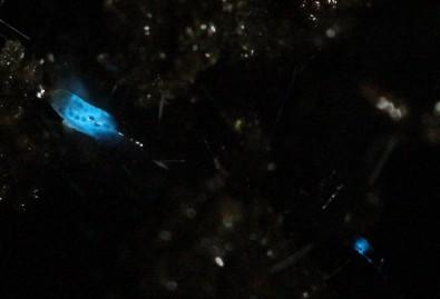
Caption: Researchers isolated molecules present in the larvae of the fungus gnat Orfelia fultoni Credit: Vadim Viviani, UFSCar

A September 9, 2020 Fundação de Amparo à Pesquisa do Estado de São Paulo (FAPESP) press release (also on EurekAlert but published Sept. 11, 2020) announces research into ‘blue’ bioluminescence,

Molecules belonging to an almost unknown bioluminescent system found in larvae of the fungus gnat Orfelia fultoni (subfamily Keroplatinae) have been isolated for the first time by researchers at the Federal University of São Carlos (UFSCar) in the state of São Paulo, Brazil. The small fly is one of the few terrestrial organisms that produce blue light. It inhabits riverbanks in the Appalachian Mountains in the eastern United States. A key part of its bioluminescent system is a molecule also present in two recently discovered Brazilian flies.

The study, supported by Paulo Research Foundation – FAPESP, is published in Scientific Reports. Five authors are affiliated with UFSCar and two with universities in the United States.

The bioluminescent systems of glow-worms, fireflies and other insects are normally made up of luciferin (a low molecular weight molecule) and luciferase, an enzyme that catalyzes the oxidation of luciferin by oxygen, producing light. While some bioluminescent systems are well known and even used in biotechnological applications, others are poorly understood, including blue light-emitting systems, such as that of O. fultoni.

“In the published paper, we describe the properties of the insect’s luciferase and luciferin and their anatomical location in its larvae. We also specify several possible proteins that are possible candidates for the luciferase. We don’t yet know what type of protein it is, but it’s likely to be a hexamerin. In insects, hexamerins are storage proteins that provide amino acids, besides having other functions, such as binding low molecular weight compounds, like luciferin,” said Vadim Viviani, a professor in UFSCar’s Sustainability Science and Technology Center (CCTS) in Sorocaba, São Paulo, and principal investigator for the study.

The study was part of the FAPESP-funded project “Arthropod bioluminescence“. The partnership with United States-based researchers dates from a previous project, supported by FAPESP and the United States National Science Foundation (NSF), in partnership with Vanderbilt University (VU), located in Nashville, Tennessee.

In addition to luciferin and luciferase, researchers began characterizing a complex found in insects of the family Keroplatidae, which, in addition to O. fultoni, also includes a Brazilian species in the genus Neoditomyia that produces only luciferin and hence does not emit light.

Because they do not use it to emit light, the luciferin in O. fultoni and the Brazilian Neoditomyia has been named keroplatin. In larvae of this subfamily, keroplatin is associated with “black bodies” – large cells containing dark granules, proteins and probably mitochondria (energy-producing organelles). Researchers are still investigating the biological significance of this association between keroplatin and mitochondria.

“It’s a mystery,” Viviani said. “This luciferin may play a role in the mitochondrial energy metabolism. At night, probably in the presence of a natural chemical reducer, the luciferin is released by these black bodies and reacts with the surrounding luciferase to produce blue light. These are possibilities we plan to study.”

Brazilian cousins

An important factor in the elucidation of the United States insect’s bioluminescent system was the discovery of a larva that lives in Intervales State Park in São Paulo in 2018. It does not emit light but produces luciferin, similar to O. fultoni (read more at: agencia.fapesp.br/29066).

In their latest study, the group injected purified luciferase from the United States species into larvae of the Brazilian species, which then produced blue light. The nonluminescent Brazilian species is more abundant in nature than the United States species, so a larger amount of the material could be obtained for study purposes, especially to characterize the luciferin (keroplatin) present in both species.

In 2019, the group discovered and described Neoceroplatus betaryensis, a new species of fungus gnat, in collaboration with Cassius Stevani, a professor at the University of São Paulo’s Institute of Chemistry (IQ-USP). It was the first blue light-emitting insect found in South America and was detected in a privately held forest reserve near the Upper Ribeira State Tourist Park (PETAR) in the southern portion of the state of São Paulo. A close relative of O. fultoni, N. betaryensis inhabits fallen tree trunks in humid places (read more at: agencia.fapesp.br/31797).

“We show that the bioluminescent system of this Brazilian species is identical to that of O. fultoni. However, the insect is very rare, and so it’s hard to obtain sufficient material for research purposes,” Viviani said.

The researchers are now cloning the insect’s luciferase and characterizing it in molecular terms. They are also analyzing the chemical structure of its luciferin and the morphology of its lanterns.

“Once all this has been determined, we’ll be able to synthesize the luciferin and luciferase in the lab and use these systems in a range of biotech applications, such as studying cells. This will help us understand more about human diseases, among other things,” Viviani said.

Here’s a link to and a citation for the paper,

A new brilliantly blue-emitting luciferin-luciferase system from Orfelia fultoni and Keroplatinae (Diptera) by Vadim R. Viviani, Jaqueline R. Silva, Danilo T. Amaral, Vanessa R. Bevilaqua, Fabio C. Abdalla, Bruce R. Branchini & Carl H. Johnson. Scientific Reports volume 10, Article number: 9608 (2020) DOI: https://doi.org/10.1038/s41598-020-66286-1 Published 15 June 2020

This paper is open access.

Blue quantum dots and your television screen

Scientists used equipment at the Canadian Light Source (CLS; synchrotron in Saskatoon, Saskatchewan, Canada) in the quest for better glowing dots on your television (maybe computers and telephones, too?) screen. From an August 20, 2020 news item on Nanowerk,

There are many things quantum dots could do, but the most obvious place they could change our lives is to make the colours on our TVs and screens more pristine. Research using the Canadian Light Source (CLS) at the University of Saskatchewan is helping to bring this technology closer to our living rooms.

An August 19, 2020 CLS news release (also received via email) by Victoria Martinez, which originated the news item, explains what quantum dots are and fills in with technical details about this research,

Quantum dots are nanocrystals that glow, a property that scientists have been working with to develop next-generation LEDs. When a quantum dot glows, it creates very pure light in a precise wavelength of red, blue or green. Conventional LEDs, found in our TV screens today, produce white light that is filtered to achieve desired colours, a process that leads to less bright and muddier colours.

Until now, blue-glowing quantum dots, which are crucial for creating a full range of colour, have proved particularly challenging for researchers to develop. However, University of Toronto (U of T) researcher Dr. Yitong Dong and collaborators have made a huge leap in blue quantum dot fluorescence, results they recently published in Nature Nanotechnology.

“The idea is that if you have a blue LED, you have everything. We can always down convert the light from blue to green and red,” says Dong. “Let’s say you have green, then you cannot use this lower-energy light to make blue.”

The team’s breakthrough has led to quantum dots that produce green light at an external quantum efficiency (EQE) of 22% and blue at 12.3%. The theoretical maximum efficiency is not far off at 25%, and this is the first blue perovskite LED reported as achieving an EQE higher than 10%.

The Science

Dong has been working in the field of quantum dots for two years in Dr. Edward Sargent’s research group at the U of T. This astonishing increase in efficiency took time, an unusual production approach, and overcoming several scientific hurdles to achieve.

CLS techniques, particularly GIWAXS [grazing incidence wide-angle X-ray scattering] on the HXMA beamline [hard X-ray micro-analysis (HXMA)], allowed the researchers to verify the structures achieved in their quantum dot films. This validated their results and helped clarify what the structural changes achieve in terms of LED performance.

“The CLS was very helpful. GIWAXS is a fascinating technique,” says Dong.

The first challenge was uniformity, important to ensuring a clear blue colour and to prevent the LED from moving towards producing green light.

“We used a special synthetic approach to achieve a very uniform assembly, so every single particle has the same size and shape. The overall film is nearly perfect and maintains the blue emission conditions all the way through,” says Dong.

Next, the team needed to tackle the charge injection needed to excite the dots into luminescence. Since the crystals are not very stable, they need stabilizing molecules to act as scaffolding and support them. These are typically long molecule chains, with up to 18 carbon-non-conductive molecules at the surface, making it hard to get the energy to produce light.

“We used a special surface structure to stabilize the quantum dot. Compared to the films made with long chain molecules capped quantum dots, our film has 100 times higher conductivity, sometimes even 1000 times higher.”

This remarkable performance is a key benchmark in bringing these nanocrystal LEDs to market. However, stability remains an issue and quantum dot LEDs suffer from short lifetimes. Dong is excited about the potential for the field and adds, “I like photons, these are interesting materials, and, well, these glowing crystals are just beautiful.”

Here’s a link to and a citation for the paper,

Bipolar-shell resurfacing for blue LEDs based on strongly confined perovskite quantum dots by Yitong Dong, Ya-Kun Wang, Fanglong Yuan, Andrew Johnston, Yuan Liu, Dongxin Ma, Min-Jae Choi, Bin Chen, Mahshid Chekini, Se-Woong Baek, Laxmi Kishore Sagar, James Fan, Yi Hou, Mingjian Wu, Seungjin Lee, Bin Sun, Sjoerd Hoogland, Rafael Quintero-Bermudez, Hinako Ebe, Petar Todorovic, Filip Dinic, Peicheng Li, Hao Ting Kung, Makhsud I. Saidaminov, Eugenia Kumacheva, Erdmann Spiecker, Liang-Sheng Liao, Oleksandr Voznyy, Zheng-Hong Lu, Edward H. Sargent. Nature Nanotechnology volume 15, pages668–674(2020) DOI: https://doi.org/10.1038/s41565-020-0714-5 Published: 06 July 2020 Issue Date: August 2020

This paper is behind a paywall.

If you search “Edward Sargent,” he’s the last author listed in the citation, here on this blog, you will find a number of postings that feature work from his laboratory at the University of Toronto.

Brighten and whiten your teeth (more safely) with nanoparticles?

This is for anyone who’s ever suspected that the all the tooth brightening and whitening might not be such a good idea after all. A July 18, 2018 news item on Nanowerk announces work on what scientists hope will be a safer way to whiten teeth (Note: A link has been removed),

In the age of Instagram and Snapchat, everyone wants to have perfect pearly whites. To get a brighter smile, consumers can opt for over the counter teeth-whitening treatments or a trip to the dentist to have their teeth bleached professionally. But both types of treatments can harm teeth.

According to an article published in ACS Biomaterials Science & Engineering (“Blue-Light -Activated Nano-TiO2@PDA for Highly Effective and Nondestructive Tooth Whitening”), researchers have now developed a new, less destructive method.

A July 18, 2018 American Chemical Society (ACS) news release (also on EurekAlert), which originated the news item expands on the theme,

Teeth can become discolored on their outer surfaces when people consume colored foods and drinks, such as coffee, tea or red wine. As a result, many people turn to non-invasive whitening treatments that bleach the teeth. Currently, the most common bleaching agent is hydrogen peroxide, which steals electrons from the pigment molecules that cause teeth discoloration, and this process can be sped up by exposing teeth to blue light. But high concentrations of hydrogen peroxide can break down a tooth’s enamel, causing sensitivity or cell death. So, Xiaolei Wang, Lan Liao and colleagues wanted to see if a different blue-light-activated compound could be a safer, but still effective, alternative.

The team modified titanium dioxide nanoparticles with polydopamine (nano-TiO2@PDA) so that they could be activated with blue light. In a proof-of-concept experiment, the nano-TiO2@PDA particles were evenly coated on the surface of a tooth and irradiated with blue light. After four hours of treatment, the whitening level was similar to that obtained with hydrogen-peroxide-based agents. The group notes that no significant enamel damage was found on the surface of the tooth, and the treatment was significantly less cytotoxic than hydrogen peroxide. In addition, the nano-TiO2@PDA therapy showed antibacterial activity against certain bacteria.

Here’s a link to and a citation for the paper,

Blue-Light -Activated Nano-TiO2@PDA for Highly Effective and Nondestructive Tooth Whitening by Feng Zhang, Chongxue Wu, Ziyu Zhou, Jiaolong Wang, Weiwei Bao, Lina Dong, Zihao Zhang, Jing Ye, Lan Liao, and Xiaolei Wang. ACS Biomater. Sci. Eng., Article ASAP DOI: 10.1021/acsbiomaterials.8b00548 Publication Date (Web): June 19, 2018

Copyright © 2018 American Chemical Society

This paper is behind a paywall.

Of course, there’s always the question of what happens as we pour more and more engineered titanium dioxide nanoparticles into our bodies and ultimately into the environment.