Tag Archives: bone regeneration

Hot nano-chisel for creating artificial bones?

If ‘chisel’ made you think of sculpting, you are correct. The researchers are alluding to the process of sculpting in their research.
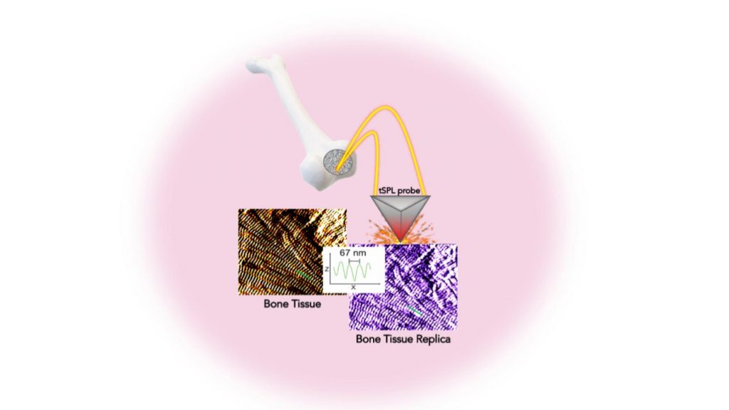
Researchers were able to replicate — with sub-15 nm resolution — bone tissue structure in a biocompatible material using thermal scanning probe lithography. This method opens up unprecedented possibilities for pioneering new stem cell studies and biomedical applications. Courtesy: New York University Tandon School of Engineering

From a February 9, 2021 news item on phys.org (Note: Links have been removed),

A holy grail for orthopedic research is a method for not only creating artificial bone tissue that precisely matches the real thing, but does so in such microscopic detail that it includes tiny structures potentially important for stem cell differentiation, which is key to bone regeneration.

Researchers at the NYU [New York University] Tandon School of Engineering and New York Stem Cell Foundation Research Institute (NYSF) have taken a major step by creating the exact replica of a bone using a system that pairs biothermal imaging with a heated “nano-chisel.” In a study, “Cost and Time Effective Lithography of Reusable Millimeter Size Bone Tissue Replicas with Sub-15 nm Feature Size on a Biocompatible Polymer,” which appears in the journal Advanced Functional Materials, the investigators detail a system allowing them to sculpt, in a biocompatible material, the exact structure of the bone tissue, with features smaller than the size of a single protein—a billion times smaller than a meter. This platform, called, bio-thermal scanning probe lithography (bio-tSPL), takes a “photograph” of the bone tissue, and then uses the photograph to produce a bona-fide replica of it.

The team, led by Elisa Riedo, professor of chemical and biomolecular engineering at NYU Tandon, and Giuseppe Maria de Peppo, a Ralph Lauren Senior Principal Investigator at the NYSF, demonstrated that it is possible to scale up bio-tSPL to produce bone replicas on a size meaningful for biomedical studies and applications, at an affordable cost. These bone replicas support the growth of bone cells derived from a patient’s own stem cells, creating the possibility of pioneering new stem cell applications with broad research and therapeutic potential. This technology could revolutionize drug discovery and result in the development of better orthopedic implants and devices.

A February 8, 2021 NYU Tandon School of Engineering news release (also on EurekAlert but published February 9, 2021), which originated the news item, explains the work in further detail,

In the human body, cells live in specific environments that control their behavior and support tissue regeneration via provision of morphological and chemical signals at the molecular scale. In particular, bone stem cells are embedded in a matrix of fibers — aggregates of collagen molecules, bone proteins, and minerals. The bone hierarchical structure consists of an assembly of micro- and nano- structures, whose complexity has hindered their replication by standard fabrication methods so far.

“tSPL is a powerful nanofabrication method that my lab pioneered a few years ago, and it is at present implemented by using a commercially available instrument, the NanoFrazor,” said Riedo. “However, until today, limitations in terms of throughput and biocompatibility of the materials have prevented its use in biological research. We are very excited to have broken these barriers and to have led tSPL into the realm of biomedical applications.”

Its time- and cost-effectiveness, as well as the cell compatibility and reusability of the bone replicas, make bio-tSPL an affordable platform for the production of surfaces that perfectly reproduce any biological tissue with unprecedented precision.

“I am excited about the precision achieved using bio-tSPL. Bone-mimetic surfaces, such as the one reproduced in this study, create unique possibilities for understanding cell biology and modeling bone diseases, and for developing more advanced drug screening platforms,” said de Peppo. “As a tissue engineer, I am especially excited that this new platform could also help us create more effective orthopedic implants to treat skeletal and maxillofacial defects resulting from injury or disease.”

Here’s a link to and a citation for the paper,

Cost and Time Effective Lithography of Reusable Millimeter Size Bone Tissue Replicas With Sub‐15 nm Feature Size on A Biocompatible Polymer by Xiangyu Liu, Alessandra Zanut, Martina Sladkova‐Faure, Liyuan Xie, Marcus Weck, Xiaorui Zheng, Elisa Riedo, Giuseppe Maria de Peppo. Advanced Functional Materials DOI: https://doi.org/10.1002/adfm.202008662 First published: 05 February 2021

This paper is behind a paywall.
Regenerating dental enamel

For anyone who’s concerned about their dental enamel, this research might prove encouraging. From a June 1, 2018 news item on Nanowerk,

Researchers at Queen Mary University of London [UK][ have developed a new way to grow mineralised materials which could regenerate hard tissues such as dental enamel and bone.

Enamel, located on the outer part of our teeth, is the hardest tissue in the body and enables our teeth to function for a large part of our lifetime despite biting forces, exposure to acidic foods and drinks and extreme temperatures. This remarkable performance results from its highly organised structure.

However, unlike other tissues of the body, enamel cannot regenerate once it is lost, which can lead to pain and tooth loss. These problems affect more than 50 per cent of the world’s population and so finding ways to recreate enamel has long been a major need in dentistry.

A June 1, 2018 Queen Mary University of London press release, which originated the news item, provides more detail,

The study, published in Nature Communications, shows that this new approach can create materials with remarkable precision and order that look and behave like dental enamel.

The materials could be used for a wide variety of dental complications such as the prevention and treatment of tooth decay or tooth sensitivity – also known as dentin hypersensitivity.

Simple and versatile

Dr Sherif Elsharkawy, a dentist and first author of the study from Queen Mary’s School of Engineering and Materials Science, said: “This is exciting because the simplicity and versatility of the mineralisation platform opens up opportunities to treat and regenerate dental tissues. For example, we could develop acid resistant bandages that can infiltrate, mineralise, and shield exposed dentinal tubules of human teeth for the treatment of dentin hypersensitivity.”

The mechanism that has been developed is based on a specific protein material that is able to trigger and guide the growth of apatite nanocrystals at multiple scales – similarly to how these crystals grow when dental enamel develops in our body. This structural organisation is critical for the outstanding physical properties exhibited by natural dental enamel.

Lead author Professor Alvaro Mata, also from Queen Mary’s School of Engineering and Materials Science, said: “A major goal in materials science is to learn from nature to develop useful materials based on the precise control of molecular building-blocks. The key discovery has been the possibility to exploit disordered proteins to control and guide the process of mineralisation at multiple scales. Through this, we have developed a technique to easily grow synthetic materials that emulate such hierarchically organised architecture over large areas and with the capacity to tune their properties.”

Mimic other hard tissues

Enabling control of the mineralisation process opens the possibility to create materials with properties that mimic different hard tissues beyond enamel such as bone and dentin. As such, the work has the potential to be used in a variety of applications in regenerative medicine. In addition, the study also provides insights into the role of protein disorder in human physiology and pathology.

Here’s a link to and a citation for the paper,

Protein disorder–order interplay to guide the growth of hierarchical mineralized structures by Sherif Elsharkawy, Maisoon Al-Jawad, Maria F. Pantano, Esther Tejeda-Montes, Khushbu Mehta, Hasan Jamal, Shweta Agarwal, Kseniya Shuturminska, Alistair Rice, Nadezda V. Tarakina, Rory M. Wilson, Andy J. Bushby, Matilde Alonso, Jose C. Rodriguez-Cabello, Ettore Barbieri, Armando del Río Hernández, Molly M. Stevens, Nicola M. Pugno, Paul Anderson, & Alvaro Mata. Nature Communicationsvolume 9, Article number: 2145 (2018) Published 01 June 2018 DOI: https://doi.org/10.1038/s41467-018-04319-0

This paper is open access.

One final comment, this work is at the ‘in vitro’ stage. More colloquially, this is being done in a petri dish or glass vial or some other container and it’s going to be a long time before there are going to be any human clinical trials, assuming the work gets that far.

Sugar in your bones might be better for you than you think

These days sugar is often  viewed as leading to health problems but there is an instance where it may be useful—bone regeneration. From a June 19, 2017 news item on Nanowerk (Note: A link has been removed),

There hasn’t been a gold standard for how orthopaedic spine surgeons promote new bone growth in patients, but now Northwestern University scientists have designed a bioactive nanomaterial that is so good at stimulating bone regeneration it could become the method surgeons prefer.

While studied in an animal model of spinal fusion, the method for promoting new bone growth could translate readily to humans, the researchers say, where an aging but active population in the U.S. is increasingly receiving this surgery to treat pain due to disc degeneration, trauma and other back problems. Many other procedures could benefit from the nanomaterial, ranging from repair of bone trauma to treatment of bone cancer to bone growth for dental implants.

“Regenerative medicine can improve quality of life by offering less invasive and more successful approaches to promoting bone growth,” said Samuel I. Stupp, who developed the new nanomaterial. “Our method is very flexible and could be adapted for the regeneration of other tissues, including muscle, tendons and cartilage.”

Stupp is director of Northwestern’s Simpson Querrey Institute for BioNanotechnology and the Board of Trustees Professor of Materials Science and Engineering, Chemistry, Medicine and Biomedical Engineering.

For the interdisciplinary study, Stupp collaborated with Dr. Wellington K. Hsu, associate professor of orthopaedic surgery, and Erin L. K. Hsu, research assistant professor of orthopaedic surgery, both at Northwestern University Feinberg School of Medicine. The husband-and-wife team is working to improve clinically employed methods of bone regeneration.

Sugar molecules on the surface of the nanomaterial provide its regenerative power. The researchers studied in vivo the effect of the “sugar-coated” nanomaterial on the activity of a clinically used growth factor, called bone morphogenetic protein 2 (BMP-2). They found the amount of protein needed for a successful spinal fusion was reduced to an unprecedented level: 100 times less of BMP-2 was needed. This is very good news, because the growth factor is known to cause dangerous side effects when used in the amounts required to regenerate high-quality bone, and it is expensive as well.

A June 19, 2017 Northwestern University news release by Megan Fellman, which originated the news item, tells the rest of the story,

Stupp’s biodegradable nanomaterial functions as an artificial extracellular matrix, which mimics what cells in the body usually interact with in their surroundings. BMP-2 activates certain types of stem cells and signals them to become bone cells. The Northwestern matrix, which consists of tiny nanoscale filaments, binds the protein by molecular design in the way that natural sugars bind it in our bodies and then slowly releases it when needed, instead of in one early burst, which can contribute to side effects.

To create the nanostructures, the research team led by Stupp synthesized a specific type of sugar that closely resembles those used by nature to activate BMP-2 when cell signaling is necessary for bone growth. Rapidly moving flexible sugar molecules displayed on the surface of the nanostructures “grab” the protein in a specific spot that is precisely the same one used in biological systems when it is time to deploy the signal. This potentiates the bone-growing signals to a surprising level that surpasses even the naturally occurring sugar polymers in our bodies.

In nature, the sugar polymers are known as sulfated polysaccharides, which have super-complex structures impossible to synthesize at the present time with chemical techniques. Hundreds of proteins in biological systems are known to have specific domains to bind these sugar polymers in order to activate signals. Such proteins include those involved in the growth of blood vessels, cell recruitment and cell proliferation, all very important biologically in tissue regeneration. Therefore, the approach of the Stupp team could be extended to other regenerative targets.

Spinal fusion is a common surgical procedure that joins adjacent vertebra together using a bone graft and growth factors to promote new bone growth, which stabilizes the spine. The bone used in the graft can come from the patient’s pelvis — an invasive procedure — or from a bone bank.

“There is a real need for a clinically efficacious, safe and cost-effective way to form bone,” said Wellington Hsu, a spine surgeon. “The success of this nanomaterial makes me excited that every spine surgeon may one day subscribe to this method for bone graft. Right now, if you poll an audience of spine surgeons, you will get 15 to 20 different answers on what they use for bone graft. We need to standardize choice and improve patient outcomes.”

In the in vivo portion of the study, the nanomaterial was delivered to the spine using a collagen sponge. This is the way surgeons currently deliver BMP-2 clinically to promote bone growth.

The Northwestern research team plans to seek approval from the Food and Drug Administration to launch a clinical trial studying the nanomaterial for bone regeneration in humans.

“We surgeons are looking for optimal carriers for growth factors and cells,” Wellington Hsu said. “With its numerous binding sites, the long filaments of this new nanomaterial is more successful than existing carriers in releasing the growth factor when the body is ready. Timing is critical for success in bone regeneration.”

In the new nanomaterial, the sugars are displayed in a scaffold built from self-assembling molecules known as peptide amphiphiles, first developed by Stupp 15 years ago. These synthetic molecules have been essential in his work on regenerative medicine.

“We focused on bone regeneration to demonstrate the power of the sugar nanostructure to provide a big signaling boost,” Stupp said. “With small design changes, the method could be used with other growth factors for the regeneration of all kinds of tissues. One day we may be able to fully do away with the use of growth factors made by recombinant biotechnology and instead empower the natural ones in our bodies.”

Here’s a link to and a citation for the paper,

Sulfated glycopeptide nanostructures for multipotent protein activation by Sungsoo S. Lee, Timmy Fyrner, Feng Chen, Zaida Álvarez, Eduard Sleep, Danielle S. Chun, Joseph A. Weiner, Ralph W. Cook, Ryan D. Freshman, Michael S. Schallmo, Karina M. Katchko, Andrew D. Schneider, Justin T. Smith, Chawon Yun, Gurmit Singh, Sohaib Z. Hashmi, Mark T. McClendon, Zhilin Yu, Stuart R. Stock, Wellington K. Hsu, Erin L. Hsu, & Samuel I. Stupp. Nature Nanotechnology 12, 821–829 (2017) doi:10.1038/nnano.2017.109 Published online 19 June 2017

This paper is behind a paywall.

Eggshell-based bioplastics

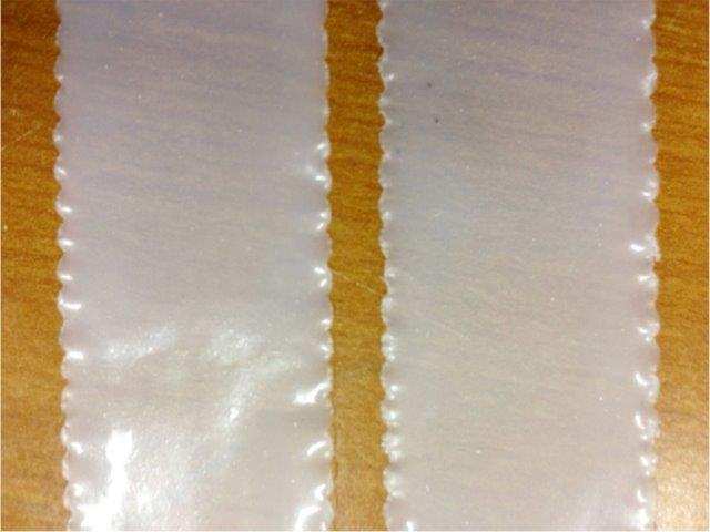
Adding eggshell nanoparticles to a bioplastic (shown above) increases the strength and flexibility of the material, potentially making it more attractive for use in the packaging industry. Credit: Vijaya Rangari/Tuskegee University

Adding eggshell nanoparticles to a bioplastic (shown above) increases the strength and flexibility of the material, potentially making it more attractive for use in the packaging industry. Credit: Vijaya Rangari/Tuskegee University

A March 15, 2016 news item on Nanowerk describes the research,

Eggshells are both marvels and afterthoughts. Placed on end, they are as strong as the arches supporting ancient Roman aqueducts. Yet they readily crack in the middle, and once that happens, we discard them without a second thought. But now scientists report that adding tiny shards of eggshell to bioplastic could create a first-of-its-kind biodegradable packaging material that bends but does not easily break.

The researchers present their work today [March 15, 2016] at the 251st National Meeting & Exposition of the American Chemical Society (ACS).

A March 15, 2016 ACS news release (also on EurekAlert), which originated the news item, describes the work further,

“We’re breaking eggshells down into their most minute components and then infusing them into a special blend of bioplastics that we have developed,” says Vijaya K. Rangari, Ph.D. “These nano-sized eggshell particles add strength to the material and make them far more flexible than other bioplastics on the market. We believe that these traits — along with its biodegradability in the soil — could make this eggshell bioplastic a very attractive alternative packaging material.”

Worldwide, manufacturers produce about 300 million tons of plastic annually. Almost 99 percent of it is made with crude oil and other fossil fuels. Once it is discarded, petroleum-based plastics can last for centuries without breaking down. If burned, these plastics release carbon dioxide into the atmosphere, which can contribute to global climate change.

As an alternative, some manufacturers are producing bioplastics — a form of plastic derived from cornstarch, sweet potatoes or other renewable plant-based sources — that readily decompose or biodegrade once they are in the ground. However, most of these materials lack the strength and flexibility needed to work well in the packaging industry. And that’s a problem since the vast majority of plastic is used to hold, wrap and encase products. So petroleum-based materials continue to dominate the market, particularly in grocery and other retail stores, where estimates suggest that up to a trillion plastic bags are distributed worldwide every year.

To find a solution, Rangari, graduate student Boniface Tiimob and colleagues at Tuskegee University experimented with various plastic polymers. Eventually, they latched onto a mixture of 70 percent polybutyrate adipate terephthalate (PBAT), a petroleum polymer, and 30 percent polylactic acid (PLA), a polymer derived from cornstarch. PBAT, unlike other oil-based plastic polymers, is designed to begin degrading as soon as three months after it is put into the soil.

This mixture had many of the traits that the researchers were looking for, but they wanted to further enhance the flexibility of the material. So they created nanoparticles made of eggshells. They chose eggshells, in part, because they are porous, lightweight and mainly composed of calcium carbonate, a natural compound that easily decays.

The shells were washed, ground up in polypropylene glycol and then exposed to ultrasonic waves that broke the shell fragments down into nanoparticles more than 350,000 times smaller than the diameter of a human hair. Then, in a laboratory study, they infused a small fraction of these particles, each shaped like a deck of cards, into the 70/30 mixture of PBAT and PLA. The researchers found that this addition made the mixture 700 percent more flexible than other bioplastic blends. They say this pliability could make it ideal for use in retail packaging, grocery bags and food containers — including egg cartons.

In addition to bioplastics, Rangari’s team is investigating using eggshell nanoparticles to enhance wound healing, bone regeneration and drug delivery.

Bone bio-patches from the University of Iowa

Let’s take a look at the bone patch developed at the University Iowa,

Researchers at the University of Iowa have created a bio patch to regenerate missing or damaged bone. The patch has been shown to nearly fully regrow missing skull, seen in the image above. Image courtesy of Satheesh Elangovan. & University of Iowa

Researchers at the University of Iowa have created a bio patch to regenerate missing or damaged bone. The patch has been shown to nearly fully regrow missing skull, seen in the image above. Image courtesy of Satheesh Elangovan. & University of Iowa

A Nov. 7, 2013 news item on Nanowerk provides information explaining the bone bio-patch,

Researchers at the University of Iowa have created a bio patch to regenerate missing or damaged bone by putting DNA into a nano-sized particle that delivers bone-producing instructions directly into cells.

The bone-regeneration kit relies on a collagen platform seeded with particles containing the genes needed for producing bone. In experiments, the gene-encoding bio patch successfully regrew bone fully enough to cover skull wounds in test animals. It also stimulated new growth in human bone marrow stromal cells in lab experiments.

The study is novel in that the researchers directly delivered bone-producing instructions (using piece of DNA that encodes for a platelet-derived growth factor called PDGF-B) to existing bone cells in vivo, allowing those cells to produce the proteins that led to more bone production. Previous attempts had relied on repeated applications from the outside, which is costly, intensive, and harder to replicate consistently.

The Nov. 6, 2013 University of Iowa news piece, which originated the news item and was written by Richard C. Lewis, provides some insight from the researchers (Note: Links have been removed),

“We delivered the DNA to the cells, so that the cells produce the protein and that’s how the protein is generated to enhance bone regeneration,” explains Aliasger Salem, professor in the College of Pharmacy and a co-corresponding author on the paper, published in the journal Biomaterials. ”If you deliver just the protein, you have keep delivering it with continuous injections to maintain the dose. With our method, you get local, sustained expression over a prolonged period of time without having to give continued doses of protein.”

The researchers believe the patch has several potential uses in dentistry. For instance, it could be used to rebuild bone in the gum area that serves as the concrete-like foundation for dental implants. That prospect would be a “life-changing experience” for patients who need implants and don’t have enough bone in the surrounding area, says Satheesh Elangovan, assistant professor in the UI’s College of Dentistry and a joint first author, as well as co-corresponding author, on the paper. It also can be used to repair birth defects where there’s missing bone around the head or face.

“We can make a scaffold in the actual shape and size of the defect site, and you’d get complete regeneration to match the shape of what should have been there,” Elangovan says.

The news article goes on to provide details about how the bio-patch was created,

The team started with a collagen scaffold. The researchers then loaded the bio patch with synthetically created plasmids, each of which is outfitted with the genetic instructions for producing bone. They then inserted the scaffold on to a 5-millimeter by 2-millimeter missing area of skull in test animals. Four weeks later, the team compared the bio patch’s effectiveness to inserting a scaffold with no plasmids or taking no action at all.

The plasmid-seeded bio patch grew 44-times more bone and soft tissue in the affected area than with the scaffold alone, and was 14-fold higher than the affected area with no manipulation. Aerial and cross-sectional scans showed the plasmid-encoded scaffolds had spurred enough new bone growth to nearly close the wound area, the researchers report.

The plasmid does its work by entering bone cells already in the body – usually those located right around the damaged area that wander over to the scaffold. The team used a polymer to shrink the particle’s size (like creating a zip file, for example) and to give the plasmid the positive electrical charge that would make it easier for the resident bone cells to take them in.

“The delivery mechanism is the scaffold loaded with the plasmid,” Salem says. “When cells migrate into the scaffold, they meet with the plasmid, they take up the plasmid, and they get the encoding to start producing PDGF-B, which enhances bone regeneration.”

The researchers also point out that their delivery system is nonviral. That means the plasmid is less likely to cause an undesired immune response and is easier to produce in mass quantities, which lowers the cost.

“The most exciting part to me is that we were able to develop an efficacious, nonviral-based gene-delivery system for treating bone loss,” says Sheetal D’mello, a graduate student in pharmacy and a joint first author on the paper.

Elangovan and Salem next hope to create a bio platform that promotes new blood vessel growth– needed for extended and sustained bone growth.

Here’s a link to and a citation for the published paper,

The enhancement of bone regeneration by gene activated matrix encoding for platelet derived growth factor by Satheesh Elangovan, Sheetal R. D’Mello, Liu Hong, Ryan D. Ross, Chantal Allamargot, Deborah V. Dawson, Clark M. Stanford, Georgia K. Johnson, D. Rick Sumnerd,& Aliasger K. Salem. Biomaterials Volume 35, Issue 2, January 2014, Pages 737–747 DOI: 10.1016/j.biomaterials.2013.10.021

This paper is behind a paywall.

A ‘glass jaw’ might turn out to be a good thing

I don’t know if the phrase ‘glass jaw’ is used much any more but it was a term for someone who couldn’t ‘take’ a punch to the jaw (i.e., the person was instantly rendered unconscious or helplessly groggy). If scientists at Missouri University of Science and Technology (Missouri S&T)  have their way, the phrase ‘glass jaw’ will have a new meaning as per the July 26, 2012 news item on ScienceDaily,

Researchers at Missouri University of Science and Technology have developed a type of glass implant that could one day be used to repair injured bones in the arms, legs and other areas of the body that are most subject to the stresses of weight.

This marks the first time researchers have shown a glass implant strong enough to bear weight can also integrate with bone and promote bone growth, says lead researcher Dr. Mohamed N. Rahaman, professor of materials science and engineering at Missouri S&T.

The July 26, 2013 Missouri S&T news release by Andrew Careaga, which originated the news item, describes the work leading to this latest research,

In previous work, the Missouri S&T researchers developed a glass implant strong enough to handle the weight and pressure of repetitive movement, such as walking or lifting. In their most recent study, published in the journal Acta Biomaterialia, the research team reported that the glass implant, in the form of a porous scaffolding, also integrates with bone and promotes bone growth.

This combination of strength and bone growth opens new possibilities for bone repair, says Rahaman, who also directs Missouri S&T’s Center for Biomedical Science and Engineering, where the research was conducted.

The news release then goes on to describe one of the problems with using synthetic materials for bone repair and explains how this latest research addresses the issue,

Conventional approaches to structural bone repair involve either the use of a porous metal, which does not reliably heal bone, or a bone allograft from a cadaver. Both approaches are costly and carry risks, Rahaman says. He thinks the type of glass implant developed in his center could provide a more feasible approach for repairing injured bones. The glass is bioactive, which means that it reacts when implanted in living tissue and convert to a bone-like material.

In their latest research, Rahaman and his colleagues implanted bioactive glass scaffolds into sections of the calvarial bones (skullcaps) of laboratory rats, then examined how well the glass integrated with the surrounding bone and how quickly new bone grew into the scaffold. The scaffolds are manufactured in Rahaman’s lab through a process known as robocasting – a computer-controlled technique to manufacture materials from ceramic slurries, layer by layer – to ensure uniform structure for the porous material.

In previous studies by the Missouri S&T researchers, porous scaffolds of the silicate glass, known as 13-93, were found to have the same strength properties as cortical bone. Cortical bones are those outer bones of the body that bear the most weight and undergo the most repetitive stress. They include the long bones of the arms and legs.

But what Rahaman and his colleagues didn’t know was how well the silicate 13-93 bioactive glass scaffolds would integrate with bone or how quickly bone would grow into the scaffolding.

“You can have the strongest material in the world, but it also must encourage bone growth in a reasonable amount of time,” says Rahaman. He considers three to six months to be a reasonable time frame for completely regenerating an injured bone into one strong enough to bear weight.

In their studies, the S&T researchers found that the bioactive glass scaffolds bonded quickly to bone and promoted a significant amount of new bone growth within six weeks.

While the skullcap is not a load-bearing bone, it is primarily a cortical bone. The purpose of this research was to demonstrate how well this type of glass scaffolding – already shown to be strong – would interact with cortical bone.

Rahaman and his fellow researchers in the Center for Biomedical Science and Engineering are now experimenting with true load-bearing bones. They are now testing the silicate 13-93 implants in the femurs (leg bones) of laboratory rats.

In the future, Rahaman plans to experiment with modified glass scaffolds to see how well they enhance certain attributes within bone. For instance, doping the glass with copper should promote the growth of blood vessels or capillaries within the new bone, while doping the glass with silver will give it antibacterial properties.

It’s exciting work but they are years from human clinical trials. Still, for those who want to explore further, here’s a link to and a citation for the published paper,

Enhanced bone regeneration in rat calvarial defects implanted with surface-modified and BMP-loaded bioactive glass (13-93) scaffolds by Xin Liua, Mohamed N. Rahaman, Yongxing Liu, B. Sonny Bal, and Lynda F. Bonewald. Acta Biomaterialia, July 2013 issue (Volume 9, Issue 7)  http://dx.doi.org/10.1016/j.actbio.2013.03.039

This paper is behind a paywall.

Tissue regeneration by injection

I’ve got two items: one from the University of Nottingham (UK) where they’re working on tissue regeneration for bones, muscles, and the heart.The second item is from Simon Fraser University (Vancouver, Canada)where the focus is on regenerating bones.

Here’s more about the work at the University of Nottingham from the [July 3, 2012] news item on Nanowerk,

The University of Nottingham has begun the search for a new class of injectable materials that will stimulate stem cells to regenerate damaged tissue in degenerative and age related disorders of the bone, muscle and heart.

The work, which is currently at the experimental stage, could lead to treatments for diseases that currently have no cure. The aim is to produce radical new treatments that will reduce the need for invasive surgery, optimise recovery and reduce the risk of undesirable scar tissue.

The research, which brings together expertise in The University of Nottingham’s Malaysia Campus (UNMC) and UK campus, is part of the Rational Bioactive Materials Design for Tissue Generation project (Biodesign). This €11m EU funded research project which involves 21 research teams from across Europe is made up of leading experts in degenerative disease and regenerative medicine.

The original July 3, 2012 news release from the University of Nottingham includes a video which offers some additional insight (sadly ,it cannot be embedded here) and more information (Note: I have removed a link),

Kevin Shakesheff, Professor of Advanced Drug Delivery and Tissue Engineering and Head of the School of Pharmacy, said: “This research heralds a step-change in approaches to tissue regeneration. Current biomaterials are poorly suited to the needs of tissue engineering and regenerative medicine. The aim of Biodesign is to develop new materials and medicines that will stimulate tissue regeneration rather than wait for the body to start the process itself. The aim is to fabricate advanced biomaterials that match the basic structure of each tissue so the cells can take over the recovery process themselves.”

The Canadian project at Simon Fraser University features a singular focus on bone regeneration, from the July 19, 2012 news release on EurekAlert,

A Simon Fraser University researcher is leading a team of scientists working to create new drugs to stimulate bone regeneration – research that will be furthered by a $2.5 million grant from the Canadian Institutes of Health Research (CIHR).

Lead researcher Robert Young heads a team of internationally recognized experts in bone disease and drug development. The researchers are focusing on developing small molecule compounds and nano-medicines that stimulate bone regeneration, and hope to identify new therapeutic approaches by improving understanding of bone renewal biology.

Their objective is to develop new therapeutic agents that promote bone repair, regeneration and renewal, and prove their efficiency in reproducing or improving bone strength.

The research involves studying the “natural controls” that guide the development of cells in the bones toward either bone forming or bone resorbing cells, setting the stage for the next generation of bone regenerative therapies.

The grant is one of three announced today by the federal government targeting bone health research and totalling $7 million. The others focus on wrist fractures management and identifying bone loss in gum disease.

The funding is through the CIHR’s Institute of Musculoskeletal Health and Arthritis and addresses priorities identified at a 2009 national Bone Health Consensus Conference.

I’ve decided to focus on tissues today so there will be something about tissue engineering and jellyfish (artificial) shortly.