Tag Archives: Boyang Zhang

Protocols for mouse-human chimeric embryos

This work on a type of species boundary-crossing could be very disturbing for some folks. That said, here’s more about the science from a July 2, 2021 news item on phys.org,

A year after University at Buffalo [in New York state] scientists demonstrated that it was possible to produce millions of mature human cells in a mouse embryo, they have published a detailed description of the method so that other laboratories can do it, too.

A July 2, 2021 University at Buffalo (UB) news release (also on EurekAlert) by Ellen Goldbaum, which originated the news item, explains why scientists have created these chimeras,

The ability to produce millions of mature human cells in a living organism, called a chimera, which contains the cells of two species, is critical if the ultimate promise of stem cells to treat or cure human disease is to be realized. But to produce those mature cells, human primed stem cells must be converted back into an earlier, less developed naive state so that the human stem cells can co-develop with the inner cell mass in a mouse blastocyst.

The protocol outlining how to do that has now been published in Nature Protocols by the UB scientists. They were invited to publish it because of the significant interest generated by the team’s initial publication describing their breakthrough last May [2020].

“This paper will enable many scientists to use this new platform to study the human disease of their interest,” said Jian Feng, PhD, professor of physiology and biophysics in the Jacobs School of Medicine and Biomedical Sciences at UB and senior author. “Over time, it will transform biomedical research toward a more effective use of the human model system to directly study virtually any inborn condition of an individual. It will stimulate unforeseen discoveries and applications that may fundamentally change our understanding of human biology and medicine.”

The protocol will allow scientists to create animal models that Feng said provide a much more realistic picture of embryonic development than has ever been possible. These more realistic animal models also will have the potential to reveal the mechaniswms behind numerous diseases, especially those that afflict individuals from birth.

Better mouse models

“This step-by-step protocol will benefit the entire field by enabling other scientists to use our methods to generate chimeras to study human diseases that they are experts in,” said Feng. “It will lead to the generation of better mouse models for various human diseases, such as sickle cell anemia, COVID-19 and many others, or various human developmental disorders.” The paper demonstrates how to generate naive human pluripotent stem cells from existing induced pluripotent stem cells that may be derived from patients with various diseases, how to generate mouse-human chimeras using these cells and how to quantify the amount of human cells in the chimeras.

“Using our method, one can now track the development of naive human pluripotent stem cells in mouse-human chimeric embryos in real-time,” said Feng. These stem cells can then be manipulated either genetically or pharmacologically, providing valuable information about human development and disease.

“For example, one can label naive human pluripotent stem cells by inserting green fluorescent protein in a hemoglobin gene to study the development of human red blood cells in mouse-human chimeras,” said Feng.

Another application is to generate humanized mouse models to study many human diseases.

“These mice contain critical human cells, tissues or even organs so that they more accurately reflect the human condition,” said Feng. “With our method, the human cells are made along with the mouse during the development of the mouse embryo. There would be better matching and no rejections, because there are ways for the human cells to be made where there is no competition from their mouse counterparts.”

Organs for transplant in the future

By allowing others to improve and adapt the method to eventually generate chimeras in larger animals, this protocol may also lead to the generation of human organs to address the dire shortage of organs available for transplant, said Feng.

“If naive human pluripotent stem cells are able to generate significant amounts of mature human cells in other larger species, it could be possible to make human tissues or even human organs in chimeric animals,” Feng explained.

This would be possible using blastocyst complementation where, Feng explained, normal pluripotent stem cells from one species can reconstitute an organ for that species in a blastocyst of another species that been genetically modified not to grow that particular organ.

Feng added: “Ultimately, a better understanding of how human cells develop and grow in chimeras may enable the generation of human cells, tissues and organs in a completely artificial system and fundamentally change how we treat many human diseases. Research using chimeras is a bridge that must be crossed to reach that possibility.”

Here’s a link to and a citation for the 2021 article,

Generation of mouse–human chimeric embryos by Boyang Zhang, Hanqin Li, Zhixing Hu, Houbo Jiang, Aimee B. Stablewski, Brandon J. Marzullo, Donald A. Yergeau & Jian Feng. Nature Protocols (2021) DOI: https://doi.org/10.1038/s41596-021-00565-7 Published 02 July 2021

This article is behind a paywall.

Here’s a link to and citation for the 2020 work, which led to the publication of the protocols,

Transient inhibition of mTOR in human pluripotent stem cells enables robust formation of mouse-human chimeric embryos by Zhixing Hu, Hanqin Li, Houbo Jiang, Yong Ren, Xinyang Yu, Jingxin Qiu, Aimee B. Stablewski, Boyang Zhang, Michael J. Buck, Jian Feng. Science Advances 13 May 2020: Vol. 6, no. 20, eaaz0298 DOI: 10.1126/sciadv.aaz0298

This paper is open access.
University of Toronto (Canada) researchers and lab-grown heart and liver tissue (person-on-a-chip)

Usually called ‘human-on-a-chip’, a team at the University of Toronto have developed a two-organ ‘person on a chip’ according to a March 7, 2016 news item on phys.org (Note: Links have been removed),

Researchers at U of T [University of Toronto] Engineering have developed a new way of growing realistic human tissues outside the body. Their “person-on-a-chip” technology, called AngioChip, is a powerful platform for discovering and testing new drugs, and could eventually be used to repair or replace damaged organs.

Professor Milica Radisic (IBBME, ChemE), graduate student Boyang Zhang and the rest of the team are among those research groups around the world racing to find ways to grow human tissues in the lab, under conditions that mimic a real person’s body. They have developed unique methods for manufacturing small, intricate scaffolds for individual cells to grow on. These artificial environments produce cells and tissues that resemble the real thing more closely than those grown lying flat in a petri dish.

The team’s recent creations have included BiowireTM—an innovative method of growing heart cells around a silk suture—as well as a scaffold for heart cells that snaps together like sheets of Velcro. But AngioChip takes tissue engineering to a whole new level. “It’s a fully three-dimensional structure complete with internal blood vessels,” says Radisic. “It behaves just like vasculature, and around it there is a lattice for other cells to attach and grow.” …

A March 7, 2016 University of Toronto news release (also on EurekAlert), which originated the news item, provides more detail about the AngioChip,

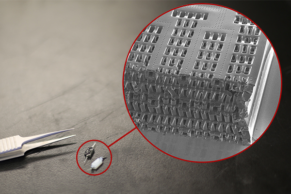
Zhang built the scaffold out of POMaC, a polymer that is both biodegradable and biocompatible. The scaffold is built out of a series of thin layers, stamped with a pattern of channels that are each about 50 to 100 micrometres wide. The layers, which resemble the computer microchips, are then stacked into a 3D structure of synthetic blood vessels. As each layer is added, UV light is used to cross-link the polymer and bond it to the layer below.

When the structure is finished, it is bathed in a liquid containing living cells. The cells quickly attach to the inside and outside of the channels and begin growing just as they would in the human body.

“Previously, people could only do this using devices that squish the cells between sheets of silicone and glass,” says Radisic. “You needed several pumps and vacuum lines to run just one chip. Our system runs in a normal cell culture dish, and there are no pumps; we use pressure heads to perfuse media through the vasculature. The wells are open, so you can easily access the tissue.”

Using the platform, the team has built model versions of both heart and liver tissues that function like the real thing. “Our liver actually produced urea and metabolized drugs,” says Radisic. They can connect the blood vessels of the two artificial organs, thereby modelling not just the organs themselves, but the interactions between them. They’ve even injected white blood cells into the vessels and watched as they squeezed through gaps in the vessel wall to reach the tissue on the other side, just as they do in the human body.

The news release also mentions potential markets and the work that needs to be accomplished before AngioChip is available for purchase,

AngioChip has great potential in the field of pharmaceutical testing. Current drug-testing methods, such as animal testing and controlled clinical trials, are costly and fraught with ethical concerns. Testing on lab-grown human tissues would provide a realistic model at a fraction of the cost, but this area of research is still in its infancy. “In the last few years, it has become possible to order cultures of human cells for testing, but they’re grown on a plate, a two-dimensional environment,” says Radisic. “They don’t capture all the functional hallmarks of a real heart muscle, for example.”

A more realistic platform like AngioChip could enable drug companies to detect dangerous side effects and interactions between organ compartments long before their products reach the market, saving countless lives. It could also be used to understand and validate the effectiveness of current drugs and even to screen libraries of chemical compounds to discover new drugs. Through TARA Biosystems Inc., a spin-off company co-founded by Radisic, the team is already working on commercializing the technology.

In future, Radisic envisions her lab-grown tissues being implanted into the body to repair organs damaged by disease. Because the cells used to seed the platform can come from anyone, the new tissues could be genetically identical to the intended host, reducing the risk of organ rejection. Even in its current form, the team has shown that the AngioChip can be implanted into a living animal, its artificial blood vessels connected to a real circulatory system. The polymer scaffolding itself simply biodegrades after several months.

The team still has much work to do. Each AngioChip is currently made by hand; if the platform is to be used industrially, the team will need to develop high-throughput manufacturing methods to create many copies at once. Still, the potential is obvious. “It really is multifunctional, and solves many problems in the tissue engineering space,” says Radisic. “It’s truly next-generation.”

Here’s a link to and a citation for the paper,

Biodegradable scaffold with built-in vasculature for organ-on-a-chip engineering and direct surgical anastomosis by Boyang Zhang, Miles Montgomery, M. Dean Chamberlain, Shinichiro Ogawa, Anastasia Korolj, Aric Pahnke, Laura A. Wells, Stéphane Massé, Jihye Kim, Lewis Reis, Abdul Momen, Sara S. Nunes, Aaron R. Wheeler, Kumaraswamy Nanthakumar, Gordon Keller, Michael V. Sefton, & Milica Radisic. Nature Materials (2016) doi:10.1038/nmat4570 Published online 07 March 2016

This paper is behind a paywall.

The researchers have made two images illustrating their work available. There’s this still image,

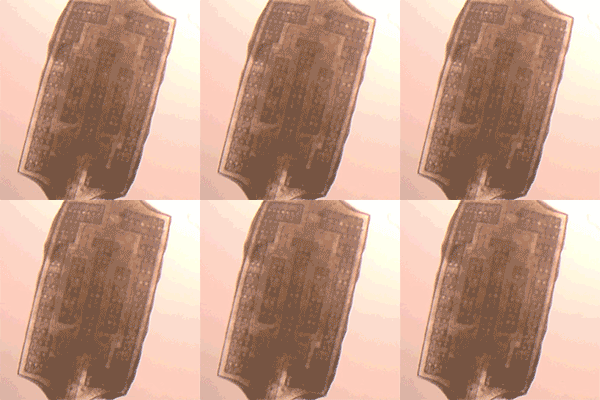
These tiny polymer scaffolds contain channels that are about 100 micrometres wide, about the same diameter as a human hair. When seeded with cells, the channels act as artificial blood vessels. By mimicking tissues in the human heart and other organs, these scaffolds provide a new way to test drugs for potentially dangerous side effects. (Image: Tyler Irving/Boyang Zhang/Kevin Soobrian)

These tiny polymer scaffolds contain channels that are about 100 micrometres wide, about the same diameter as a human hair. When seeded with cells, the channels act as artificial blood vessels. By mimicking tissues in the human heart and other organs, these scaffolds provide a new way to test drugs for potentially dangerous side effects. (Image: Tyler Irving/Boyang Zhang/Kevin Soobrian)

Perhaps more intriguing is this one,

UofT_AngioChipMoving

When seeded with heart cells, the flexible polymer scaffold contracts with a regular rhythm, just like real heart tissue. (Image: Boyang Zhang)

I have mentioned ‘human-on-a-chip’ projects many times here and as the news release writer notes, there is an international race. My July 1, 2015 posting (cross-posted from the June 30, 2015 posting [Testing times: the future of animal alternatives] on the International Innovation blog [a CORDIS-listed project dissemination partner for FP7 and H2020 projects]) notes a couple of those projects,

Organ-on-a-chip projects use stem cells to create human tissues that replicate the functions of human organs. Discussions about human-on-a-chip activities – a phrase used to describe 10 interlinked organ chips – were a highlight of the 9th World Congress on Alternatives to Animal Testing held in Prague, Czech Republic, last year. One project highlighted at the event was a joint US National Institutes of Health (NIH), US Food and Drug Administration (FDA) and US Defense Advanced Research Projects Agency (DARPA) project led by Dan Tagle that claimed it would develop functioning human-on-a-chip by 2017. However, he and his team were surprisingly close-mouthed and provided few details making it difficult to assess how close they are to achieving their goal.

By contrast, Uwe Marx – Leader of the ‘Multi-Organ-Chip’ programme in the Institute of Biotechnology at the Technical University of Berlin and Scientific Founder of TissUse, a human-on-a-chip start-up company – claims to have sold two-organ chips. He also claims to have successfully developed a four-organ chip and that he is on his way to building a human-on-a-chip. Though these chips remain to be seen, if they are, they will integrate microfluidics, cultured cells and materials patterned at the nanoscale to mimic various organs, and will allow chemical testing in an environment that somewhat mirrors a human.

As for where the University of Toronto efforts fit into the race, I don’t know for sure. It’s the first time I’ve come across a reference to liver tissue producing urea but I believe there’s at least one other team in China which has achieved a three-dimensional, more lifelike aspect for liver tissue in my Jan. 29, 2016 posting ‘Constructing a liver’.