A Nov. 18, 2016 Arizona State University (ASU) news release on EurekAlert describes an initiative drawing together academics to examine the social and ethical implications of the new neuroscience work,
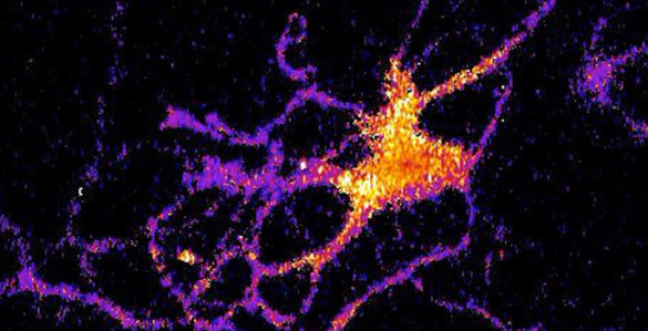
Repairing traumatic brain injury, staving off dementia, improving memory, regulating emotion, controlling devices with mere thoughts, plus a multitude of other means of interacting with or influencing fundamental brain functions are here or will be soon.
A breathtaking array of new technologies to diagnose, treat or influence human brains have been landing on society’s doorstep — with no signs of slowing. Yet regulations and accepted practices guide the development and use of neurotechnology, especially those relating to broader societal issues, appear to be rudimentary in comparison.
The flood of emerging brain technologies is raising ethical, social and legal questions: Who has access? Who has control? Who benefits? And who doesn’t? What are the potential risks and costs?
As diagnostic, treatment and enhancement technologies with potential to alter the way we perceive and interact with our environment pour in from diverse sources, there is a struggle for policy to keep pace with rapid advancements. A group of key stakeholders are seeking to draw from sundry efforts dealing with these issues and establish guidance materials that proactively address concerns around neurotechnologies, and steer research and development activities toward positive outcomes.
Arizona State University collaborated with the Organization for Economic Co-operation and Development (OECP), and the National Academies of Sciences, Engineering and Medicine to convene a 1.5 day workshop in September [2016] that specifically focused on the ethical, legal and social issues related to swiftly developing fields of neuroscience and research into the human brain.
“The goal was to bring together a diverse set of actors, from across the globe, in order to identify key areas of research activities, and then consider how broader ethical, legal and social issues can be brought into the research agenda at an early stage in the technology’s development. In doing so, we hope to identify, and develop, core approaches and guidance materials that can help ensure that developments which affect brain function and treat disease are not only safe and effective, but also are in the best interest of patients and society more generally,” said Diana Bowman of ASU’s School for the Future of Innovation in Society (SFIS) and Sandra Day O’Connor College of Law and co-author of a recent article reporting on the workshop’s accomplishments.
The article, “Neurotechnology and Society: Strengthening Responsible Innovation in Brain Science” published in the journal Neuron emphasized the need for having discussions “now, rather than waiting until the technology has been proven to be safe and effective for therapeutic applications.”
Drawing from the experience of previous initiatives in areas such as genomics and nanotechnology, the workshop participants, representing a broad array of backgrounds and disciplines, produced a series of key points for use in formulating policy, designing best practices and frameworks for integrating brain science and society.
Included in the discussion was the director of SFIS, David Guston, whose work in “anticipatory governance” points to the need for considering social values and managing ethical inputs during early development stages of advancements rather than waiting until technologies are entering markets. The article describes this as an approach that “combines techniques for anticipation, the development of realistic scenarios and public engagement to help inform policy and the development of an appropriate governance framework.” But it also notes that costs and political risks are associated with meaningful public engagement.
The article summarized five “key policy implications emanating from the OECD Workshop” and called for increases in transparency, collaboration, mechanisms to enable responsible innovation and participation on a global scale to reflect broad social needs and values in the creation of guidelines and evidentiary standards.
There is a Sept. 15, 2016 interview with lead investigator, Diana Bowman, in ASU Now. It was conducted just prior to the Sept. 2016 OECD workshop,
Question: What could go wrong if research and development in brain science goes unchecked?
[DB] Answer: The human brain is considered the last frontier for medical research; its complexity, and importance to defining the individual self, has ensured that the brain has received substantial attention from scientists, physicians, philosophers and ethicists alike.
Large-scale investment into brain research by the public and private sectors promises to unlock new therapies and novel approaches for treating costly illnesses and disease including, for example, dementia, Alzheimer’s and traumatic brain injuries.
But as we begin to unlock, and better understand, how the human brain functions there is the potential for abuse, or for ethical and legal concerns to go unchecked. Think Minority Report. Think functional magnetic resonance imaging of criminals, in an effort to predict impulsivity and recidivism and where such approaches to preventing criminal activities, and potentially limiting freedom in order to prevent crime, could infringe fundamental human rights.
Q: How have we gotten to this point? And what ethical dilemmas have come up, so far?
[DB] A: Different technologies and their applications are already giving rise to a myriad of ethical and moral dilemmas.
Of these, some of the most pressing related to human enhancement and the ways and extent to which we should pursue human enhancement technologies: Do the benefits of invasive neuromodulation outweigh potential risks? And what are the privacy and data security risks associated with innovations with brain-computer interface? And what does this convergence mean for ‘the self’?
Theses types of questions are being considered by different groups and individuals around the world. Greater coordination is needed and we need to speed up the pace at which these types of questions are considered and acted upon.
Q: How quickly has the technology advanced?
[DB] A: Research on the human brain is not, in itself, new. Brain science research and advances in neurotechnology has already resulted in numerous medical devices already entering the market.
However, the convergence of other technologies such as nanotechnology, synthetic biology and additive manufacturing, combined with enhanced imaging technologies and more powerful infrastructure, will accelerate the pace at which new neurotechnologies evolve, and the entry of new drugs and products into the market.
So too will the aging population, and the pressures that meeting their physical and mental health needs place on governments and other stakeholders.
Q: Do we expect regulations to be put in place? Are there any now? Should there be?
[DB] A: Existing regulatory regimes will capture the types of research being undertaken by scientists involved in these large-scale brain projects, and the products that the projects give rise to — whether that is in the United States, Australia or Israel to name just a few.
The more important question, though, is whether these existing regimes and instruments shall be adequate and effective for the types of technologies and products that they give rise to? And this question we cannot answer at this time given that we simply do not know what this exciting field or research shall give rise to.
The fact that we cannot answer this question makes an event such as the Neurotechnology and Society workshop all the more important.
Bringing together key stakeholders across all relevant fields in order to better understand the trajectory of the research and technologies shall allow us to begin the process of evaluating the effectiveness of the current regimes, and, where necessary, to proactively explore other regulatory and governance models so as to ensure that the potential benefits outweigh potential risks.
Q: Who is this science for?
[DB] A: One can reasonably assume that the therapies and products that are initially developed as a result of this fundamental research will, at least in the first instance, benefit a small segment of the population (i.e., those who are most able to afford them), the majority of whom are likely to reside in developed economies.
This alone presents a myriad of social and ethical challenges, and while not new in themselves, need to be incorporated into funding decisions including the prioritization of research.
For this to be meaningful, different stakeholders need to be at the table and difficult conversations will need to take place. Such conversations need to take place now, ahead of the science, and alongside the development of the science and technologies.
Q: What would be going too far?
[DB] A: Going too far, in my view, can be simply described as follows: not learning the lessons from previous technologies, including their entry into the market and consumer acceptance thereof. For society to be able to take full advantage of the benefits offered by neurotechnologies, we need to ensure that broader ethical, social and legal questions are addressed in real time, and that society is actively engaged in the development, and deployment, of the technologies.
In any event, here’s a link to the paper/report resulting from the Sept. 2016 workshop on brain technologies and their social and ethical implications,
Neurotechnology and Society: Strengthening Responsible Innovation in Brain Science by
Hermann Garden, Diana M. Bowman, Sebastian Haesler, David E. Winickoff. Neuron Volume 92, Issue 3, p642–646, 2 November 2016 DOI: http://dx.doi.org/10.1016/j.neuron.2016.10.053
This paper appears to be open access.