Tag Archives: calcium

Good for your bones and good for art conservation: calcium

The statues on Easter Island, the Great Wall of China, Egyptian pyramids, MesoAmerican pyramids, castles in Europe and other structures made of stone are deteriorating and now comes another approach to halting the destruction. (I have covered other approaches to the problem in two previous postings, a December 5, 2017 posting, Europe’s cathedrals get a ‘lift’ with nanoparticles, and an October 21, 2014 posting, Heart of stone.)

An August 7, 2019 news item on ScienceDaily announces the latest in conserving stone monuments and structures,

When it comes to cultural heritage sites, there are few things historians wouldn’t do to preserve them for future generations. In particular, stone buildings and sculptures made of plaster and marble are increasingly at risk of damage from air pollution, acid rain and other factors. Researchers now report a new, calcium-based conservation treatment inspired by nature that overcomes many drawbacks of currently used methods.

An August 7, 2019 American Chemical Society (ACS) news release, which originated the news item, provides a bit more technical detail,

Historically, conservation scientists have turned to alkoxysilanes, silicon-based molecules used to consolidate stone and other artworks, in their preservation efforts. However, alkoxysilane treatments do not bond properly with non-silicate surfaces, are prone to cracking and are limited in their ability to repel water. Adding other compounds to this treatment have helped overcome these effects, but only to a point. Instead Encarnación Ruiz Agudo and colleagues looked to nature for inspiration, and found that calcium could be the answer. As a major element of strong, natural structures like bone and kidney stones, the researchers theorized that nanoparticles made from calcium could bolster alkoxysilanes and provide the desired protective effects to conserve historical artifacts.

The researchers made calcium carbonate and calcium oxalate nanoparticles and included polydimethylsiloxane (PDMS) as a stabilizer. PDMS also likely helps the nanoparticles bond to surfaces. The team added the nanoparticles to traditional alkoxysilane treatments, then applied them to samples of three different building materials: white marble, calcarenite limestone and gypsum plaster, and put the samples through a battery of tests. Overall, the results showed enhanced hydrophobicity, less cracking and improved surface adhesion compared to alkoxysilane treatments alone, with calcium oxalate providing a marked improvement in acid resistance. A minimal color effect was observed, but the researchers say this change was within acceptable values for conservation efforts.

The authors acknowledge funding from the European Regional Development Fund, the Regional Government of Andalusia, the Spanish Ministry of Economy and Finance and the University of Granada.

Here’s a link to and a citation for the paper,

Bioinspired Alkoxysilane Conservation Treatments for Building Materials Based on Amorphous Calcium Carbonate and Oxalate Nanoparticles by A. Burgos-Cara, C. Rodríguez-Navarro, M. Ortega-Huertas, E. Ruiz-Agudo. ACS Appl. Nano Mater.2019XXXXXXXXXX-XXX DOI: https://doi.org/10.1021/acsanm.9b00905 Publication Date:July 18, 2019 Copyright © 2019 American Chemical Society

This paper is behind a paywall.

Rijksmuseum’s ‘live’ restoration of Rembrandt’s masterpiece: The Nightwatch: is it or isn’t it like watching paint dry?

Somewhere in my travels, I saw ‘like watching paint dry’ as a description for the experience of watching researchers examining Rembrandt’s Night Watch. Granted it’s probably not that exciting but there has to be something to be said for being present while experts undertake an extraordinary art restoration effort. The Night Watch is not only a masterpiece—it’s huge.

This posting was written closer to the time the ‘live’ restoration first began. I have an update at the end of this posting.

A July 8, 2019 news item on the British Broadcasting Corporation’s (BBC) news online sketches in some details,

The masterpiece, created in 1642, has been placed inside a specially designed glass chamber so that it can still be viewed while being restored.

Enthusiasts can follow the latest on the restoration work online.

The celebrated painting was last restored more than 40 years ago after it was slashed with a knife.

The Night Watch is considered Rembrandt’s most ambitious work. It was commissioned by the mayor and leader of the civic guard of Amsterdam, Frans Banninck Cocq, who wanted a group portrait of his militia company.

The painting is nearly 4m tall and 4.5m wide (12.5 x 15 ft) and weighs 337kg (743lb) [emphasis mine]. As well as being famous for its size, the painting is acclaimed for its use of dramatic lighting and movement.

But experts at Amsterdam’s Rijksmuseum are concerned that aspects of the masterpiece are changing, pointing as an example to the blanching of the figure of a small dog. The museum said the multi-million euro research and restoration project under way would help staff gain a better understanding of the painting’s condition.

An October 16, 2018 Rijksmuseum press release announced the restoration work months prior to the start (Note: Some of the information is repetitive;),

Before the restoration begins, The Night Watch will be the centrepiece of the Rijksmuseum’s display of their entire collection of more than 400 works by Rembrandt in an exhibition to mark the 350th anniversary of the artist’s death opening on 15 February 2019.

Commissioned in 1642 by the mayor and leader of the civic guard of Amsterdam, Frans Banninck Cocq, to create a group portrait of his shooting company, The Night Watch is recognised as one of the most important works of art in the world today and hangs in the specially designed “Gallery of Honour” at the Rijksmuseum. It is more than 40 years since The Night Watch underwent its last major restoration, following an attack on the painting in 1975.

The Night Watch will be encased in a state-of-the-art clear glass chamber designed by the French architect Jean Michel Wilmotte. This will ensure that the painting can remain on display for museum visitors. A digital platform will allow viewers from all over the world to follow the entire process online [emphasis mine] continuing the Rijksmuseum innovation in the digital field.

Taco Dibbits, General Director Rijksmuseum: The Night Watch is one of the most famous paintings in the world. It belongs to us all, and that is why we have decided to conduct the restoration within the museum itself – and everyone, wherever they are, will be able to follow the process online.

The Rijksmuseum continually monitors the condition of The Night Watch, and it has been discovered that changes are occurring, such as the blanching [emphasis mine] on the dog figure at the lower right of the painting. To gain a better understanding of its condition as a whole, the decision has been taken to conduct a thorough examination. This detailed study is necessary to determine the best treatment plan, and will involve imaging techniques, high-resolution photography and highly advanced computer analysis. Using these and other methods, we will be able to form a very detailed picture of the painting – not only of the painted surface, but of each and every layer, from varnish to canvas.

A great deal of experience has been gained in the Rijksmuseum relating to the restoration of Rembrandt’s paintings. Last year saw the completion of the restoration of Rembrandt’s spectacular portraits of Marten Soolmans and Oopjen Coppit. The research team working on The Night Watch is made up of researchers, conservators and restorers from the Rijksmuseum, which will conduct this research in close collaboration with museums and universities in the Netherlands and abroad.

The Night Watch

The group portrait of the officers and other members of the militia company of District II, under the command of Captain Frans Banninck Cocq and Lieutenant Willem van Ruytenburch, now known as The Night Watch, is Rembrandt’s most ambitious painting. This 1642 commission by members of Amsterdam’s civic guard is Rembrandt’s first and only painting of a militia group. It is celebrated particularly for its bold and energetic composition, with the musketeers being depicted ‘in motion’, rather than in static portrait poses. The Night Watch belongs to the city of Amsterdam, and it been the highlight of the Rijksmuseum collection since 1808. The architect of the Rijksmuseum building Pierre Cuypers (1827-1921) even created a dedicated gallery of honour for The Night Watch, and it is now admired there by more than 2.2 million people annually.

2019, The Year of Rembrandt

The Year of Rembrandt, 2019, marks the 350th anniversary of the artist’s death with two major exhibitions honouring the great master painter. All the Rembrandts of the Rijksmuseum (15 February to 10 June 2019) will bring together the Rijksmuseum’s entire collection of Rembrandt’s paintings, drawings and prints, for the first time in history. The second exhibition, Rembrandt-Velázquez (11 October 2019 to 19 January 2020), will put the master in international context by placing 17th-century Spanish and Dutch masterpieces in dialogue with each another.

First, the restoration work is not being livestreamed; the digital platform Operation Night Watch is a collection of resources, which are being updated constantly, For example, the first scan was placed online in Operation Night Watch on July 16, 2019.

Second, ‘blanching’ reminded me of a June 22, 2017 posting where I featured research into why masterpieces were turning into soap, (Note: The second paragraph should be indented to indicated that it’s an excerpt fro the news release. Unfortunately, the folks at WordPress appear to have removed the tools that would allow me to do that and more),

This piece of research has made a winding trek through the online science world. First it was featured in an April 20, 2017 American Chemical Society news release on EurekAlert

A good art dealer can really clean up in today’s market, but not when some weird chemistry wreaks havoc on masterpieces. Art conservators started to notice microscopic pockmarks forming on the surfaces of treasured oil paintings that cause the images to look hazy. It turns out the marks are eruptions of paint caused, weirdly, by soap that forms via chemical reactions. Since you have no time to watch paint dry, we explain how paintings from Rembrandts to O’Keefes are threatened by their own compositions — and we don’t mean the imagery.

….

Getting back to the Night Watch, there’s a July 8, 2019 Rijksmuseum press release which provides some technical details,

On 8 July 2019 the Rijksmuseum starts Operation Night Watch. It will be the biggest and most wide-ranging research and conservation project in the history of Rembrandt’s masterpiece. The goal of Operation Night Watch is the long-term preservation of the painting. The entire operation will take place in a specially designed glass chamber so the visiting public can watch.

Never before has such a wide-ranging and thorough investigation been made of the condition of The Night Watch. The latest and most advanced research techniques will be used, ranging from digital imaging and scientific and technical research, to computer science and artificial intelligence. The research will lead to a better understanding of the painting’s original appearance and current state, and provide insight into the many changes that The Night Watch has undergone over the course of the last four centuries. The outcome of the research will be a treatment plan that will form the basis for the restoration of the painting.

Operation Night Watch can also be followed online from 8 July 2019 at rijksmuseum.nl/nightwatch

From art historical research to artificial intelligence

Operation Night Watch will look at questions regarding the original commission, Rembrandt’s materials and painting technique, the impact of previous treatments and later interventions, as well as the ageing, degradation and future of the painting. This will involve the newest and most advanced research methods and technologies, including art historical and archival research, scientific and technical research, computer science and artificial intelligence.

During the research phase The Night Watch will be unframed and placed on a specially designed easel. Two platform lifts will make it possible to study the entire canvas, which measures 379.5 cm in height and 454.5 cm in width.

Advanced imaging techniques

Researchers will make use of high resolution photography, as well as a variety of advanced imaging techniques, such as macro X-ray fluorescence scanning (macro-XRF) and hyperspectral imaging, also called infrared reflectance imaging spectroscopy (RIS), to accurately determine the condition of the painting.

56 macro-XRF scans

The Night Watch will be scanned millimetre by millimetre using a macro X-ray fluorescence scanner (macro-XRF scanner). This instrument uses X-rays to analyse the different chemical elements in the paint, such as calcium, iron, potassium and cobalt. From the resulting distribution maps of the various chemical elements in the paint it is possible to determine which pigments were used. The macro-XRF scans can also reveal underlying changes in the composition, offering insights into Rembrandt’s painting process. To scan the entire surface of the The Night Watch it will be necesary to make 56 scans, each one of which will take 24 hours.

12,500 high-resolution photographs

A total of some 12,500 photographs will be taken at extremely high resolution, from 180 to 5 micrometres, or a thousandth of a millimetre. Never before has such a large painting been photographed at such high resolution. In this way it will be possible to see details such as pigment particles that normally would be invisible to the naked eye. The cameras and lamps will be attached to a dynamic imaging frame designed specifically for this purpose.

Glass chamber

Operation Night Watch is for everyone to follow and will take place in full view of the visiting public in an ultra-transparent glass chamber designed by the French architect Jean Michel Wilmotte.

Research team

The Rijksmuseum has extensive experience and expertise in the investigation and treatment of paintings by Rembrandt. The conservation treatment of Rembrandt’s portraits of Marten Soolmans and Oopjen Coppit was completed in 2018. The research team working on The Night Watch is made up of more than 20 Rijksmuseum scientists, conservators, curators and photographers. For this research, the Rijksmuseum is also collaborating with museums and universities in the Netherlands and abroad, including the Dutch Cultural Heritage Agency (RCE), Delft University of Technology (TU Delft), the University of Amsterdam (UvA), Amsterdam University Medical Centre (AUMC), University of Antwerp (UA) and National Gallery of Art, Washington DC.

The Night Watch

Rembrandt’s Night Watch is one of the world’s most famous works of art. The painting is the property of the City of Amsterdam, and it is the heart of Amsterdam’s Rijksmuseum, where it is admired by more than two million visitors each year. The Night Watch is the Netherland’s foremost national artistic showpiece, and a must-see for tourists.

Rembrandt’s group portrait of officers and other civic guardsmen of District 2 in Amsterdam under the command of Captain Frans Banninck Cocq and Lieutenant Willem van Ruytenburch has been known since the 18th century as simply The Night Watch. It is the artist’s most ambitious painting. One of Amsterdam’s 20 civic guard companies commissioned the painting for its headquarters, the Kloveniersdoelen, and Rembrandt completed it in 1642. It is Rembrandt’s only civic guard piece, and it is famed for the lively and daring composition that portrays the troop in active poses rather than the traditional static ones.

Donors and partners

AkzoNobel is main partner of Operation Night Watch.

Operation Night Watch is made possible by The Bennink Foundation, PACCAR Foundation, Piet van der Slikke & Sandra Swelheim, American Express Foundation, Familie De Rooij, Het AutoBinck Fonds, Segula Technologies, Dina & Kjell Johnsen, Familie D. Ermia, Familie M. van Poecke, Henry M. Holterman Fonds, Irma Theodora Fonds, Luca Fonds, Piek-den Hartog Fonds, Stichting Zabawas, Cevat Fonds, Johanna Kast-Michel Fonds, Marjorie & Jeffrey A. Rosen, Stichting Thurkowfonds and the Night Watch Fund.

With the support of the Ministry of Education, Culture and Science, the City of Amsterdam, Founder Philips and main sponsors ING, BankGiro Loterij and KPN every year more than 2 million people visit the Rijksmuseum and The Night Watch.

Details:
Rembrandt van Rijn (1606-1669)
The Night Watch, 1642
oil on canvas
Rijksmuseum, on loan from the Municipality of Amsterdam

Update as of November 22, 2019

I just clicked on the Operation Night Watch link and found a collection of resources including videos of live updates from October 2019. As noted earlier, they’re not livestreaming the restoration. The October 29, 2019 ‘live update’ features a host speaking in Dutch (with English subtitles in the version I was viewing) and interviews with the scientists conducting the research necessary before they start actually restoring the painting.
Growing shells atom-by-atom

The University of California at Davis (UC Davis) and the University of Washington (state) collaborated in research into fundamental questions on how aquatic animals grow. From an Oct. 24, 2016 news item on ScienceDaily,

For the first time scientists can see how the shells of tiny marine organisms grow atom-by-atom, a new study reports. The advance provides new insights into the mechanisms of biomineralization and will improve our understanding of environmental change in Earth’s past.

An Oct. 24, 2016 UC Davis news release by Becky Oskin, which originated the news item, provides more detail,

Led by researchers from the University of California, Davis and the University of Washington, with key support from the U.S. Department of Energy’s Pacific Northwest National Laboratory, the team examined an organic-mineral interface where the first calcium carbonate crystals start to appear in the shells of foraminifera, a type of plankton.

“We’ve gotten the first glimpse of the biological event horizon,” said Howard Spero, a study co-author and UC Davis geochemistry professor. …

Foraminifera’s Final Frontier

The researchers zoomed into shells at the atomic level to better understand how growth processes may influence the levels of trace impurities in shells. The team looked at a key stage — the interaction between the biological ‘template’ and the initiation of shell growth. The scientists produced an atom-scale map of the chemistry at this crucial interface in the foraminifera Orbulina universa. This is the first-ever measurement of the chemistry of a calcium carbonate biomineralization template, Spero said.

Among the new findings are elevated levels of sodium and magnesium in the organic layer. This is surprising because the two elements are not considered important architects in building shells, said lead study author Oscar Branson, a former postdoctoral researcher at UC Davis who is now at the Australian National University in Canberra. Also, the greater concentrations of magnesium and sodium in the organic template may need to be considered when investigating past climate with foraminifera shells.

Calibrating Earth’s Climate

Most of what we know about past climate (beyond ice core records) comes from chemical analyses of shells made by the tiny, one-celled creatures called foraminifera, or “forams.” When forams die, their shells sink and are preserved in seafloor mud. The chemistry preserved in ancient shells chronicles climate change on Earth, an archive that stretches back nearly 200 million years.

The calcium carbonate shells incorporate elements from seawater — such as calcium, magnesium and sodium — as the shells grow. The amount of trace impurities in a shell depends on both the surrounding environmental conditions and how the shells are made. For example, the more magnesium a shell has, the warmer the ocean was where that shell grew.

“Finding out how much magnesium there is in a shell can allow us to find out the temperature of seawater going back up to 150 million years,” Branson said.

But magnesium levels also vary within a shell, because of nanometer-scale growth bands. Each band is one day’s growth (similar to the seasonal variations in tree rings). Branson said considerable gaps persist in understanding what exactly causes the daily bands in the shells.

“We know that shell formation processes are important for shell chemistry, but we don’t know much about these processes or how they might have changed through time,” he said. “This adds considerable uncertainty to climate reconstructions.”

Atomic Maps

The researchers used two cutting-edge techniques: Time-of-Flight Secondary Ionization Mass Spectrometry (ToF-SIMS) and Laser-Assisted Atom Probe Tomography (APT). ToF-SIMS is a two-dimensional chemical mapping technique which shows the elemental composition of the surface of a polished sample. The technique was developed for the elemental analysis of complex polymer materials, and is just starting to be applied to natural samples like shells.

APT is an atomic-scale three-dimensional mapping technique, developed for looking at internal structures in advanced alloys, silicon chips and superconductors. The APT imaging was performed at the Environmental Molecular Sciences Laboratory, a U.S. Department of Energy Office of Science User Facility at the Pacific Northwest National Laboratory.

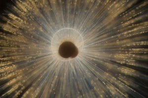
This foraminifera is just starting to form its adult spherical shell. The calcium carbonate spherical shell first forms on a thin organic template, shown here in white, around the dark juvenile skeleton. Calcium carbonate spines then extend from the juvenile skeleton through the new sphere and outward. The bright flecks are algae that the foraminifera “farm” for sustenance.Howard Spero/University of California, Davis

This foraminifera is just starting to form its adult spherical shell. The calcium carbonate spherical shell first forms on a thin organic template, shown here in white, around the dark juvenile skeleton. Calcium carbonate spines then extend from the juvenile skeleton through the new sphere and outward. The bright flecks are algae that the foraminifera “farm” for sustenance.Howard Spero/University of California, Davis

An Oct. 24, 2016 University of Washington (state) news release (also on EurekAlert) adds more information (there is a little repetition),

Unseen out in the ocean, countless single-celled organisms grow protective shells to keep them safe as they drift along, living off other tiny marine plants and animals. Taken together, the shells are so plentiful that when they sink they provide one of the best records for the history of ocean chemistry.

Oceanographers at the University of Washington and the University of California, Davis, have used modern tools to provide an atomic-scale look at how that shell first forms. Results could help answer fundamental questions about how these creatures grow under different ocean conditions, in the past and in the future. …

“There’s this debate among scientists about whether shelled organisms are slaves to the chemistry of the ocean, or whether they have the physiological capacity to adapt to changing environmental conditions,” said senior author Alex Gagnon, a UW assistant professor of oceanography.

The new work shows, he said, that they do exert some biologically-based control over shell formation.

“I think it’s just incredible that we were able to peer into the intricate details of those first moments that set how a seashell forms,” Gagnon said. “And that’s what sets how much of the rest of the skeleton will grow.”

The results could eventually help understand how organisms at the base of the marine food chain will respond to more acidic waters. And while the study looked at one organism, Orbulina universa, which is important for understanding past climate, the same method could be used for other plankton, corals and shellfish.

The study used tools developed for materials science and semiconductor research to view the shell formation in the most detail yet to see how the organisms turn seawater into solid mineral.

“We’re interested more broadly in the question ‘How do organisms make shells?'” said first author Oscar Branson, a former postdoctoral researcher at the University of California, Davis who is now at Australian National University in Canberra. “We’ve focused on a key stage in mineral formation — the interaction between biological template materials and the initiation of shell growth by an organism.”

These tiny single-celled animals, called foraminifera, can’t reproduce anywhere but in their natural surroundings, which prevents breeding them in captivity. The researchers caught juvenile foraminifera by diving in deep water off Southern California. Then they then raised them in the lab, using tiny pipettes to feed them brine shrimp during their weeklong lives.

Marine shells are made from calcium carbonate, drawing the calcium and carbon from surrounding seawater. But the animal first grows a soft template for the mineral to grow over. Because this template is trapped within the growing skeleton, it acts as a snapshot of the chemical conditions during the first part of skeletal growth.

To see this chemical picture, the authors analyzed tiny sections of foraminifera template with a technique called atom probe tomography at the Pacific Northwest National Laboratory. This tool creates an atom-by-atom picture of the organic template, which was located using a chemical tag.

Results show that the template contains more magnesium and sodium atoms than expected, and that this could influence how the mineral in the shell begins to grow around it.

“One of the key stages in growing a skeleton is when you make that first bit, when you build that first bit of structure. Anything that changes that process is a key control point,” Gagnon said.

The clumping suggests that magnesium and sodium play a role in the first stages of shell growth. If their availability changes for any reason, that could influence how the shell grows beyond what simple chemistry would predict.

“We can say who the players are — further experiments will have to tell us exactly how important each of them is,” Gagnon said.

Follow-up work will try to grow the shells and create models of their formation to see how the template affects growth under different conditions, such as more acidic water.

“Translating that into, ‘Can these forams survive ocean acidification?’ is still many steps down the line,” Gagnon cautioned. “But you can’t do that until you have a picture of what that surface actually looks like.”

The researchers also hope that by better understanding the exact mechanism of shell growth they could tease apart different aspects of seafloor remains so the shells can be used to reconstruct more than just the ocean’s past temperature. In the study, they showed that the template was responsible for causing fine lines in the shells — one example of the rich chemical information encoded in fossil shells.

“There are ways that you could separate the effects of temperature from other things and learn much more about the past ocean,” Gagnon said.

Here’s a link to and a citation for the paper,

Nanometer-Scale Chemistry of a Calcite Biomineralization Template: Implications for Skeletal Composition and Nucleation, Proceedings of the National Academy of Sciences, www.pnas.org/cgi/doi/10.1073/pnas.1522864113

This paper is behind a paywall.

Nanoparticles in baby formula

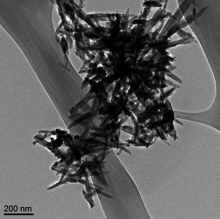
Needle-like particles of hydroxyapatite found in infant formula by ASU researchers. Westerhoff and Schoepf/ASU, CC BY-ND

Needle-like particles of hydroxyapatite found in infant formula by ASU [Arizona State University] researchers. Westerhoff and Schoepf/ASU, CC BY-ND

Nanowerk is featuring an essay about hydroxyapatite nanoparticles in baby formula written by Dr. Andrew Maynard in a May 17, 2016 news item (Note: A link has been removed),

There’s a lot of stuff you’d expect to find in baby formula: proteins, carbs, vitamins, essential minerals. But parents probably wouldn’t anticipate finding extremely small, needle-like particles. Yet this is exactly what a team of scientists here at Arizona State University [ASU] recently discovered.

The research, commissioned and published by Friends of the Earth (FoE) – an environmental advocacy group – analyzed six commonly available off-the-shelf baby formulas (liquid and powder) and found nanometer-scale needle-like particles in three of them. The particles were made of hydroxyapatite – a poorly soluble calcium-rich mineral. Manufacturers use it to regulate acidity in some foods, and it’s also available as a dietary supplement.

Andrew’s May 17, 2016 essay first appeared on The Conversation website,

Looking at these particles at super-high magnification, it’s hard not to feel a little anxious about feeding them to a baby. They appear sharp and dangerous – not the sort of thing that has any place around infants. …

… questions like “should infants be ingesting them?” make a lot of sense. However, as is so often the case, the answers are not quite so straightforward.

Andrew begins by explaining about calcium and hydroxyapatite (from The Conversation),

Calcium is an essential part of a growing infant’s diet, and is a legally required component in formula. But not necessarily in the form of hydroxyapatite nanoparticles.

Hydroxyapatite is a tough, durable mineral. It’s naturally made in our bodies as an essential part of bones and teeth – it’s what makes them so strong. So it’s tempting to assume the substance is safe to eat. But just because our bones and teeth are made of the mineral doesn’t automatically make it safe to ingest outright.

The issue here is what the hydroxyapatite in formula might do before it’s digested, dissolved and reconstituted inside babies’ bodies. The size and shape of the particles ingested has a lot to do with how they behave within a living system.

He then discusses size and shape, which are important at the nanoscale,

Size and shape can make a difference between safe and unsafe when it comes to particles in our food. Small particles aren’t necessarily bad. But they can potentially get to parts of our body that larger ones can’t reach. Think through the gut wall, into the bloodstream, and into organs and cells. Ingested nanoscale particles may be able to interfere with cells – even beneficial gut microbes – in ways that larger particles don’t.

These possibilities don’t necessarily make nanoparticles harmful. Our bodies are pretty well adapted to handling naturally occurring nanoscale particles – you probably ate some last time you had burnt toast (carbon nanoparticles), or poorly washed vegetables (clay nanoparticles from the soil). And of course, how much of a material we’re exposed to is at least as important as how potentially hazardous it is.

Yet there’s a lot we still don’t know about the safety of intentionally engineered nanoparticles in food. Toxicologists have started paying close attention to such particles, just in case their tiny size makes them more harmful than otherwise expected.

Currently, hydroxyapatite is considered safe at the macroscale by the US Food and Drug Administration (FDA). However, the agency has indicated that nanoscale versions of safe materials such as hydroxyapatite may not be safe food additives. From Andrew’s May 17, 2016 essay,

Hydroxyapatite is a tough, durable mineral. It’s naturally made in our bodies as an essential part of bones and teeth – it’s what makes them so strong. So it’s tempting to assume the substance is safe to eat. But just because our bones and teeth are made of the mineral doesn’t automatically make it safe to ingest outright.

The issue here is what the hydroxyapatite in formula might do before it’s digested, dissolved and reconstituted inside babies’ bodies. The size and shape of the particles ingested has a lot to do with how they behave within a living system. Size and shape can make a difference between safe and unsafe when it comes to particles in our food. Small particles aren’t necessarily bad. But they can potentially get to parts of our body that larger ones can’t reach. Think through the gut wall, into the bloodstream, and into organs and cells. Ingested nanoscale particles may be able to interfere with cells – even beneficial gut microbes – in ways that larger particles don’t.These possibilities don’t necessarily make nanoparticles harmful. Our bodies are pretty well adapted to handling naturally occurring nanoscale particles – you probably ate some last time you had burnt toast (carbon nanoparticles), or poorly washed vegetables (clay nanoparticles from the soil). And of course, how much of a material we’re exposed to is at least as important as how potentially hazardous it is.Yet there’s a lot we still don’t know about the safety of intentionally engineered nanoparticles in food. Toxicologists have started paying close attention to such particles, just in case their tiny size makes them more harmful than otherwise expected.

Putting particle size to one side for a moment, hydroxyapatite is classified by the US Food and Drug Administration (FDA) as “Generally Regarded As Safe.” That means it considers the material safe for use in food products – at least in a non-nano form. However, the agency has raised concerns that nanoscale versions of food ingredients may not be as safe as their larger counterparts.Some manufacturers may be interested in the potential benefits of “nanosizing” – such as increasing the uptake of vitamins and minerals, or altering the physical, textural and sensory properties of foods. But because decreasing particle size may also affect product safety, the FDA indicates that intentionally nanosizing already regulated food ingredients could require regulatory reevaluation.In other words, even though non-nanoscale hydroxyapatite is “Generally Regarded As Safe,” according to the FDA, the safety of any nanoscale form of the substance would need to be reevaluated before being added to food products.Despite this size-safety relationship, the FDA confirmed to me that the agency is unaware of any food substance intentionally engineered at the nanoscale that has enough generally available safety data to determine it should be “Generally Regarded As Safe.”Casting further uncertainty on the use of nanoscale hydroxyapatite in food, a 2015 report from the European Scientific Committee on Consumer Safety (SCCS) suggests there may be some cause for concern when it comes to this particular nanomaterial.Prompted by the use of nanoscale hydroxyapatite in dental products to strengthen teeth (which they consider “cosmetic products”), the SCCS reviewed published research on the material’s potential to cause harm. Their conclusion?

The available information indicates that nano-hydroxyapatite in needle-shaped form is of concern in relation to potential toxicity. Therefore, needle-shaped nano-hydroxyapatite should not be used in cosmetic products.

This recommendation was based on a handful of studies, none of which involved exposing people to the substance. Researchers injected hydroxyapatite needles directly into the bloodstream of rats. Others exposed cells outside the body to the material and observed the effects. In each case, there were tantalizing hints that the small particles interfered in some way with normal biological functions. But the results were insufficient to indicate whether the effects were meaningful in people.

As Andrew also notes in his essay, none of the studies examined by the SCCS OEuropean Scientific Committee on Consumer Safety) looked at what happens to nano-hydroxyapatite once it enters your gut and that is what the researchers at Arizona State University were considering (from the May 17, 2016 essay),

The good news is that, according to preliminary studies from ASU researchers, hydroxyapatite needles don’t last long in the digestive system.

This research is still being reviewed for publication. But early indications are that as soon as the needle-like nanoparticles hit the highly acidic fluid in the stomach, they begin to dissolve. So fast in fact, that by the time they leave the stomach – an exceedingly hostile environment – they are no longer the nanoparticles they started out as.

These findings make sense since we know hydroxyapatite dissolves in acids, and small particles typically dissolve faster than larger ones. So maybe nanoscale hydroxyapatite needles in food are safer than they sound.

This doesn’t mean that the nano-needles are completely off the hook, as some of them may get past the stomach intact and reach more vulnerable parts of the gut. But the findings do suggest these ultra-small needle-like particles could be an effective source of dietary calcium – possibly more so than larger or less needle-like particles that may not dissolve as quickly.

Intriguingly, recent research has indicated that calcium phosphate nanoparticles form naturally in our stomachs and go on to be an important part of our immune system. It’s possible that rapidly dissolving hydroxyapatite nano-needles are actually a boon, providing raw material for these natural and essential nanoparticles.

While it’s comforting to know that preliminary research suggests that the hydroxyapatite nanoparticles are likely safe for use in food products, Andrew points out that more needs to be done to insure safety (from the May 17, 2016 essay),

And yet, even if these needle-like hydroxyapatite nanoparticles in infant formula are ultimately a good thing, the FoE report raises a number of unresolved questions. Did the manufacturers knowingly add the nanoparticles to their products? How are they and the FDA ensuring the products’ safety? Do consumers have a right to know when they’re feeding their babies nanoparticles?

Whether the manufacturers knowingly added these particles to their formula is not clear. At this point, it’s not even clear why they might have been added, as hydroxyapatite does not appear to be a substantial source of calcium in most formula. …

And regardless of the benefits and risks of nanoparticles in infant formula, parents have a right to know what’s in the products they’re feeding their children. In Europe, food ingredients must be legally labeled if they are nanoscale. In the U.S., there is no such requirement, leaving American parents to feel somewhat left in the dark by producers, the FDA and policy makers.

As far as I’m aware, the Canadian situation is much the same as the US. If the material is considered safe at the macroscale, there is no requirement to indicate that a nanoscale version of the material is in the product.

I encourage you to read Andrew’s essay in its entirety. As for the FoE report (Nanoparticles in baby formula: Tiny new ingredients are a big concern), that is here.

Could nanoparticles in your mouthwash affect for your cells?

The first news item I’m going to highlight was posted on Nanowerk, March 8, 2012 and is focused on the use of silver nanoparticles in mouthwashes and dentures to prevent yeast infections,

Yeasts which cause hard-to-treat mouth infections are killed using silver nanoparticles in the laboratory, scientists have found. These yeast infections, caused by Candida albicans and Candida glabrata target the young, old and immuno-compromised. Professor Mariana Henriques, University of Minho [Portugal], and her colleagues hope to test silver nanoparticles in mouthwash and dentures as a potential preventative measure against these infections.

Professor Henriques and her team, who published their research in the Society for Applied Microbiology’s journal Letters in Applied Microbiology(“Silver nanoparticles: influence of stabilizing agent and diameter on antifungal activity against Candida albicans and Candida glabrata biofilms”), looked at the use of different sizes of silver nanoparticles to determine their anti-fungal properties …

The scientists used artificial biofilms in conditions which mimic those of saliva as closely as possible. They then added different sizes and concentrations of silver nanoparticles and found that different sizes of nanoparticles were equally effective at killing the yeasts. Due to the diversity of the sizes of nanoparticles demonstrating anti-fungal properties the researchers hope this will enable the nanoparticles to be used in many different applications.

Some researchers have expressed concerns around the safety of nanoparticle use but the authors stress this research is at an early stage and extensive safety trials will be carried out before any product reaches the market. [emphasis mine]

Following on the notion of safety and gargling silver nanoparticles, coincidentally, there was another news item also dated March 8, 2012 on Nanowerk, this one about the impact that nanoparticles may have on nutrient uptake,

Nanoparticles are everywhere. From cosmetics and clothes, to soda and snacks. But as versatile as they are, nanoparticles also have a downside, say researchers at Binghamton University and Cornell University in a recent paper published in the journal Nature Nanotechnology (“Oral exposure to polystyrene nanoparticles affects iron absorption”). These tiny particles, even in low doses, could have a big impact on our long-term health.

According to lead author of the article, Gretchen Mahler, assistant professor of bioengineering at Binghamton University, much of the existing research on the safety of nanoparticles has been on the direct health effects. But what Mahler, Michael L. Shuler of Cornell University and a team of researchers really wanted to know was what happens when someone gets constant exposure in small doses – the kind you’d get if you were taken a drug or supplement that included nanoparticles in some form. [e.g. silver nanoparticles in your mouthwash or on your dentures]

“We thought that the best way to measure the more subtle effects of this kind of intake was to monitor the reaction of intestinal cells,” said Mahler. “And we did this in two ways: in vitro, through human intestinal-lining cells that we had cultured in the lab; and in vivo, through the intestinal linings of live chickens. Both sets of results pointed to the same thing – that exposure to nanoparticles influences the absorption of nutrients into the bloodstream.”

As for why the researchers focused on iron and tested polystyrene nanoparticles (from the news item),

The uptake of iron, an essential nutrient, was of particular interest due to the way it is absorbed and processed through the intestines. The way Mahler and the team tested this was to use polystyrene nanoparticles because of its easily traceable fluorescent properties.

“What we found was that for brief exposures, iron absorption dropped by about 50 percent,” said Mahler. “But when we extended that period of time, absorption actually increased by about 200 percent. It was very clear – nanoparticles definitely affects iron uptake and transport.”

While acute oral exposure caused disruptions to intestinal iron transport, chronic exposure caused a remodeling of the intestinal villi – the tiny, finger-like projections that are vital to the intestine’s ability to absorb nutrients – making them larger and broader, thus allowing iron to enter the bloodstream much faster.

As to whether these changes are good or bad the researchers don’t speculate. They do have plans for more testing,

calcium,
copper,
zinc, and
fat-soluble vitamins A, D, E and K

They don’t mention any changes in the types of nanoparticles they might be testing in future.

In any event, our bodies have changed a lot over the centuries, you just have to visit a pyramid in Egypt or a museum that holds medieval armour to observe that humans were once much shorter than we are today.