Tag Archives: carbon nanoparticles

A new graphene-based contrast agent for magnetic resonance imaging (MRI)

After teaching a continuing studies course on bioelectronics for Simon Fraser University (Vancouver, Canada), I’ve developed a mild interest in magnetic resonance imaging and contrast agents which this Nov. 11, 2016 news item on phys.org satisfies,

Graphene, the atomically thin sheets of carbon that materials scientists are hoping to use for everything from nanoelectronics and aircraft de-icers to batteries and bone implants, may also find use as contrast agents for magnetic resonance imaging (MRI), according to new research from Rice University.

“They have a lot of advantages compared with conventionally available contrast agents,” Rice researcher Sruthi Radhakrishnan said of the graphene-based quantum dots she has studied for the past two years. “Virtually all of the widely used contrast agents contain toxic metals, but our material has no metal. It’s just carbon, hydrogen, oxygen and fluorine, and in all of our tests so far it has shown no signs of toxicity.”

The initial findings for Rice’s nanoparticles—disks of graphene that are decorated with fluorine atoms and simply organic molecules that make them magnetic—are described in a new paper in the journal Particle and Particle Systems characterization.

A Nov. 10, 2016 Rice University (Texas, US) news release, which originated the news item, describes the work in more detail,

Pulickel Ajayan, the Rice materials scientist who is directing the work, said the fluorinated graphene oxide quantum dots could be particularly useful as MRI contrast agents because they could be targeted to specific kinds of tissues.

“There are tried-and-true methods for attaching biomarkers to carbon nanoparticles, so one could easily envision using these quantum dots to develop tissue-specific contrast agents,” Ajayan said. “For example, this method could be used to selectively target specific types of cancer or brain lesions caused by Alzheimer’s disease. That kind of specificity isn’t available with today’s contrast agents.”

MRI scanners make images of the body’s internal structures using strong magnetic fields and radio waves. As diagnostic tests, MRIs often provide greater detail than X-rays without the harmful radiation, and as a result, MRI usage has risen sharply over the past decade. More than 30 million MRIs are performed annually in the U.S.

Radhakrishnan said her work began in 2014 after Ajayan’s research team found that adding fluorine to either graphite or graphene caused the materials to show up well on MRI scans.

All materials are influenced by magnetic fields, including animal tissues. In MRI scanners, a powerful magnetic field causes individual atoms throughout the body to become magnetically aligned. A pulse of radio energy is used to disrupt this alignment, and the machine measures how long it takes for the atoms in different parts of the body to become realigned. Based on these measures, the scanner can build up a detailed image of the body’s internal structures.

MRI contrast agents shorten the amount of time it takes for tissues to realign and significantly improve the resolution of MRI scans. Almost all commercially available contrast agents are made from toxic metals like gadolinium, iron or manganese.

“We worked with a team from MD Anderson Cancer Center to assess the cytocompatibility of fluorinated graphene oxide quantum dots,” Radhakrishnan said. “We used a test that measures the metabolic activity of cell cultures and detects toxicity as a drop in metabolic activity. We incubated quantum dots in kidney cell cultures for up to three days and found no significant cell death in the cultures, even at the highest concentrations.”

The fluorinated graphene oxide quantum dots Radhakrishnan studies can be made in less than a day, but she spent two years perfecting the recipe for them. She begins with micron-sized sheets of graphene that have been fluorinated and oxidized. When these are added to a solvent and stirred for several hours, they break into smaller pieces. Making the material smaller is not difficult, but the process for making small particles with the appropriate magnetic properties is exacting. Radhakrishnan said there was no “eureka moment” in which she suddenly achieved the right results by stumbling on the best formula. Rather, the project was marked by incremental improvements through dozens of minor alterations.

“It required a lot of optimization,” she said. “The recipe matters a lot.”

Radhakrishnan said she plans to continue studying the material and hopes to eventually have a hand in proving that it is safe and effective for clinical MRI tests.

“I would like to see it applied commercially in clinical ways because it has a lot of advantages compared with conventionally available agents,” she said.

Here’s a link to and a citation for the paper,

Metal-Free Dual Modal Contrast Agents Based on Fluorographene Quantum Dots by Sruthi Radhakrishnan, Atanu Samanta, Parambath M. Sudeep, Kiersten L. Maldonado, Sendurai A. Mani, Ghanashyam Acharya, Chandra Sekhar Tiwary, Abhishek K. Singh, and Pulickel M. Ajayan. Particle & Particle Systems Characterization DOI: 10.1002/ppsc.201600221 Version of Record online: 21 OCT 2016

This paper is behind a paywall.
Tattoo therapy for chronic disease?

It’s good to wake up to something truly new. In this case, it’s using tattoos and nanoparticles for medical applications. From a Sept. 22, 2016 news item on ScienceDaily,

A temporary tattoo to help control a chronic disease might someday be possible, according to scientists at Baylor College of Medicine [Texas, US] who tested antioxidant nanoparticles created at Rice University [Texas, US].

A Sept. 22, 2016 Rice University news release, which originated the news item, provides more information and some good explanations of the terms used (Note: Links have been removed),

A proof-of-principle study led by Baylor scientist Christine Beeton published today by Nature’s online, open-access journal Scientific Reports shows that nanoparticles modified with polyethylene glycol are conveniently choosy as they are taken up by cells in the immune system.

That could be a plus for patients with autoimmune diseases like multiple sclerosis, one focus of study at the Beeton lab. “Placed just under the skin, the carbon-based particles form a dark spot that fades over about one week as they are slowly released into the circulation,” Beeton said.

T and B lymphocyte cells and macrophages are key components of the immune system. However, in many autoimmune diseases such as multiple sclerosis, T cells are the key players. One suspected cause is that T cells lose their ability to distinguish between invaders and healthy tissue and attack both.

In tests at Baylor, nanoparticles were internalized by T cells, which inhibited their function, but ignored by macrophages. “The ability to selectively inhibit one type of cell over others in the same environment may help doctors gain more control over autoimmune diseases,” Beeton said.

“The majority of current treatments are general, broad-spectrum immunosuppressants,” said Redwan Huq, lead author of the study and a graduate student in the Beeton lab. “They’re going to affect all of these cells, but patients are exposed to side effects (ranging) from infections to increased chances of developing cancer. So we get excited when we see something new that could potentially enable selectivity.” Since the macrophages and other splenic immune cells are unaffected, most of a patient’s existing immune system remains intact, he said.

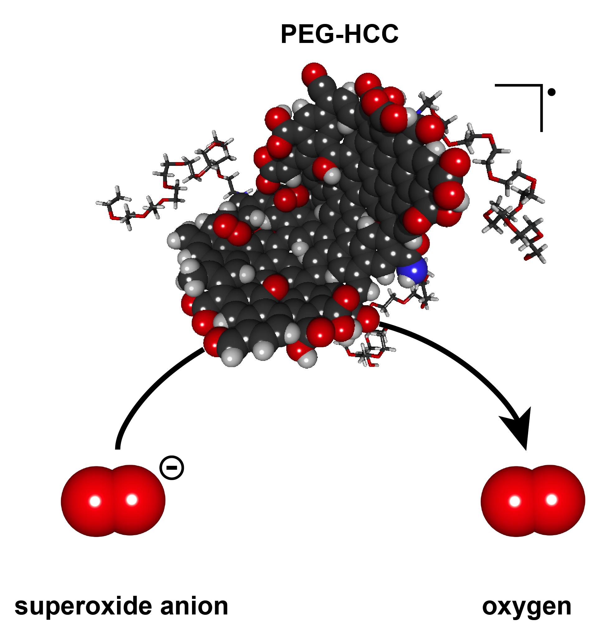
The soluble nanoparticles synthesized by the Rice lab of chemist James Tour have shown no signs of acute toxicity in prior rodent studies, Huq said. They combine polyethylene glycol with hydrophilic carbon clusters, hence their name, PEG-HCCs. The carbon clusters are 35 nanometers long, 3 nanometers wide and an atom thick, and bulk up to about 100 nanometers in globular form with the addition of PEG. They have proven to be efficient scavengers of reactive oxygen species called superoxide molecules, which are expressed by cells the immune system uses to kill invading microorganisms.

T cells use superoxide in a signaling step to become activated. PEG-HCCs remove this superoxide from the T cells, preventing their activation without killing the cells.

Beeton became aware of PEG-HCCs during a presentation by former Baylor graduate student Taeko Inoue, a co-author of the new study. “As she talked, I was thinking, ‘That has to work in models of multiple sclerosis,’” Beeton said. “I didn’t have a good scientific rationale, but I asked for a small sample of PEG-HCCs to see if they affected immune cells.

“We found they affected the T lymphocytes and not the other splenic immune cells, like the macrophages. It was completely unexpected,” she said.

The Baylor lab’s tests on animal models showed that small amounts of PEG-HCCs injected under the skin are slowly taken up by T lymphocytes, where they collect and inhibit the cell’s function. They also found the nanoparticles did not remain in T cells and dispersed within days after uptake by the cells.

“That’s an issue because you want a drug that’s in the system long enough to be effective, but not so long that, if you have a problem, you can’t remove it,” Beeton said. “PEG-HCCs can be administered for slow release and don’t stay in the system for long. This gives us much better control over the circulating half-life.”

“The more we study the abilities of these nanoparticles, the more surprised we are at how useful they could be for medical applications,” Tour said. The Rice lab has published papers with collaborators at Baylor and elsewhere on using functionalized nanoparticles to deliver cancer drugs to tumors and to quench the overproduction of superoxides after traumatic brain injuries.

Beeton suggested delivering carbon nanoparticles just under the skin rather than into the bloodstream would keep them in the system longer, making them more available for uptake by T cells. And the one drawback – a temporary but visible spot on the skin that looks like a tattoo – could actually be a perk to some.

“We saw it made a black mark when we injected it, and at first we thought that’s going to be a real problem if we ever take it into the clinic,” Beeton said. “But we can work around that. We can inject into an area that’s hidden, or use micropattern needles and shape it.

“I can see doing this for a child who wants a tattoo and could never get her parents to go along,” she said. “This will be a good way to convince them.”

The research was supported by Baylor College of Medicine, the National Multiple Sclerosis Society, National Institutes of Health, the Dan L. Duncan Cancer Center, John S. Dunn Gulf Coast Consortium for Chemical Genomics and the U.S. Army-funded Traumatic Brain Injury Consortium.

That’s an interesting list of funders at the end of the news release.

Here’s a link to and a citation for the paper,

Preferential uptake of antioxidant carbon nanoparticles by T lymphocytes for immunomodulation by Redwan Huq, Errol L. G. Samuel, William K. A. Sikkema, Lizanne G. Nilewski, Thomas Lee, Mark R. Tanner, Fatima S. Khan, Paul C. Porter, Rajeev B. Tajhya, Rutvik S. Patel, Taeko Inoue, Robia G. Pautler, David B. Corry, James M. Tour, & Christine Beeton. Scientific Reports 6, Article number: 33808 (2016) doi:10.1038/srep33808 Published online: 22 September 2016

This paper is open access.

Here’s an image provided by the researchers,

Polyethylene glycol-hydrophilic carbon clusters developed at Rice University were shown to be selectively taken up by T cells, which inhibits their function, in tests at Baylor College of Medicine. The researchers said the nanoparticles could lead to new strategies for controlling autoimmune diseases like multiple sclerosis. (Credit: Errol Samuel/Rice University) - See more at: http://news.rice.edu/2016/09/22/tattoo-therapy-could-ease-chronic-disease/#sthash.sIfs3b0S.dpuf

Polyethylene glycol-hydrophilic carbon clusters developed at Rice University were shown to be selectively taken up by T cells, which inhibits their function, in tests at Baylor College of Medicine. The researchers said the nanoparticles could lead to new strategies for controlling autoimmune diseases like multiple sclerosis. (Credit: Errol Samuel/Rice University)

Improve car performance with graphene balls

Lubrication is vital for car engines and it can be expensive when you get it wrong or when it’s not as effective as it could be. A Jan. 25, 2016 news item on Nanowerk highlights some research focused on improving the quality of engine lubrication,

When an automobile’s engine is improperly lubricated, it can be a major hit to the pocketbook and the environment.

For the average car, 15 percent of the fuel consumption is spent overcoming friction in the engine and transmission. When friction is high, gears have to work harder to move. This means the car burns more fuel and emits more carbon dioxide into the atmosphere.

“Every year, millions of tons of fuel are wasted because of friction,” said Northwestern Engineering’s Jiaxing Huang, associate professor of materials science and engineering. “It’s a serious problem.”

While oil helps reduce this friction, people have long searched for additives that enhance oil’s performance. Huang and his collaborators discovered that crumpled graphene balls are an extremely promising lubricant additive. In a series of tests, oil modified with crumpled graphene balls outperformed some commercial lubricants by 15 percent, both in terms of reducing friction and the degree of wear on steel surfaces.

A Jan. 25, 2015 McCormick School of Engineering at Northwestern University news release, which originated the news item, provides more information about the team’s work,

About five years ago, Huang discovered crumpled graphene balls — a novel type of ultrafine particles that resemble crumpled paper balls. The particles are made by drying tiny water droplets with graphene-based sheets inside. “Capillary force generated by the evaporation of water crumples the sheets into miniaturized paper balls,” Huang said. “Just like how we crumple a piece of paper with our hands.”

Shortly after making this discovery, Huang explained it to Chung [Yip-Wah Chung, professor of materials science and engineering] during a lunch in Hong Kong by crumpling a napkin and juggling it. “When the ball landed on the table, it rolled,” Chung recalled. “It reminded me of ball bearings that roll between surfaces to reduce friction.”

That “a-ha!” moment led to a collaboration among the two professors and Wang, who was in the middle of editing a new Encyclopedia of Tribology with Chung.

Nanoparticles, particularly carbon nanoparticles, previously have been studied to help increase the lubrication of oil. The particles, however, do not disperse well in oil and instead tend to clump together, which makes them less effective for lubrication. The particles may jam between the gear’s surfaces causing severe aggregation that increases friction and wear. To overcome this problem, past researchers have modified the particles with extra chemicals, called surfactants, to make them disperse. But this still doesn’t entirely solve the problem.

“Under friction, the surfactant molecules can rub off and decompose,” Chung said. “When that happens, the particles clump up again.”

Because of their unique shape, crumpled graphene balls self-disperse without needing surfactants that are attracted to oil. With their pointy surfaces, they are unable to make close contact with the other graphene balls. Even when they are squeezed together, they easily separate again when disturbed.

Huang and his team also found that performance of crumpled graphene balls is not sensitive to their concentrations in the oil. “A few are already sufficient, and if you increase the concentration by 10 times, performance is about the same,” Huang said. “For all other carbon additives, such performance is very sensitive to concentration. You have to find the sweet spot.”

“The problem with finding a sweet spot is that, during operation, the local concentration of particles near the surfaces under lubrication could fluctuate,” Wang [Q. Jane Wang, professor of mechanical engineering] added. “This leads to unstable performance for most other additive particles.”

Next, the team plans to explore the additional benefit of using crumpled graphene balls in oil: they can also be used as carriers. Because the ball-like particles have high surface area and open spaces, they are good carriers for materials with other functions, such as corrosion inhibition.

Here’s a link to and a citation for the paper,

Self-dispersed crumpled graphene balls in oil for friction and wear reduction by Xuan Dou, Andrew R. Koltonow, Xingliang He, Hee Dong Jang, Qian Wang, Yip-Wah Chung, and Jiaxing Huang. PNAS 2016 doi:10 .1038/srep03863 Published ahead of print January 25, 2016

This paper is behind a paywall.

One final comment, it’s a bit unusual to see the term ‘carbon nanoparticle’. Generally speaking, carbon nanoparticles seem to have their own names, graphene, carbon nanotubes, and buckminsterfullerenes come to mind.

Making carbon nanoparticles at home with honey or molasses

No need to rush and buy any ingredients as the University of Illinois at Urbana-Champaign researchers do not provide a recipe for cooking up carbon nanoparticles. However, it is diverting to think that one day we might be able to make these items at home. From a June 19, 2015 news item by Stuart Milne on the Azonano website,

Researchers at the University of Illinois have discovered an easy method to produce carbon nanoparticles for biomedical applications. These carbon nanoparticles can be made at home within a couple of hours using easily available ingredients and molasses.

A June 19 (?), 2015 University of Illinois at Urbana-Champaign news release (also on EurekAlert) provides more detail about the research,

“If you have a microwave and honey or molasses, you can pretty much make these particles at home,” Pan [professor Dipanjan Pan] said. “You just mix them together and cook it for a few minutes, and you get something that looks like char, but that is nanoparticles with high luminescence. This is one of the simplest systems that we can think of. It is safe and highly scalable for eventual clinical use.”

These “next-generation” carbon spheres have several attractive properties, the researchers found. They naturally scatter light in a manner that makes them easy to differentiate from human tissues, eliminating the need for added dyes or fluorescing molecules to help detect them in the body.

The nanoparticles are coated with polymers that fine-tune their optical properties and their rate of degradation in the body. The polymers can be loaded with drugs that are gradually released.

The nanoparticles also can be made quite small, less than eight nanometers in diameter (a human hair is 80,000 to 100,000 nanometers thick).

“Our immune system fails to recognize anything under 10 nanometers,” Pan said. “So, these tiny particles are kind of camouflaged, I would say; they are hiding from the human immune system.”

The team tested the therapeutic potential of the nanoparticles by loading them with an anti-melanoma drug and mixing them in a topical solution that was applied to pig skin.

Bhargava’s [professor Rohit Bhargava] laboratory used vibrational spectroscopic techniques to identify the molecular structure of the nanoparticles and their cargo.

“Raman and infrared spectroscopy are the two tools that one uses to see molecular structure,” Bhargava said. “We think we coated this particle with a specific polymer and with specific drug-loading – but did we really? We use spectroscopy to confirm the formulation as well as visualize the delivery of the particles and drug molecules.”

The team found that the nanoparticles did not release the drug payload at room temperature, but at body temperature began to release the anti-cancer drug. The researchers also determined which topical applications penetrated the skin to a desired depth.

In further experiments, the researchers found they could alter the infusion of the particles into melanoma cells by adjusting the polymer coatings. Imaging confirmed that the infused cells began to swell, a sign of impending cell death.

“This is a versatile platform to carry a multitude of drugs – for melanoma, for other kinds of cancers and for other diseases,” Bhargava said. “You can coat it with different polymers to give it a different optical response. You can load it with two drugs, or three, or four, so you can do multidrug therapy with the same particles.”

“By using defined surface chemistry, we can change the properties of these particles,” Pan said. “We can make them glow at a certain wavelength and also we can tune them to release the drugs in the presence of the cellular environment. That is, I think, the beauty of the work.”

Here’s a link to and a citation for the paper,

Tunable Luminescent Carbon Nanospheres with Well-Defined Nanoscale Chemistry for Synchronized Imaging and Therapy by Prabuddha Mukherjee, Santosh K. Misra, Mark C. Gryka, Huei-Huei Chang, Saumya Tiwari, William L. Wilson, John W. Scott, Rohit Bhargava, and Dipanjan Pan. Small
DOI: 10.1002/smll.201500728 Article first published online: 20 MAY 2015

© 2015 WILEY-VCH Verlag GmbH & Co. KGaA, Weinheim

This paper is behind a paywall.

Antioxidant-like carbon nanoparticles could help heal traumatic brain injuries

The research sounds exciting but all of the testing has taken place in laboratories on animal models (rats). The Oct. 18, 2012 news item on Azonano describes why the research team wanted to test  antioxidant-like carbon nanotubes for use with traumatic brain injury (TBI) patients,

Thomas Kent, James Tour and colleagues explain that TBI disrupts the supply of oxygen-rich blood to the brain. With the brain so oxygen-needy — accounting for only 2 percent of a person’s weight, but claiming 20 percent of the body’s oxygen supply — even a mild injury, such as a concussion, can have serious consequences. Reduced blood flow and resuscitation result in a build-up of free-radicals, which can kill brain cells. Despite years of far-ranging efforts, no effective treatment has emerged for TBI. That’s why the scientists tried a new approach, based on nanoparticles so small that 1000 would fit across the width of a human hair.

The American Chemical Society (ACS) Oct. 17, 2912 news release, which originated the news item, provides a few details about the research,

They [the research team]  describe development and successful laboratory tests of nanoparticles, called PEG-HCCs. In laboratory rats, the nanoparticles acted like antioxidants, rapidly restoring blood flow to the brain following resuscitation after TBI. “This finding is of major importance for improving patient health under clinically relevant conditions during resuscitative care, and it has direct implications for the current [TBI] war-fighter victims in the Afghanistan and Middle East theaters,” they say.

The abstract for the paper gives more insight,

Injury to the neurovasculature is a feature of brain injury and must be addressed to maximize opportunity for improvement. Cerebrovascular dysfunction, manifested by reduction in cerebral blood flow (CBF), is a key factor that worsens outcome after traumatic brain injury (TBI), most notably under conditions of hypotension. We report here that a new class of antioxidants, poly(ethylene glycol)-functionalized hydrophilic carbon clusters (PEG-HCCs), which are nontoxic carbon particles, rapidly restore CBF in a mild TBI/hypotension/resuscitation rat model when administered during resuscitation—a clinically relevant time point. Along with restoration of CBF, there is a concomitant normalization of superoxide and nitric oxide levels. Given the role of poor CBF in determining outcome, this finding is of major importance for improving patient health under clinically relevant conditions during resuscitative care, and it has direct implications for the current TBI/hypotension war-fighter victims in the Afghanistan and Middle East theaters. The results also have relevancy in other related acute circumstances such as stroke and organ transplantation.

I notice this treatment has shown some success for mildTBI/hypotension if applied in the resuscitation phase and the testing, as I mentioned earlier, has been done on rats. For anyone who wants more information about this promising treatment,

Antioxidant Carbon Particles Improve Cerebrovascular Dysfunction Following Traumatic Brain Injury by Brittany R. Bitner, Daniela C. Marcano, Jacob M. Berlin, Roderic H. Fabian, Leela Cherian, James C. Culver, Mary E. Dickinson, Claudia S. Robertson, Robia G. Pautler, Thomas A. Kent, and James M. Tour. ACS Nano, 2012, 6 (9), pp 8007–8014 DOI: 10.1021/nn302615f

The article is behind a paywall and I notice it was published online Aug. 6, 2012. It looks like the ACS may may have tried to publicize this at the time of publication and decided to try again now in the hope of getting more publicity for this work.