Tag Archives: cartilage

Lab-made cartilage gel for stiff, achy knees

Researchers claim their lab-made cartilage is better than the real thing in an August 11, 2022 news item on phys.org, Note: Links have been removed,

Over-the-counter pain relievers, physical therapy, steroid injections—some people have tried it all and are still dealing with knee pain.

Often knee pain comes from the progressive wear and tear of cartilage known as osteoarthritis, which affects nearly one in six adults—867 million people—worldwide. For those who want to avoid replacing the entire knee joint, there may soon be another option that could help patients get back on their feet fast, pain-free, and stay that way.

Writing in the journal Advanced Functional Materials, a Duke University-led team says they have created the first gel-based cartilage substitute that is even stronger and more durable than the real thing.

Caption: Duke researchers have developed a gel-based cartilage substitute to relieve achy knees that’s even stronger and more durable than the real thing. Clinical trials to start next year. Credit: Canva Credit: Benjamin Wiley, Duke University

Here’s the August 11, 2022 Duke University news release (also on EurekAlert), which originated the news item, where you’ll find more details about the research, Note: Links have been removed,

Mechanical testing reveals that the Duke team’s hydrogel — a material made of water-absorbing polymers — can be pressed and pulled with more force than natural cartilage, and is three times more resistant to wear and tear.

Implants made of the material are currently being developed by Sparta Biomedical and tested in sheep. Researchers are gearing up to begin clinical trials in humans next year.

“If everything goes according to plan, the clinical trial should start as soon as April 2023,” said Duke chemistry professor Benjamin Wiley, who led the research along with Duke mechanical engineering and materials science professor Ken Gall.

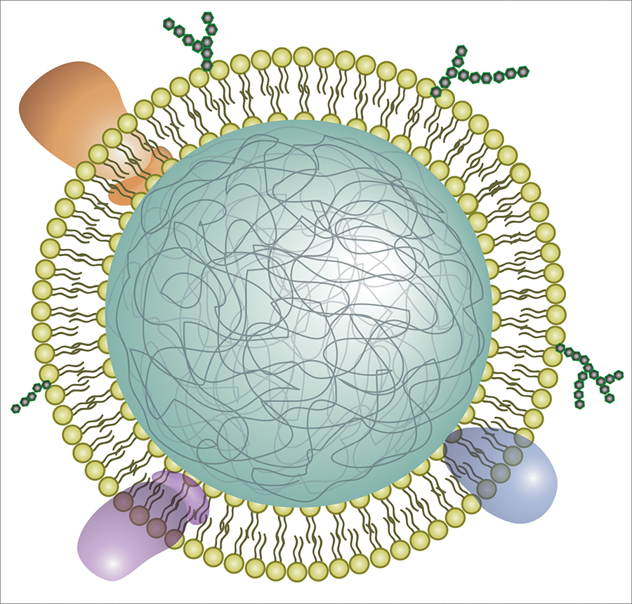
To make this material, the Duke team took thin sheets of cellulose fibers and infused them with a polymer called polyvinyl alcohol — a viscous goo consisting of stringy chains of repeating molecules — to form a gel.

The cellulose fibers act like the collagen fibers in natural cartilage, Wiley said — they give the gel strength when stretched. The polyvinyl alcohol helps it return to its original shape. The result is a Jello-like material, 60% water, which is supple yet surprisingly strong.

Natural cartilage can withstand a whopping 5,800 to 8,500 pounds per inch of tugging and squishing, respectively, before reaching its breaking point. Their lab-made version is the first hydrogel that can handle even more. It is 26% stronger than natural cartilage in tension, something like suspending seven grand pianos from a key ring, and 66% stronger in compression — which would be like parking a car on a postage stamp.

“It’s really off the charts in terms of hydrogel strength,” Wiley said.

The team has already made hydrogels with remarkable properties. In 2020, they reported that they had created the first hydrogel strong enough for knees, which feel the force of two to three times body weight with each step.

Putting the gel to practical use as a cartilage replacement, however, presented additional design challenges. One was achieving the upper limits of cartilage’s strength. Activities like hopping, lunging, or climbing stairs put some 10 Megapascals of pressure on the cartilage in the knee, or about 1,400 pounds per square inch. But the tissue can take up to four times that before it breaks.

“We knew there was room for improvement,” Wiley said.

In the past, researchers attempting to create stronger hydrogels used a freeze-thaw process to produce crystals within the gel, which drive out water and help hold the polymer chains together. In the new study, instead of freezing and thawing the hydrogel, the researchers used a heat treatment called annealing to coax even more crystals to form within the polymer network.

By increasing the crystal content, the researchers were able to produce a gel that can withstand five times as much stress from pulling and nearly twice as much squeezing relative to freeze-thaw methods.

The improved strength of the annealed gel also helped solve a second design challenge: securing it to the joint and getting it to stay put.

Cartilage forms a thin layer that covers the ends of bones so they don’t grind against one another. Previous studies haven’t been able to attach hydrogels directly to bone or cartilage with sufficient strength to keep them from breaking loose or sliding off. So the Duke team came up with a different approach.

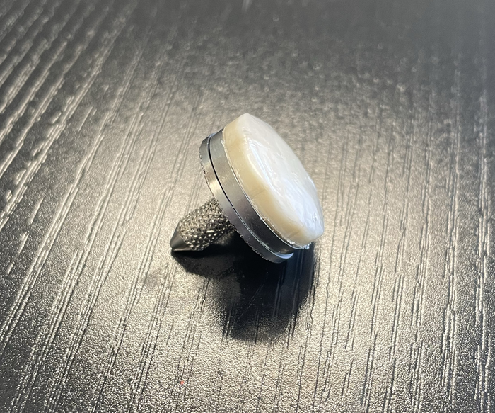
Their method of attachment involves cementing and clamping the hydrogel to a titanium base. This is then pressed and anchored into a hole where the damaged cartilage used to be. Tests show the design stays fastened 68% more firmly than natural cartilage on bone.

“Another concern for knee implants is wear over time, both of the implant itself and the opposing cartilage,” Wiley said.

Other researchers have tried replacing damaged cartilage with knee implants made of metal or polyethylene, but because these materials are stiffer than cartilage they can chafe against other parts of the knee.

In wear tests, the researchers took artificial cartilage and natural cartilage and spun them against each other a million times, with a pressure similar to what the knee experiences during walking. Using a high-resolution X-ray scanning technique called micro-computed tomography (micro-CT), the scientists found that the surface of their lab-made version held up three times better than the real thing. Yet because the hydrogel mimics the smooth, slippery, cushiony nature of real cartilage, it protects other joint surfaces from friction as they slide against the implant.

Natural cartilage is remarkably durable stuff. But once damaged, it has limited ability to heal because it doesn’t have any blood vessels, Wiley said.

In the United States, osteoarthritis is twice as common today than it was a century ago. Surgery is an option when conservative treatments fail. Over the decades surgeons have developed a number of minimally invasive approaches, such as removing loose cartilage, or making holes to stimulate new growth, or transplanting healthy cartilage from a donor. But all of these methods require months of rehab, and some percentage of them fail over time.

Generally considered a last resort, total knee replacement is a proven way to relieve pain. But artificial joints don’t last forever, either. Particularly for younger patients who want to avoid major surgery for a device that will only need to be replaced again down the line, Wiley said, “there’s just not very good options out there.”

“I think this will be a dramatic change in treatment for people at this stage,” Wiley said.

This work was supported in part by Sparta Biomedical and by the Shared Materials Instrumentation Facility at Duke University. Wiley and Gall are shareholders in Sparta Biomedical.

Here’s a link to and a citation for the paper,

A Synthetic Hydrogel Composite with a Strength and Wear Resistance Greater than Cartilage by Jiacheng Zhao, Huayu Tong, Alina Kirillova, William J. Koshut, Andrew Malek, Natasha C. Brigham, Matthew L. Becker, Ken Gall, Benjamin J. Wiley. Advanced Functional Materials DOI: https://doi.org/10.1002/adfm.202205662 First published: 04 August 2022

This paper is behind a paywall.

You can find Sparta Biomedical here.

Alleviating joint damage and inflammation from arthritis with neutrophil nanosponges

Assuming you’d be happy with limiting the damage for rheumatoid arthritis, at some point in the future, this research looks promisin. Right now it appears the researchers aren’t anywhere close to a clinical trial. From a Sept. 3, 2018 news item on ScienceDaily,

Engineers at the University of California San Diego [UCSD] have developed neutrophil “nanosponges” that can safely absorb and neutralize a variety of proteins that play a role in the progression of rheumatoid arthritis. Injections of these nanosponges effectively treated severe rheumatoid arthritis in two mouse models. Administering the nanosponges early on also prevented the disease from developing.

A Sept. 3, 2018 UCSD press release (also on EurekAlert), which originated the news item, provides more detail,

“Nanosponges are a new paradigm of treatment to block pathological molecules from triggering disease in the body,” said senior author Liangfang Zhang, a nanoengineering professor at the UC San Diego Jacobs School of Engineering. “Rather than creating treatments to block a few specific types of pathological molecules, we are developing a platform that can block a broad spectrum of them, and this way we can treat and prevent disease more effectively and efficiently.”

This work is one of the latest examples of therapeutic nanosponges developed by Zhang’s lab. Zhang, who is affiliated with the Institute of Engineering in Medicine and Moores Cancer Center at UC San Diego, and his team previously developed red blood cell nanosponges to combat and prevent MRSA infections and macrophage nanosponges to treat and manage sepsis.

neutrophil nanosponge cartoon
Illustration of a neutrophil cell membrane-coated nanoparticle.

The new nanosponges are nanoparticles of biodegradable polymer coated with the cell membranes of neutrophils, a type of white blood cell.

Neutrophils are among the immune system’s first responders against invading pathogens. They are also known to play a role in the development of rheumatoid arthritis, a chronic autoimmune disease that causes painful inflammation in the joints and can ultimately lead to damage of cartilage and bone tissue.

When rheumatoid arthritis develops, cells in the joints produce inflammatory proteins called cytokines. Release of cytokines signals neutrophils to enter the joints. Once there, cytokines bind to receptors on the neutrophil surfaces, activating them to release more cytokines, which in turn draws more neutrophils to the joints and so on.

The nanosponges essentially nip this inflammatory cascade in the bud. By acting as tiny neutrophil decoys, they intercept cytokines and stop them from signaling even more neutrophils to the joints, reducing inflammation and joint damage.

These nanosponges offer a promising alternative to current treatments for rheumatoid arthritis. Some monoclonal antibody drugs, for example, have helped patients manage symptoms of the disease, but they work by neutralizing only specific types of cytokines. This is not sufficient to treat the disease, said Zhang, because there are so many different types of cytokines and pathological molecules involved.

“Neutralizing just one or two types might not be as effective. So our approach is to take neutrophil cell membranes, which naturally have receptors to bind all these different types of cytokines, and use them to manage an entire population of inflammatory molecules,” said Zhang.

“This strategy removes the need to identify specific cytokines or inflammatory signals in the process. Using entire neutrophil cell membranes, we’re cutting off all these inflammatory signals at once,” said first author Qiangzhe Zhang, a Ph.D. student in Professor Liangfang Zhang’s research group at UC San Diego.

To make the neutrophil nanosponges, the researchers first developed a method to separate neutrophils from whole blood. They then processed the cells in a solution that causes them to swell and burst, leaving the membranes behind. The membranes were then broken up into much smaller pieces. Mixing them with ball-shaped nanoparticles made of biodegradable polymer fused the neutrophil cell membranes onto the nanoparticle surfaces.

“One of the major challenges of this work was streamlining this entire process, from isolating neutrophils from blood to removing the membranes, and making this process repeatable. We spent a lot of time figuring this out and eventually created a consistent neutrophil nanosponge production line,” said Qiangzhe Zhang.

In mouse models of severe rheumatoid arthritis, injecting nanosponges in inflamed joints led to reduced swelling and protected cartilage from further damage. The nanosponges performed just as well as treatments in which mice were administered a high dose of monoclonal antibodies.

The nanosponges also worked as a preventive treatment when administered prior to inducing the disease in another group of mice.

Professor Liangfang Zhang cautions that the nanosponge treatment does not eliminate the disease. “We are basically able to manage the disease. It’s not completely gone. But swelling is greatly reduced and cartilage damage is minimized,” he said.

The team hopes to one day see their work in clinical trials.

Here’s a link to and a citation for the paper,

Neutrophil membrane-coated nanoparticles inhibit synovial inflammation and alleviate joint damage in inflammatory arthritis by Qiangzhe Zhang, Diana Dehaini, Yue Zhang, Julia Zhou, Xiangyu Chen, Lifen Zhang, Ronnie H. Fang, Weiwei Gao, & Liangfang Zhang. Nature Nanotechnology (2018) DOI: https://doi.org/10.1038/s41565-018-0254-4 Published 03 September 2018

This paper is behind a paywall.

3D bioprinting: a conference about the latest trends (May 3 – 5, 2017 at the University of British Columbia, Vancouver)

The University of British Columbia’s (UBC) Peter Wall Institute for Advanced Studies (PWIAS) is hosting along with local biotech firm, Aspect Biosystems, a May 3 -5, 2017 international research roundtable known as ‘Printing the Future of Therapeutics in 3D‘.

A May 1, 2017 UBC news release (received via email) offers some insight into the field of bioprinting from one of the roundtable organizers,

This week, global experts will gather [4] at the University of British
Columbia to discuss the latest trends in 3D bioprinting—a technology
used to create living tissues and organs.

In this Q&A, UBC chemical and biological engineering professor
Vikramaditya Yadav [5], who is also with the Regenerative Medicine
Cluster Initiative in B.C., explains how bioprinting could potentially
transform healthcare and drug development, and highlights Canadian
innovations in this field.

WHY IS 3D BIOPRINTING SIGNIFICANT?

Bioprinted tissues or organs could allow scientists to predict
beforehand how a drug will interact within the body. For every
life-saving therapeutic drug that makes its way into our medicine
cabinets, Health Canada blocks the entry of nine drugs because they are
proven unsafe or ineffective. Eliminating poor-quality drug candidates
to reduce development costs—and therefore the cost to consumers—has
never been more urgent.

In Canada alone, nearly 4,500 individuals are waiting to be matched with
organ donors. If and when bioprinters evolve to the point where they can
manufacture implantable organs, the concept of an organ transplant
waiting list would cease to exist. And bioprinted tissues and organs
from a patient’s own healthy cells could potentially reduce the risk
of transplant rejection and related challenges.

HOW IS THIS TECHNOLOGY CURRENTLY BEING USED?

Skin, cartilage and bone, and blood vessels are some of the tissue types
that have been successfully constructed using bioprinting. Two of the
most active players are the Wake Forest Institute for Regenerative
Medicine in North Carolina, which reports that its bioprinters can make
enough replacement skin to cover a burn with 10 times less healthy
tissue than is usually needed, and California-based Organovo, which
makes its kidney and liver tissue commercially available to
pharmaceutical companies for drug testing.

Beyond medicine, bioprinting has already been commercialized to print
meat and artificial leather. It’s been estimated that the global
bioprinting market will hit $2 billion by 2021.

HOW IS CANADA INVOLVED IN THIS FIELD?

Canada is home to some of the most innovative research clusters and
start-up companies in the field. The UBC spin-off Aspect Biosystems [6]
has pioneered a bioprinting paradigm that rapidly prints on-demand
tissues. It has successfully generated tissues found in human lungs.

Many initiatives at Canadian universities are laying strong foundations
for the translation of bioprinting and tissue engineering into
mainstream medical technologies. These include the Regenerative Medicine
Cluster Initiative in B.C., which is headed by UBC, and the University
of Toronto’s Institute of Biomaterials and Biomedical Engineering.

WHAT ETHICAL ISSUES DOES BIOPRINTING CREATE?

There are concerns about the quality of the printed tissues. It’s
important to note that the U.S. Food and Drug Administration and Health
Canada are dedicating entire divisions to regulation of biomanufactured
products and biomedical devices, and the FDA also has a special division
that focuses on regulation of additive manufacturing – another name
for 3D printing.

These regulatory bodies have an impressive track record that should
assuage concerns about the marketing of substandard tissue. But cost and
pricing are arguably much more complex issues.

Some ethicists have also raised questions about whether society is not
too far away from creating Replicants, à la _Blade Runner_. The idea is
fascinating, scary and ethically grey. In theory, if one could replace
the extracellular matrix of bones and muscles with a stronger substitute
and use cells that are viable for longer, it is not too far-fetched to
create bones or muscles that are stronger and more durable than their
natural counterparts.

WILL DOCTORS BE PRINTING REPLACEMENT BODY PARTS IN 20 YEARS’ TIME?

This is still some way off. Optimistically, patients could see the
technology in certain clinical environments within the next decade.
However, some technical challenges must be addressed in order for this
to occur, beginning with faithful replication of the correct 3D
architecture and vascularity of tissues and organs. The bioprinters
themselves need to be improved in order to increase cell viability after
printing.

These developments are happening as we speak. Regulation, though, will
be the biggest challenge for the field in the coming years.

There are some events open to the public (from the international research roundtable homepage),

OPEN EVENTS

You’re invited to attend the open events associated with Printing the Future of Therapeutics in 3D.

Café Scientifique

Thursday, May 4, 2017
Telus World of Science
5:30 – 8:00pm [all tickets have been claimed as of May 2, 2017 at 3:15 pm PT]

3D Bioprinting: Shaping the Future of Health

Imagine a world where drugs are developed without the use of animals, where doctors know how a patient will react to a drug before prescribing it and where patients can have a replacement organ 3D-printed using their own cells, without dealing with long donor waiting lists or organ rejection. 3D bioprinting could enable this world. Join us for lively discussion and dessert as experts in the field discuss the exciting potential of 3D bioprinting and the ethical issues raised when you can print human tissues on demand. This is also a rare opportunity to see a bioprinter live in action!

Open Session

Friday, May 5, 2017
Peter Wall Institute for Advanced Studies
2:00 – 7:00pm

A Scientific Discussion on the Promise of 3D Bioprinting

The medical industry is struggling to keep our ageing population healthy. Developing effective and safe drugs is too expensive and time-consuming to continue unchanged. We cannot meet the current demand for transplant organs, and people are dying on the donor waiting list every day.  We invite you to join an open session where four of the most influential academic and industry professionals in the field discuss how 3D bioprinting is being used to shape the future of health and what ethical challenges may be involved if you are able to print your own organs.

ROUNDTABLE INFORMATION

The University of British Columbia and the award-winning bioprinting company Aspect Biosystems, are proud to be co-organizing the first “Printing the Future of Therapeutics in 3D” International Research Roundtable. This event will congregate global leaders in tissue engineering research and pharmaceutical industry experts to discuss the rapidly emerging and potentially game-changing technology of 3D-printing living human tissues (bioprinting). The goals are to:

Highlight the state-of-the-art in 3D bioprinting research
Ideate on disruptive innovations that will transform bioprinting from a novel research tool to a broadly adopted systematic practice
Formulate an actionable strategy for industry engagement, clinical translation and societal impact
Present in a public forum, key messages to educate and stimulate discussion on the promises of bioprinting technology

The Roundtable will bring together a unique collection of industry experts and academic leaders to define a guiding vision to efficiently deploy bioprinting technology for the discovery and development of new therapeutics. As the novel technology of 3D bioprinting is more broadly adopted, we envision this Roundtable will become a key annual meeting to help guide the development of the technology both in Canada and globally.

We thank you for your involvement in this ground-breaking event and look forward to you all joining us in Vancouver for this unique research roundtable.

Kind Regards,
The Organizing Committee
Christian Naus, Professor, Cellular & Physiological Sciences, UBC
Vikram Yadav, Assistant Professor, Chemical & Biological Engineering, UBC
Tamer Mohamed, CEO, Aspect Biosystems
Sam Wadsworth, CSO, Aspect Biosystems
Natalie Korenic, Business Coordinator, Aspect Biosystems

I’m glad to see this event is taking place—and with public events too! (Wish I’d seen the Café Scientifique announcement earlier when I first checked for tickets  yesterday. I was hoping there’d been some cancellations today.) Finally, for the interested, you can find Aspect Biosystems here.

Hydrogels and cartilage; repurposing vehicles in space; big bang has ‘fingerprints’

The American Institute of Physics (AIP) has made a selection of four articles freely available (h/t Mar. 9, 2015 news item on Azonano).

From a March 6, 2015 AIP news release,

WASHINGTON D.C., March 6, 2015 — The following articles are freely available online from Physics Today (www.physicstoday.org), the world’s most influential and closely followed magazine devoted to physics and the physical science community.

You are invited to read, share, blog about, link to, or otherwise enjoy:

1) STIFF AND SUPPLE CARTILAGE SUBSTITUTE

Physics Today‘s Ashley Smart reports on hydrogels that mimic the tricky nature of cartilage thanks to magnetically aligned nanosheets.

“In the realm of bioengineering, hydrogels are something of an all-purpose material. Made up of networks of interlinked, hydrophilic polymers, they tend to be soft, biocompatible, and highly absorbent…. The new material mimics the articular cartilage that lubricates our joints: It can support a heavy load along one direction while stretching and shearing with ease in the others.”

MORE: http://dx.doi.org/10.1063/PT.3.2707

2) GIVING SPACECRAFT A SECOND LEASE ON LIFE WHILE HURTLING THROUGH THE COSMOS

Physics Today‘s Toni Feder reports on the innovative processes undertaken to repurpose various spacecraft in flight, including Kepler, Voyager, Deep Impact, Spitzer, and the Hubble Space Telescope.

“A comeback like Kepler’s is ‘not unique, but it’s unusual,’ says Derek Buzasi of Florida Gulf Coast University, who reinvented the Wide-Field Infrared Explorer (WIRE) after it failed following its 1999 launch. ‘Spacecraft are built for a specialized purpose, so they are hard to repurpose. You have to come up with something they are capable of at the same time they are incapable of their original mission.’

Deep Impact’s original mission was to hurl a copper ball at a comet and watch the impact. In its continued form as EPOXI, the spacecraft went on to visit another comet and, on the way, served as an observatory for user- proposed targets.”

MORE: http://dx.doi.org/10.1063/PT.3.2713

3) CONGRESSMAN & FUSION RESEARCHER REFLECTS ON SCIENCE POLICY

Physics Today‘s David Kramer interviews Rush Holt, the New Jersey congressman who retired from office and this past December took the helm of the American Association for the Advancement of Science.

“PT: What do you consider to be your accomplishments in Congress?

HOLT: I focused a lot on science education. Our real problem is not that we’re failing to produce excellent scientists, because we are [producing them], but rather that we have failed to maintain an appreciation for and understanding of science in the general population. I was able to keep a spotlight on the need but wasn’t able to accomplish as much as I wanted. We got science included in the subjects emphasized by federal law. But we haven’t really improved teacher professional development and other things we need to do.”

MORE: http://dx.doi.org/10.1063/PT.3.2714

4) PARTICLE PHYSICS AND THE COSMIC MICROWAVE BACKGROUND

In this article, physics researchers John Carlstrom, Tom Crawford and Lloyd Knox discuss the fingerprints of the Big Bang and quantum fluctuations in the early universe, which may soon reveal physics at unprecedented energy scales.

“With its empirical successes, inflation is by consensus the best paradigm—notwithstanding some notable dissenting views—for the mechanism that generated the primordial density fluctuations that led to all structure in the universe. Its success has motivated physicists to search for the siblings of those fluctuations, the gravitational waves, via their signature in the polarization of the CMB. If discovered, that gravitational imprint would open up an observational window onto quantum gravitational effects, extremely early times, and extremely high energies.”

MORE: http://dx.doi.org/10.1063/PT.3.2718

I have checked; all of the links do lead to the articles.

Cartilage; the ‘official tissue’ of tissue engineering

What is this fascination with cartilage? For the second time this week (see yesterday’s [April 30, 2014] posting: Replacement cartilage grown on laboratory chip)  there’s a news item about a team, this time from Columbia University School of Engineering and Applied Sciences (aka Columbia Engineering), growing cartilage. From an April 30, 2014 news item on ScienceDaily,

Researchers at Columbia Engineering announced today that they have successfully grown fully functional human cartilage in vitro from human stem cells derived from bone marrow tissue. Their study, which demonstrates new ways to better mimic the enormous complexity of tissue development, regeneration, and disease, is published in the April 28 Early Online edition of Proceedings of the National Academy of Sciences (PNAS).

“We’ve been able — for the first time — to generate fully functional human cartilage from mesenchymal stem cells by mimicking in vitro the developmental process of mesenchymal condensation,” says Gordana Vunjak-Novakovic, who led the study and is the Mikati Foundation Professor of Biomedical Engineering at Columbia Engineering and professor of medical sciences. “This could have clinical impact, as this cartilage can be used to repair a cartilage defect, or in combination with bone in a composite graft grown in lab for more complex tissue reconstruction.”

An April 30, 2014 Columbia Engineering news release by Holly Evans, which originated the news item, provides some insight into the issues associated with tissue engineering and cartilage,

For more than 20 years, researchers have unofficially called cartilage the “official tissue of tissue engineering,” Vunjak-Novakovic observes. [emphasis mine] Many groups studied cartilage as an apparently simple tissue: one single cell type, no blood vessels or nerves, a tissue built for bearing loads while protecting bone ends in the joints. While there has been great success in engineering pieces of cartilage using young animal cells, no one has, until now, been able to reproduce these results using adult human stem cells from bone marrow or fat, the most practical stem cell source. Vunjak-Novakovic’s team succeeded in growing cartilage with physiologic architecture and strength by radically changing the tissue-engineering approach.

The general approach to cartilage tissue engineering has been to place cells into a hydrogel and culture them in the presence of nutrients and growth factors and sometimes also mechanical loading. But using this technique with adult human stem cells has invariably produced mechanically weak cartilage. So Vunjak-Novakovic and her team, who have had a longstanding interest in skeletal tissue engineering, wondered if a method resembling the normal development of the skeleton could lead to a higher quality of cartilage.

(I love the combination of “unofficially” with “official.”) Getting back to the cartilage research, the news release goes on to describe a new technique for engineering cartilage,

Sarindr Bhumiratana, postdoctoral fellow in Vunjak-Novakovic’s Laboratory for Stem Cells and Tissue Engineering, came up with a new approach: inducing the mesenchymal stem cells to undergo a condensation stage as they do in the body before starting to make cartilage. He discovered that this simple but major departure from how things were usually being done resulted in a quality of human cartilage not seen before.

Gerard Ateshian, Andrew Walz Professor of Mechanical Engineering, professor of biomedical engineering, and chair of the Department of Mechanical Engineering, and his PhD student, Sevan Oungoulian, helped perform measurements showing that the lubricative property and compressive strength—the two important functional properties—of the tissue-engineered cartilage approached those of native cartilage. The researchers then used their method to regenerate large pieces of anatomically shaped and mechanically strong cartilage over the bone, and to repair defects in cartilage.

“Our whole approach to tissue engineering is biomimetic in nature, which means that our engineering designs are defined by biological principles,” Vunjak-Novakovic notes. “This approach has been effective in improving the quality of many engineered tissues—from bone to heart. Still, we were really surprised to see that our cartilage, grown by mimicking some aspects of biological development, was as strong as ‘normal’ human cartilage.”

The team plans next to test whether the engineered cartilage tissue maintains its structure and long-term function when implanted into a defect.

Here’s a link to and a citation for the research paper,

Large, stratified, and mechanically functional human cartilage grown in vitro by mesenchymal condensation by Sarindr Bhumiratana, Ryan E. Eton, Sevan R. Oungoulian, Leo Q. Wan, Gerard A. Ateshian, and Gordana Vunjak-Novakovic. Proceedings of the National Academy of Sciences, 2014; DOI: 10.1073/pnas.1324050111

This paper is behind a paywall.

I have an observation about both this and the other cartilage story (Replacement cartilage grown on laboratory chip) featured here. It looks to me as if these two areas of research could be complementary. The ‘laboratory chip’ story is about a new way to use 3D printing to produce cartilage more quickly where this Columbia Engineering story is about better mimicking processes in the body to engineer stronger, more resilient cartilage. Taken separately or together cartilage tissue engineering has had an exciting week.

Replacement cartilage grown on laboratory chip

Most of us don’t think too much about cartilage (soft, flexible connective tissue found in the body) unless it’s damaged in which case it’s importance becomes immediately apparent. There is no substitute for cartilage although scientists are working on that problem and it seems that one team may have made a significant breakthrough according to an April 27, 2014 news item on ScienceDaily,

In a significant step toward reducing the heavy toll of osteoarthritis around the world, scientists have created the first example of living human cartilage grown on a laboratory chip. The researchers ultimately aim to use their innovative 3-D printing approach to create replacement cartilage for patients with osteoarthritis or soldiers with battlefield injuries.

“Osteoarthritis has a severe impact on quality of life, and there is an urgent need to understand the origin of the disease and develop effective treatments” said Rocky Tuan, Ph.D., director of the Center for Cellular and Molecular Engineering at the University of Pittsburgh School of Medicine, member of the American Association of Anatomists and the study’s senior investigator. “We hope that the methods we’re developing will really make a difference, both in the study of the disease and, ultimately, in treatments for people with cartilage degeneration or joint injuries.”

Osteoarthritis is marked by a gradual disintegration of cartilage, a flexible tissue that provides padding where bones come together in a joint. Causing severe pain and loss of mobility in joints such as knees and fingers, osteoarthritis is one of the leading causes of physical disability in the United States. It is estimated that up to 1 in 2 Americans will develop some form of the disease in their lifetime.

Although some treatments can help relieve arthritis symptoms, there is no cure. Many patients with severe arthritis ultimately require a joint replacement.

An April 27,2014 Experimental Biology (EB) 2014 news release provides more insight,

Tuan said artificial cartilage built using a patient’s own stem cells could offer enormous therapeutic potential. “Ideally we would like to be able to regenerate this tissue so people can avoid having to get a joint replacement, which is a pretty drastic procedure and is unfortunately something that some patients have to go through multiple times,” said Tuan.

In addition to offering relief for people with osteoarthritis, Tuan said replacement cartilage could also be a game-changer for people with debilitating joint injuries, such as soldiers with battlefield injuries. “We really want these technologies to help wounded warriors return to service or pursue a meaningful post-combat life,” said Tuan, who co-directs the Armed Forces Institute of Regenerative Medicine, a national consortium focused on developing regenerative therapies for injured soldiers. “We are on a mission.”

Creating artificial cartilage requires three main elements: stem cells, biological factors to make the cells grow into cartilage, and a scaffold to give the tissue its shape. Tuan’s 3-D printing approach achieves all three by extruding thin layers of stem cells embedded in a solution that retains its shape and provides growth factors. “We essentially speed up the development process by giving the cells everything they need, while creating a scaffold to give the tissue the exact shape and structure that we want,” said Tuan.

The ultimate vision is to give doctors a tool they can thread through a catheter to print new cartilage right where it’s needed in the patient’s body. Although other researchers have experimented with 3-D printing approaches for cartilage, Tuan’s method represents a significant step forward because it uses visible light, while others have required UV light, which can be harmful to living cells.

In another significant step, Tuan has successfully used the 3-D printing method to produce the first “tissue-on-a-chip” replica of the bone-cartilage interface. Housing 96 blocks of living human tissue 4 millimeters across by 8 millimeters deep, the chip could serve as a test-bed for researchers to learn about how osteoarthritis develops and develop new drugs. “With more testing, I think we’ll be able to use our platform to simulate osteoarthritis, which would be extremely useful since scientists really know very little about how the disease develops,” said Tuan.

As a next step, the team is working to combine their 3-D printing method with a nanofiber spinning technique they developed previously. They hope combining the two methods will provide a more robust scaffold and allow them to create artificial cartilage that even more closely resembles natural cartilage.

Rocky Tuan presented the research during the Experimental Biology 2014 meeting on Sunday, April 27 [2014].

I haven’t been able to find any papers published on this work but you can find Rocky Tuan’s faculty page (along with a list of publications) here and you may have more luck with the EB 2014 conference website than I did.

Repairing joints with nanoscale scaffolds and stem cells

Cartilage damage is a major problem for millions of people and chondroitin supplements are widely used to counteract the pain and damage since cartilage does not regrow. Until now.

Researchers at Johns Hopkins University have used chondroitin sulfate to create nanoscaffolds for growing new cartilage. From the July 17, 2012 news release on EurekAlert,

Unlike skin, cartilage can’t repair itself when damaged. For the last decade, Elisseeff’s [Jennifer Elisseeff, Ph.D., Jules Stein Professor of Ophthalmology and director of the Translational Tissue Engineering Center at the Johns Hopkins University School of Medicine] team has been trying to better understand the development and growth of cartilage cells called chondrocytes, while also trying to build scaffolding that mimics the cartilage cell environment and generates new cartilage tissue. This environment is a 3-dimensional mix of protein fibers and gel that provides support to connective tissue throughout the body, as well as physical and biological cues for cells to grow and differentiate.

In the laboratory, the researchers created a nanofiber-based network using a process called electrospinning, which entails shooting a polymer stream onto a charged platform, and added chondroitin sulfate—a compound commonly found in many joint supplements—to serve as a growth trigger. After characterizing the fibers, they made a number of different scaffolds from either spun polymer or spun polymer plus chondroitin. They then used goat bone marrow-derived stem cells (a widely used model) and seeded them in various scaffolds to see how stem cells responded to the material.

Elisseeff  and her team watched the cells grow and found that compared to cells growing without scaffold, these cells developed into more voluminous, cartilage-like tissue. “The nanofibers provided a platform where a larger volume of tissue could be produced,” says Elisseeff, adding that 3-dimensional nanofiber scaffolds were more useful than the more common nanofiber sheets for studying cartilage defects in humans.

They’ve also experimented with animal models,

The investigators then tested their system in an animal model. They implanted the nanofiber scaffolds into damaged cartilage in the knees of rats, and compared the results to damaged cartilage in knees left alone.

They found that the use of the nanofiber scaffolds improved tissue development and repair as measured by the production of collagen, a component of cartilage. The nanofiber scaffolds resulted in greater production of a more durable type of collagen, which is usually lacking in surgically repaired cartilage tissue. In rats, for example, they found that the limbs with damaged cartilage treated with nanofiber scaffolds generated a higher percentage of the more durable collagen (type 2) than those damaged areas that were left untreated.

“Whereas scaffolds are generally pretty good at regenerating cartilage protein components in cartilage repair, there is often a lot of scar tissue-related type 1 collagen produced, which isn’t as strong,” says Elisseeff. “We found that our system generated more type 2 collagen, which ensures that cartilage lasts longer.”

“Creating a nanofiber network that enables us to more equally distribute cells and more closely mirror the actual cartilage extracellular environment are important advances in our work and in the field. These results are very promising,” she says.

I wouldn’t rush out yet for new cartilage . It’s likely to be several years before this is available to people.