Tag Archives: Centre National de la Recherche Scientifique

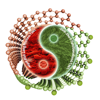
The yin and the yang of carbon nanotubes and toxicity


Illustration courtesy of the University College of London (UCL). Downloaded from http://www.ucl.ac.uk/news/news-articles/0113/130115-chemistry-resolves-toxic-concerns-about-carbon-nanotubes

Illustration courtesy of the University College of London (UCL). Downloaded from http://www.ucl.ac.uk/news/news-articles/0113/130115-chemistry-resolves-toxic-concerns-about-carbon-nanotubes

Researchers at the University College of London (UCL), France’s Centre national de la recherche scientifique (CNRS), and Italy’s University of Trieste have determined that carbon nanotube toxicity issues can be addressed be reducing their length and treating them chemically. From the Jan. 15,2013 news item on ScienceDaily,

In a new study, published January 15 [2013] in the journal Angewandte Chemie, evidence is provided that the asbestos-like reactivity and pathogenicity reported for long, pristine nanotubes can be completely alleviated if their surface is modified and their effective length is reduced as a result of chemical treatment.

First atomically described in the 1990s, carbon nanotubes are sheets of carbon atoms rolled up into hollow tubes just a few nanometres in diameter. Engineered carbon nanotubes can be chemically modified, with the addition of chemotherapeutic drugs, fluorescent tags or nucleic acids — opening up applications in cancer and gene therapy.

Furthermore, these chemically modified carbon nanotubes can pierce the cell membrane, acting as a kind of ‘nano-needle’, allowing the possibility of efficient transport of therapeutic and diagnostic agents directly into the cytoplasm of cells.

Among their downsides however, have been concerns about their safety profile. One of the most serious concerns, highlighted in 2008, involves the carcinogenic risk from the exposure and persistence of such fibres in the body. Some studies indicate that when long untreated carbon nanotubes are injected to the abdominal cavity of mice they can induce unwanted responses resembling those associated with exposure to certain asbestos fibres.

In this paper, the authors describe two different reactions which ask if any chemical modification can render the nanotubes non-toxic. They conclude that not all chemical treatments alleviate the toxicity risks associated with the material. Only those reactions that are able to render carbon nanotubes short and stably suspended in biological fluids without aggregation are able to result in safe, risk-free material.

Here’s a citation and link for this latest  research, from the ScienceDaily news item where you can also read the lead researcher’s comments about carbon nanotubes, safety, and unreasonable proposals to halt production,

Hanene Ali-Boucetta, Antonio Nunes, Raquel Sainz, M. Antonia Herrero, Bowen Tian, Maurizio Prato, Alberto Bianco, Kostas Kostarelos. Asbestos-like Pathogenicity of Long Carbon Nanotubes Alleviated by Chemical Functionalization. Angewandte Chemie International Edition, 2013; DOI: 10.1002/anie.201207664

The article is behind a paywall. I have mentioned long carbon nanotubes and their resemblance to asbestos fibres in several posts. The  Oct. 26, 2009 posting [scroll down about 1/3 of the way] highlights research which took place after the study where mice had carbon nanotubes injected into their bellies; in this second piece of research they inhaled the nanotubes.

ETA Jan. 21, 2013: Dexter Johnson gives context and commentary about this latest research into long multiwalled nanotubes (MWNTs) which he sums up as the answer to this question “What if you kept the MWNTs short?”  in a Jan. 18, 2013 posting on his Nanoclast blog (on the IEEE [Institute of Electrical and Electronics Engineers] website)
Where do buckyballs come from?

I’ve always wondered where buckyballs come from (as have scientists for the last 25 years) and now there’s an answer of sorts  (from the July 31, 2012 Florida State University news release Note: I have removed some links),

“We started with a paste of pre-existing fullerene molecules mixed with carbon and helium, shot it with a laser, and instead of destroying the fullerenes we were surprised to find they’d actually grown,” they wrote. The fullerenes were able to absorb and incorporate carbon from the surrounding gas.

By using fullenes  that contained heavy metal atoms in their centers, the scientists showed that the carbon cages remained closed throughout the process.

“If the cages grew by splitting open, we would have lost the metal atoms, but they always stayed locked inside,” Dunk [Paul Dunk, a doctoral student in chemistry and biochemistry at Florida State and lead author of the study published in Nature Communications] noted.

The researchers worked with a team of MagLab chemists using the lab’s 9.4-tesla Fourier transform ion cyclotron resonance mass spectrometer to analyze the dozens of molecular species produced when they shot the fullerene paste with the laser. The instrument works by separating molecules according to their masses, allowing the researchers to identify the types and numbers of atoms in each molecule. The process is used for applications as diverse as identifying oil spills, biomarkers and protein structures.

Dexter Johnson in his Aug. 6, 2012 posting on the Nanoclast blog on the IEEE (Institute of Electrical and Electronics Engineers) provides some context and commentary (Note: I have removed a link),

When Richard Smalley, Robert Curl, James Heath, Sean O’Brien, and Harold Kroto prepared the first buckminsterfullerene (C60) (or buckyball), they kicked off the next 25 years of nanomaterial science.

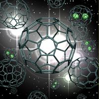
Here’s an artist’s illustration of  what these scientists have achieved, fullerene cage growth,

An artist’s representation of fullerene cage growth via carbon absorption from surrounding hot gases. Some of the cages contain lanthanum metal atoms. (Image courtesy National Science Foundation) [downloaded from Florida State University website]

 As I noted earlier I’m not alone in my fascination (from the news release),

Many people know the buckyball, also known by scientists as buckminsterfullerene, carbon 60 or C60, from the covers of their school chemistry textbooks. Indeed, the molecule represents the iconic image of “chemistry.” But how these often highly symmetrical, beautiful molecules with  fascinating properties form in the first place has been a mystery for a quarter-century. Despite worldwide investigation since the 1985 discovery of C60, buckminsterfullerene and other, non-spherical C60 molecules — known collectively as fullerenes — have kept their secrets. How? They’re born under highly energetic conditions and grow ultra-fast, making them difficult to analyze.

“The difficulty with fullerene formation is that the process is literally over in a flash — it’s next to impossible to see how the magic trick of their growth was performed,” said Paul Dunk, a doctoral student in chemistry and biochemistry at Florida State and lead author of the work.

There’s more than just idle curiosity at work (from the news release),

The buckyball research results will be important for understanding fullerene formation in extraterrestrial environments. Recent reports by NASA showed that crystals of C60 are in orbit around distant suns. This suggests that fullerenes may be more common in the universe than previously thought.

“The results of our study will surely be extremely valuable in deciphering fullerene formation in extraterrestrial environments,” said Florida State’s Harry Kroto, a Nobel Prize winner for the discovery of C60 and co-author of the current study.

The results also provide fundamental insight into self-assembly of other technologically important carbon nanomaterials such as nanotubes and the new wunderkind of the carbon family, graphene.

H/T to Nanowerk’s July 31, 2012 news item titled, Decades-old mystery how buckyballs form has been solved. In addition to Florida State University, National High Magnetic Field Laboratory (or MagLab), the CNRS  (Centre National de la Recherche Scientifique)Institute of Materials in France and Nagoya University in Japan were also involved in the research.

French scientists focus optical microscopes down to 30 nm

In fact, the French scientists are using two different imaging techniques to arrive at a resolution of 30 nm for their optical microscopes, according to the May 18, 2012 news item on Nanowerk.

Researchers from the Institut Pasteur and CNRS [Centre national de la recherche scientifique] have set up a new optical microscopy approach that combines two recent imaging techniques in order to visualize molecular assemblies without affecting their biological functions, at a resolution 10 times better than that of traditional microscopes. Using this approach, they were able to observe the AIDS virus and its capsids (containing the HIV genome) within cells at a scale of 30 nanometres, for the first time with light.

More specifically,

A study coordinated by Dr Christophe Zimmer (Institut Pasteur/CNRS), in collaboration with Dr Nathalie Arhel within the lab headed by Pr Pierre Charneau (Institut Pasteur/CNRS), shows that the association of two recent imaging techniques helps obtain unique images of molecular assemblies of HIV-1 capsids, with a resolution around 10 times better than that of traditional microscopes. This new approach, which uses super-resolution imaging and FlAsH labeling, does not affect the virus’ ability to self-replicate. It represents a major step forward in molecular biology studies, enabling the visualisation of microbial complexes at a scale of 30 nm without affecting their function.

The newly developed approach combines super-resolution PALM imaging and fluorescent FlAsH labeling. PALM imaging relies on the acquisition of thousands of low-resolution images, each of which showing only a few fluorescent molecules. The molecular positions are then calculated with high accuracy by computer programs and compiled into a single high-resolution image. FlAsH labeling involves the insertion of a 6-amino-acid peptide into the protein of interest. The binding of the FlAsH fluorophore to the peptide generates a fluorescent signal, thereby enabling the visualization of the protein. For the first time, researchers have combined these two methods in order to obtain high-resolution images of molecular structures in either fixed or living cells.

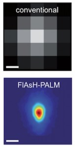
The researchers have supplied an image illustrating the difference between the conventional and new techniques will allow them to view (from the May 16, 2012 press release  [communiqué de presses] on the CNRS website),

© Institut Pasteur Reconstruction optique super-résolutive de la morphologie du VIH. L'image du dessous montre la distribution moyenne de l'enzyme intégrase observée par FlAsH-PALM. La résolution de cette technique (~30 nm) permet de retrouver la taille et la forme conique de la capside. Pour comparaison, la résolution de la microscopie conventionnelle (~200-300 nm), illustrée par l'image du dessus, ne permet pas une description détaillée de cette structure.

The conventional 200 – 300 nm resolution is shown at the top while the new 30 nm resolution achieved by combining the new techniques is shown below. This new technique has already allowed scientists to disprove a popular theory about the AIDS virus, from the May 18, 2012 news item on Nanowerk,

This new method has helped researchers visualise the AIDS Virus and localise its capsids in human cells, at a scale of 30 nm. Capsids are conical structures which contain the HIV genome. These structures must dismantle in order for the viral genome to integrate itself into the host cell’s genome. However, the timing of this disassembly has long been debated. According to a prevailing view, capsids disassemble right after infection of the host cell and, therefore, do not play an important role in the intracellular transport of the virus to the host cell’s nucleus. However, the results obtained by the researchers of the Institut Pasteur and CNRS indicate that numerous capsids remain unaltered until entry of the virus into the nucleus, confirming and strengthening earlier studies based on electron microscopy. Hence, capsids could play a more important role than commonly assumed in the replication cycle of HIV.

I gather excitement about this development is high as the scientists are suggesting that ‘microscopy’ could be known as ‘nanoscopy’ in the future.

Could science funding in the European Union have an impact on Canadian nanotechnology?

Unexpectedly they’re upping the research budget in the European Union. According to the item online at  BBC News,

The EU has announced 6.4bn euros (£5.4bn) of funding for scientific research and innovation next year – a 12% increase on this year’s allocation.

The programme is aimed at creating more than 165,000 jobs and developing “a more competitive and greener Europe”, the European Commission says.

The focus is on tackling climate change, energy projects, food security, health and Europe’s ageing population.

Grants will be awarded to about 16,000 research bodies and businesses.

“Research and innovation are the only smart and lasting route out of crisis and towards sustainable and socially equitable growth,” said the EU Commissioner for Research and Innovation, Maire Geoghegan-Quinn.

“There is no other way of creating good and well-paid jobs that will withstand the pressures of globalisation.”

EU-funded research currently accounts for about 5% of the total public funding for research in the EU, she said.

The investment includes more than 600m euros for health research, about 206m euros of which will go into clinical trials for new drugs.

Nanotechnologies will get 270m euros, while about 600m euros is earmarked for advanced computer technologies. [emphasis mine]

Another 400m euros is to be spent on computer applications that address the challenges of building a low-carbon economy and managing ageing populations.

I was inclined to view it as a piece of delightful news without really analyzing it, then David Bruggeman (Pasco Phronesis) made a salient comment,

I suspect that the European spending will be insufficient even if individual nations hold the line on their own science funding. Because even those nations are looking at significant cuts to their universities, which affect both the training of the next generation of researchers and a certain amount of research. At best the funding boosts and cuts will be a wash, but the future doesn’t look like the best. What might happen is a greater shift in attention to European Union level research compared to country level research.

David also provides a brief description of the  ‘framework programme’ that the European Union uses to fund science research so that readers (such as me) have a better understanding of the bigger picture. If you’re interested in this kind of thing, do check out his posting.

David’s commentary was particularly timely as, this morning, I came across an article about the French government funding nanotechnology research in Canada (Sherbrooke, Québec to be precise). Since the article is in French, I’m going to be relying on my translation skills (Note: I will reproduce at least some of the French, so do let me know if you spot any errors.)

There is an abbreviated version of the article (Nanotechnologies: un petit bout de France à L’UdeS) by Jonathan Custeau for the Sherbrooke Tribune here (fyi, somebody sent me a copy of the full article).

The University of Sherbrooke’s current nanotechnology laboratory (Laboratoire international associé en nanotechnologies et nanosystèmes [LIA-LN2]) is about to receiving funding to the tune of ! million Euros over three years from France’s CNRS (Centre National de la Recherche Scientifique) putting  it in a category occupied by only eight other labs in the world.

I gather the lab’s current LIA-LN2 status is a consequence of previous French funding since the university’s vice-president of research describes the current bonanza as ‘jumping to a new level’, i.e. jumping to Unité mixte international (UMI) status,

“Nous étions tellement en avance que nous sautons à un autre niveau”, fait valoir Jacques Beauvais, vice-recteur à la recherche de l’Université de Sherbrooke.

L’autre niveau, c’est l’Unité mixte internationale, un laboratoire financé par le Centre national de la recherche scientifique (CNRS français. Il n’en existe que huit à travers le monde.

“Une UMI coûte très cher, parce que c’est un vrai laboratoire, avec des chercheurs financés par le CNRS, des fonds de recherches français et européens. C’est comme s’il y avait un bout de France sur le campus de l’Université de Sherbrooke”, fait valoir Vincent Aimez, codirecteur du LIA-LN2.

The nanotechnology researchers at the University of Sherbrooke (L’UdeS) have been liaising and collaborating with researchers in Varennes, Lyon, and Grenoble, France for over two years,  so this new funding is an acknowledgment of the quality of their work.

Bravo—the award is all the more extraordinary given the concerns about science and university funding in Europe.

January 2012 is the launch date for the University of Sherbrooke’s UMI which will have a focus on bringing at least some of the academic research to the market. Miniaturized integrated circuit boards are mentioned specifically and my translation skills failed a bit here,

Les applications des recherches pourraient notamment permettre de relever le défi de la miniaturisation des puces électroniques [integrated circuit boards?]. “Nous cherchons à faire des puces avec plus de fonctions, mais qui consomment moins d’énergie, pour qu’elles restent efficaces pendant un mois par exemple. Nous voulons aussi développer des biocapteurs [?] pour des contrôles environnementaux [?] ou des analyses médicales [medical diagnostics?]”, précise Abdelkader Souifi, également codirecteur du LIA-LN2.

I found the comments regarding products quite interesting in light of the Québec government’s recent moves to improve innovation in that province as per the article (June 30, 2010) by Peter Hadekel in the Montréal Gazette. (Idle thought: This casts a new light on the recent Domtar-FPInnovations collaboration on nanocrystalline cellulose (my July 16, 2010 posting).