Tag Archives: Chad A. Mirkin

Cloaking devices made from DNA and gold nanoparticles using top-down lithography

This new technique seems promising but there’ve been a lot of ‘cloaking’ devices announced in the years I’ve been blogging and, in all likelihood, I was late to the party so I’m exercising a little caution before getting too excited. For the latest development in cloaking devices, there’s a January 18, 2018 news item on Nanowerk,

Northwestern University researchers have developed a first-of-its-kind technique for creating entirely new classes of optical materials and devices that could lead to light bending and cloaking devices — news to make the ears of Star Trek’s Spock perk up.

Using DNA [deoxyribonucleic acid] as a key tool, the interdisciplinary team took gold nanoparticles of different sizes and shapes and arranged them in two and three dimensions to form optically active superlattices. Structures with specific configurations could be programmed through choice of particle type and both DNA-pattern and sequence to exhibit almost any color across the visible spectrum, the scientists report.

A January 18, 2018 Northwestern University news release (also on EurekAlert) by Megan Fellman, which originated the news item, delves into more detail (Note: Links have been removed),

“Architecture is everything when designing new materials, and we now have a new way to precisely control particle architectures over large areas,” said Chad A. Mirkin, the George B. Rathmann Professor of Chemistry in the Weinberg College of Arts and Sciences at Northwestern. “Chemists and physicists will be able to build an almost infinite number of new structures with all sorts of interesting properties. These structures cannot be made by any known technique.”

The technique combines an old fabrication method — top-down lithography, the same method used to make computer chips — with a new one — programmable self-assembly driven by DNA. The Northwestern team is the first to combine the two to achieve individual particle control in three dimensions.

The study was published online by the journal Science today (Jan. 18). Mirkin and Vinayak P. Dravid and Koray Aydin, both professors in Northwestern’s McCormick School of Engineering, are co-corresponding authors.

Scientists will be able to use the powerful and flexible technique to build metamaterials — materials not found in nature — for a range of applications including sensors for medical and environmental uses.

The researchers used a combination of numerical simulations and optical spectroscopy techniques to identify particular nanoparticle superlattices that absorb specific wavelengths of visible light. The DNA-modified nanoparticles — gold in this case — are positioned on a pre-patterned template made of complementary DNA. Stacks of structures can be made by introducing a second and then a third DNA-modified particle with DNA that is complementary to the subsequent layers.

In addition to being unusual architectures, these materials are stimuli-responsive: the DNA strands that hold them together change in length when exposed to new environments, such as solutions of ethanol that vary in concentration. The change in DNA length, the researchers found, resulted in a change of color from black to red to green, providing extreme tunability of optical properties.

“Tuning the optical properties of metamaterials is a significant challenge, and our study achieves one of the highest tunability ranges achieved to date in optical metamaterials,” said Aydin, assistant professor of electrical engineering and computer science at McCormick.

“Our novel metamaterial platform — enabled by precise and extreme control of gold nanoparticle shape, size and spacing — holds significant promise for next-generation optical metamaterials and metasurfaces,” Aydin said.

The study describes a new way to organize nanoparticles in two and three dimensions. The researchers used lithography methods to drill tiny holes — only one nanoparticle wide — in a polymer resist, creating “landing pads” for nanoparticle components modified with strands of DNA. The landing pads are essential, Mirkin said, since they keep the structures that are grown vertical.

The nanoscopic landing pads are modified with one sequence of DNA, and the gold nanoparticles are modified with complementary DNA. By alternating nanoparticles with complementary DNA, the researchers built nanoparticle stacks with tremendous positional control and over a large area. The particles can be different sizes and shapes (spheres, cubes and disks, for example).

“This approach can be used to build periodic lattices from optically active particles, such as gold, silver and any other material that can be modified with DNA, with extraordinary nanoscale precision,” said Mirkin, director of Northwestern’s International Institute for Nanotechnology.

Mirkin also is a professor of medicine at Northwestern University Feinberg School of Medicine and professor of chemical and biological engineering, biomedical engineering and materials science and engineering in the McCormick School.

The success of the reported DNA programmable assembly required expertise with hybrid (soft-hard) materials and exquisite nanopatterning and lithographic capabilities to achieve the requisite spatial resolution, definition and fidelity across large substrate areas. The project team turned to Dravid, a longtime collaborator of Mirkin’s who specializes in nanopatterning, advanced microscopy and characterization of soft, hard and hybrid nanostructures.

Dravid contributed his expertise and assisted in designing the nanopatterning and lithography strategy and the associated characterization of the new exotic structures. He is the Abraham Harris Professor of Materials Science and Engineering in McCormick and the founding director of the NUANCE center, which houses the advanced patterning, lithography and characterization used in the DNA-programmed structures.

Here’s a link to and a citation for the paper,

Building superlattices from individual nanoparticles via template-confined DNA-mediated assembly by Qing-Yuan Lin, Jarad A. Mason, Zhongyang, Wenjie Zhou, Matthew N. O’Brien, Keith A. Brown, Matthew R. Jones, Serkan Butun, Byeongdu Lee, Vinayak P. Dravid, Koray Aydin, Chad A. Mirkin. Science 18 Jan 2018: eaaq0591 DOI: 10.1126/science.aaq0591

This paper is behind a paywall.

As noted earlier, it could be a while before cloaking devices are made available. In the meantime, you may find this image inspiring,

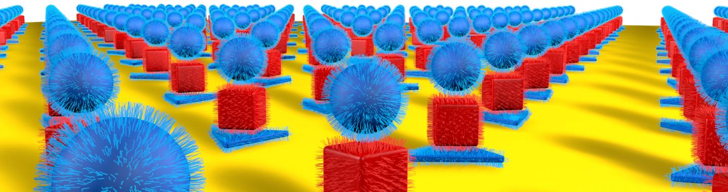
Caption: Northwestern University researchers have developed a new method to precisely arrange nanoparticles of different sizes and shapes in two and three dimensions, resulting in optically active superlattices. Credit: Northwestern University

Nominations open for Kabiller Prizes in Nanoscience and Nanomedicine ($250,000 for visionary researcher and $10,000 for young investigator)

For a change I can publish something that doesn’t have a deadline in three days or less! Without more ado (from a Feb. 20, 2017 Northwestern University news release by Megan Fellman [h/t Nanowerk’s Feb. 20, 2017 news item]),

Northwestern University’s International Institute for Nanotechnology (IIN) is now accepting nominations for two prestigious international prizes: the $250,000 Kabiller Prize in Nanoscience and Nanomedicine and the $10,000 Kabiller Young Investigator Award in Nanoscience and Nanomedicine.

The deadline for nominations is May 15, 2017. Details are available on the IIN website.

“Our goal is to recognize the outstanding accomplishments in nanoscience and nanomedicine that have the potential to benefit all humankind,” said David G. Kabiller, a Northwestern trustee and alumnus. He is a co-founder of AQR Capital Management, a global investment management firm in Greenwich, Connecticut.

The two prizes, awarded every other year, were established in 2015 through a generous gift from Kabiller. Current Northwestern-affiliated researchers are not eligible for nomination until 2018 for the 2019 prizes.

The Kabiller Prize — the largest monetary award in the world for outstanding achievement in the field of nanomedicine — celebrates researchers who have made the most significant contributions to the field of nanotechnology and its application to medicine and biology.

The Kabiller Young Investigator Award recognizes young emerging researchers who have made recent groundbreaking discoveries with the potential to make a lasting impact in nanoscience and nanomedicine.

“The IIN at Northwestern University is a hub of excellence in the field of nanotechnology,” said Kabiller, chair of the IIN executive council and a graduate of Northwestern’s Weinberg College of Arts and Sciences and Kellogg School of Management. “As such, it is the ideal organization from which to launch these awards recognizing outstanding achievements that have the potential to substantially benefit society.”

Nanoparticles for medical use are typically no larger than 100 nanometers — comparable in size to the molecules in the body. At this scale, the essential properties (e.g., color, melting point, conductivity, etc.) of structures behave uniquely. Researchers are capitalizing on these unique properties in their quest to realize life-changing advances in the diagnosis, treatment and prevention of disease.

“Nanotechnology is one of the key areas of distinction at Northwestern,” said Chad A. Mirkin, IIN director and George B. Rathmann Professor of Chemistry in Weinberg. “We are very grateful for David’s ongoing support and are honored to be stewards of these prestigious awards.”

An international committee of experts in the field will select the winners of the 2017 Kabiller Prize and the 2017 Kabiller Young Investigator Award and announce them in September.

The recipients will be honored at an awards banquet Sept. 27 in Chicago. They also will be recognized at the 2017 IIN Symposium, which will include talks from prestigious speakers, including 2016 Nobel Laureate in Chemistry Ben Feringa, from the University of Groningen, the Netherlands.

2015 recipient of the Kabiller Prize

The winner of the inaugural Kabiller Prize, in 2015, was Joseph DeSimone the Chancellor’s Eminent Professor of Chemistry at the University of North Carolina at Chapel Hill and the William R. Kenan Jr. Distinguished Professor of Chemical Engineering at North Carolina State University and of Chemistry at UNC-Chapel Hill.

DeSimone was honored for his invention of particle replication in non-wetting templates (PRINT) technology that enables the fabrication of precisely defined, shape-specific nanoparticles for advances in disease treatment and prevention. Nanoparticles made with PRINT technology are being used to develop new cancer treatments, inhalable therapeutics for treating pulmonary diseases, such as cystic fibrosis and asthma, and next-generation vaccines for malaria, pneumonia and dengue.

2015 recipient of the Kabiller Young Investigator Award

Warren Chan, professor at the Institute of Biomaterials and Biomedical Engineering at the University of Toronto, was the recipient of the inaugural Kabiller Young Investigator Award, also in 2015. Chan and his research group have developed an infectious disease diagnostic device for a point-of-care use that can differentiate symptoms.

BTW, Warren Chan, winner of the ‘Young Investigator Award’, and/or his work have been featured here a few times, most recently in a Nov. 1, 2016 posting, which is mostly about another award he won but also includes links to some his work including my April 27, 2016 post about the discovery that fewer than 1% of nanoparticle-based drugs reach their destination.

Ultimate discovery tool?

For anyone familiar with the US nanomedicine scene, Chad Mirkin’s appearance in this announcement from Northwestern University isn’t much of a surprise.  From a June 23, 2016 news item on ScienceDaily,

The discovery power of the gene chip is coming to nanotechnology. A Northwestern University research team is developing a tool to rapidly test millions and perhaps even billions or more different nanoparticles at one time to zero in on the best particle for a specific use.

When materials are miniaturized, their properties—optical, structural, electrical, mechanical and chemical—change, offering new possibilities. But determining what nanoparticle size and composition are best for a given application, such as catalysts, biodiagnostic labels, pharmaceuticals and electronic devices, is a daunting task.

“As scientists, we’ve only just begun to investigate what materials can be made on the nanoscale,” said Northwestern’s Chad A. Mirkin, a world leader in nanotechnology research and its application, who led the study. “Screening a million potentially useful nanoparticles, for example, could take several lifetimes. Once optimized, our tool will enable researchers to pick the winner much faster than conventional methods. We have the ultimate discovery tool.”

A June 23, 2016 Northwestern University news release (also on EurekAlert), which originated the news item, describes the work in more detail,

Using a Northwestern technique that deposits materials on a surface, Mirkin and his team figured out how to make combinatorial libraries of nanoparticles in a very controlled way. (A combinatorial library is a collection of systematically varied structures encoded at specific sites on a surface.) Their study will be published June 24 by the journal Science.

The nanoparticle libraries are much like a gene chip, Mirkin says, where thousands of different spots of DNA are used to identify the presence of a disease or toxin. Thousands of reactions can be done simultaneously, providing results in just a few hours. Similarly, Mirkin and his team’s libraries will enable scientists to rapidly make and screen millions to billions of nanoparticles of different compositions and sizes for desirable physical and chemical properties.

“The ability to make libraries of nanoparticles will open a new field of nanocombinatorics, where size — on a scale that matters — and composition become tunable parameters,” Mirkin said. “This is a powerful approach to discovery science.”

“I liken our combinatorial nanopatterning approach to providing a broad palette of bold colors to an artist who previously had been working with a handful of dull and pale black, white and grey pastels,” said co-author Vinayak P. Dravid, the Abraham Harris Professor of Materials Science and Engineering in the McCormick School of Engineering.

Using five metallic elements — gold, silver, cobalt, copper and nickel — Mirkin and his team developed an array of unique structures by varying every elemental combination. In previous work, the researchers had shown that particle diameter also can be varied deliberately on the 1- to 100-nanometer length scale.

Some of the compositions can be found in nature, but more than half of them have never existed before on Earth. And when pictured using high-powered imaging techniques, the nanoparticles appear like an array of colorful Easter eggs, each compositional element contributing to the palette.

To build the combinatorial libraries, Mirkin and his team used Dip-Pen Nanolithography, a technique developed at Northwestern in 1999, to deposit onto a surface individual polymer “dots,” each loaded with different metal salts of interest. The researchers then heated the polymer dots, reducing the salts to metal atoms and forming a single nanoparticle. The size of the polymer dot can be varied to change the size of the final nanoparticle.

This control of both size and composition of nanoparticles is very important, Mirkin stressed. Having demonstrated control, the researchers used the tool to systematically generate a library of 31 nanostructures using the five different metals.

To help analyze the complex elemental compositions and size/shape of the nanoparticles down to the sub-nanometer scale, the team turned to Dravid, Mirkin’s longtime friend and collaborator. Dravid, founding director of Northwestern’s NUANCE Center, contributed his expertise and the advanced electron microscopes of NUANCE to spatially map the compositional trajectories of the combinatorial nanoparticles.

Now, scientists can begin to study these nanoparticles as well as build other useful combinatorial libraries consisting of billions of structures that subtly differ in size and composition. These structures may become the next materials that power fuel cells, efficiently harvest solar energy and convert it into useful fuels, and catalyze reactions that take low-value feedstocks from the petroleum industry and turn them into high-value products useful in the chemical and pharmaceutical industries.

Here’s a diagram illustrating the work,

 Caption: A combinatorial library of polyelemental nanoparticles was developed using Dip-Pen Nanolithography. This novel nanoparticle library opens up a new field of nanocombinatorics for rapid screening of nanomaterials for a multitude of properties. Credit: Peng-Cheng Chen/James Hedrick

Caption: A combinatorial library of polyelemental nanoparticles was developed using Dip-Pen Nanolithography. This novel nanoparticle library opens up a new field of nanocombinatorics for rapid screening of nanomaterials for a multitude of properties. Credit: Peng-Cheng Chen/James Hedrick

Here’s a link to and a citation for the paper,

Polyelemental nanoparticle libraries by Peng-Cheng Chen, Xiaolong Liu, James L. Hedrick, Zhuang Xie, Shunzhi Wang, Qing-Yuan Lin, Mark C. Hersam, Vinayak P. Dravid, Chad A. Mirkin. Science  24 Jun 2016: Vol. 352, Issue 6293, pp. 1565-1569 DOI: 10.1126/science.aaf8402

This paper is behind a paywall.

Sticky-flares nanotechnology to track and observe RNA (ribonucleic acid) regulation

I like the name ‘sticky-flares’ and had hoped there was an amusing story about its origins. Ah well, perhaps I’ll have better luck next time.

This work comes out of Chad Mirkin’s lab at Northwestern University (Chicago, US) according to a July 21, 2015 news item on Azonano,

RNA [ribonucleic acid] is a fundamental ingredient in all known forms of life — so when RNA goes awry, a lot can go wrong. RNA misregulation plays a critical role in the development of many disorders, such as mental disability, autism and cancer.

A new technology — called “Sticky-flares” — developed by nanomedicine experts at Northwestern University offers the first real-time method to track and observe the dynamics of RNA distribution as it is transported inside living cells.

A July 20, 2015 Northwestern University news release by Erin Spain, which originated the news item, describes the research in a little more detail also including information about predecessor technology,

Sticky-flares have the potential to help scientists understand the complexities of RNA better than any analytical technique to date and observe and study the biological and medical significance of RNA misregulation.

Previous technologies made it possible to attain static snapshots of RNA location, but that isn’t enough to understand the complexities of RNA transport and localization within a cell. Instead of analyzing snapshots of RNA to try to understand functioning, Sticky-flares help create an experience that is more like watching live-streaming video.

“This is very exciting because much of the RNA in cells has very specific quantities and localization, and both are critical to the cell’s function, but until this development it has been very difficult, and often impossible, to probe both attributes of RNA in a live cell,” said Chad A. Mirkin, a nanomedicine expert and corresponding author of the study. “We hope that many more researchers will be able to use this platform to increase our understanding of RNA function inside cells.”

Sticky-flares are tiny spherical nucleic acid gold nanoparticle conjugates that can enter living cells and target and transfer a fluorescent reporter or “tracking device” to RNA transcripts. This fluorescent labeling can be tracked via fluorescence microscopy as it is transported throughout the cell, including the nucleus.

In the … paper, the scientists explain how they used Sticky-flares to quantify β–actin mRNA in HeLa cells (the oldest and most commonly used human cell line) as well as to follow the real-time transport of β–actin mRNA in mouse embryonic fibroblasts.

Sticky-flares are built upon another technology from Mirkin’s group called NanoFlares, which was the first genetic-based approach that is able to detect live circulating tumor cells out of the complex matrix that is human blood.

NanoFlares have been very useful for researchers that operate in the arena of quantifying gene expression. AuraSense, Inc., a biotechnology company that licensed the NanoFlare technology from Northwestern University, and EMD-Millipore, another biotech company, have commercialized NanoFlares. There are now more than 1,700 commercial forms of NanoFlares sold under the SmartFlareä name in more than 230 countries.

The Sticky-flare is designed to address limitations of SmartFlares, most notably their inability to track RNA location and enter the nucleus. The Northwestern team believes Sticky-flares are poised to become a valuable tool for researchers who desire to understand the function of RNA in live cells.

Based on the paragraph about the precursor technology’s commercial success , I gather they are excited about similar possibilities for sticky-flares.

Here’s a link to and a citation for the paper,

Quantification and real-time tracking of RNA in live cells using Sticky-flares by William E. Briley, Madison H. Bondy, Pratik S. Randeria, Torin J. Dupper, and Chad A. Mirkin. Published online before print July 20, 2015, doi: 10.1073/pnas.1510581112 PNAS July 20, 2015

This paper is behind a paywall.

Northwestern University’s (US) International Institute for Nanotechnology (IIN) rakes in some cash

Within less than a month Northwestern University’s International Institute for Nanotechnology (IIN) has been granted awarded two grants by the US Department of Defense.

4D printing

The first grant, for 4D printing, was announced in a June 11, 2015 Northwestern news release by Megan Fellman (Note: A link has been removed),

Northwestern University’s International Institute for Nanotechnology (IIN) has received a five-year, $8.5 million grant from the U.S. Department of Defense’s competitive Multidisciplinary University Research Initiative (MURI) program to develop a “4-dimensional printer” — the next generation of printing technology for the scientific world.

Once developed, the 4-D printer, operating on the nanoscale, will be used to construct new devices for research in chemistry, materials sciences and U.S. defense-related areas that could lead to new chemical and biological sensors, catalysts, microchip designs and materials designed to respond to specific materials or signals.

“This research promises to bring transformative advancement to the development of biosensors, adaptive optics, artificially engineered tissues and more by utilizing nanotechnology,” said IIN director and chemist Chad A. Mirkin, who is leading the multi-institution project. Mirkin is the George B. Rathmann Professor of Chemistry in the Weinberg College of Arts and Sciences.

The award, issued by the Air Force Office of Scientific Research, supports a team of experts from Northwestern, the University of Miami, the University of California, San Diego, and the University of Maryland.

In science, “printing” encodes information at specific locations on a material’s surface, similar to how we print words on paper with ink. The 4-dimensional printer will consist of millions of tiny elastomeric “pens” that can be used individually and independently to create nanometer-size features composed of hard or soft materials.

The information encoded can be in the form of materials with a defined set of chemical and physical properties. The printing speed and resolution determine the amount and complexity of the information that can be encoded.

Progress in fields ranging from biology to chemical sensing to computing currently are limited by the lack of low-cost equipment that can perform high-resolution printing and 3-dimensional patterning on hard materials (e.g., metals and semiconductors) and soft materials (e.g., organic and biological materials) at nanometer resolution (approximately 1,000 times smaller than the width of a human hair).

“Ultimately, the 4-D printer will provide a foundation for a new generation of tools to develop novel architectures, wherein the hard materials that form the functional components of electronics can be merged with biological or soft materials,” said Milan Mrksich, a co-principal investigator on the grant.

Mrksich is the Henry Wade Rogers Professor of Biomedical Engineering, Chemistry and Cell and Molecular Biology, with appointments in the McCormick School of Engineering and Applied Science, Weinberg and Northwestern University Feinberg School of Medicine.

A July 10, 2015 article about the ‘4D printer’ grant  by Madeline Fox for the Daily Northwestern features a description of 4D printing from Milan Mrksich, a co-principal investigator on the grant,

Milan Mrksich, one of the project’s five senior participants, said that while most people are familiar with the three dimensions of length, width and depth, there are often misconceptions about the fourth property of a four-dimensional object. Mrksich used Legos as an analogy to describe 4D printing technology.

“If you take Lego blocks, you can basically build any structure you want by controlling which Lego is connected to which Lego and controlling all their dimensions in space,” Mrksich said. “Within an object made up of nanoparticles, we’re controlling the placement — as we use a printer to control the placement of every particle, our fourth dimension lets us choose which nanoparticle with which property would be at each position.”

Thank you Dr. Mrksich and Ms. Fox for that helpful analogy.

Designing advanced bioprogrammable nanomaterials

The second grant, announced in a July 6, 2015 Northwestern news release by Megan Fellman, is apparently the only one of its kind in the US (Note: A link has been removed),

Northwestern University’s International Institute for Nanotechnology (IIN) has been awarded a U.S. Air Force Center of Excellence grant to design advanced bioprogrammable nanomaterials for solutions to challenging problems in the areas of energy, the environment, security and defense, as well as for developing ways to monitor and mitigate human stress.

The five-year, $9.8 million grant establishes the Center of Excellence for Advanced Bioprogrammable Nanomaterials (C-ABN), the only one of its kind in the country. After the initial five years, the grant potentially could be renewed for an additional five years.

“Northwestern University was chosen to lead this Center of Excellence because of its investment in infrastructure development, including new facilities and instrumentation; its recruitment of high-caliber faculty members and students; and its track record in bio-nanotechnology and cognitive sciences,” said Timothy Bunning, chief scientist at the U.S. Air Force Research Laboratory (AFRL) Materials and Manufacturing Directorate.

Led by IIN director Chad A. Mirkin, C-ABN will support collaborative, discovery-based research projects aimed at developing bioprogrammable nanomaterials that will meet both military and civilian needs and facilitate the efficient transition of these new technologies from the laboratory to marketplace.

Bioprogrammable nanomaterials are structures that typically contain a biomolecular component, such as nucleic acids or proteins, which give the materials a variety of novel capabilities. [emphasis mine] Nanomaterials can be designed to assemble into large 3-D structures, to interface with biological structures inside cells or tissues, or to interface with existing macroscale devices, for example. These new bioprogrammable nanomaterials and the fundamental knowledge gained through their development will ultimately lead to the creation of wearable, portable and/or human-interactive devices with extraordinary capabilities that will significantly impact both civilian and Air Force needs.

In one research area, scientists will work to understand the molecular underpinnings of vulnerability and resilience to stress. They will use bioprogrammable nanomaterials to develop ultrasensitive sensors capable of detecting and quantifying biomarkers for human stress in biological fluids (e.g., saliva, perspiration or blood), providing means to easily monitor the soldier during times of extreme stress. Ultimately, these bioprogrammable materials may lead to methods to increase human cellular resilience to the effects of stress and/or to correct genetic mutations that decrease cellular resilience of susceptible individuals.

Other research projects, encompassing a wide variety of nanotechnology-enabled goals, include:

Developing hybrid wearable energy-storage devices;
Developing devices to identify chemical and biological targets in a field environment;
Developing flexible bio-electronic circuits;
Designing a new class of flat optics; and
Advancing understanding of design rules between 2-D and 3-D architectures.

The analysis of these nanostructures also will extend fundamental knowledge in the fields of materials science and engineering, human performance, chemistry, biology and physics.

The center will be housed under the IIN, providing researchers with access to IIN’s strong entrepreneurial community and its close ties with Northwestern’s renowned Kellogg School of Management.

This second news release provides an interesting contrast to a recent news release from Sweden’s Karolinska Intitute where the writer was careful to note that the enzymes and organic electronic ion pumps were not living as noted in my June 26, 2015 posting. It seems nucleic acids (as in RNA and DNA) can be mentioned without a proviso in the US. as there seems to be little worry about anti-GMO (genetically modified organisms) and similar backlashes affecting biotechnology research.

Bejweled and bedazzled but not bewitched, bothered, or bewildered at Northwestern University

When discussing DNA (deoxyribonucleic acid) one doesn’t usually expect to encounter gems as one does in a Nov. 28, 2013 news item on Azonano,

Nature builds flawless diamonds, sapphires and other gems. Now a Northwestern University [located in Chicago, Illinois, US] research team is the first to build near-perfect single crystals out of nanoparticles and DNA, using the same structure favored by nature.

The Nov. 27, 2013 Northwestern University news release by Megan Fellman (also on EurekAlert), which originated the news item,, explains why single crystals are of such interest,

“Single crystals are the backbone of many things we rely on — diamonds for beauty as well as industrial applications, sapphires for lasers and silicon for electronics,” said nanoscientist Chad A. Mirkin. “The precise placement of atoms within a well-defined lattice defines these high-quality crystals.

“Now we can do the same with nanomaterials and DNA, the blueprint of life,” Mirkin said. “Our method could lead to novel technologies and even enable new industries, much as the ability to grow silicon in perfect crystalline arrangements made possible the multibillion-dollar semiconductor industry.”

His research group developed the “recipe” for using nanomaterials as atoms, DNA as bonds and a little heat to form tiny crystals. This single-crystal recipe builds on superlattice techniques Mirkin’s lab has been developing for nearly two decades.

(I wrote about Mirkin’s nanoparticle DNA work in the context of his proposed periodic table of modified nucleic acid nanoparticles in a July 5, 2013 posting.) The news release goes on to describe Mirkin’s most recent work,

In this recent work, Mirkin, an experimentalist, teamed up with Monica Olvera de la Cruz, a theoretician, to evaluate the new technique and develop an understanding of it. Given a set of nanoparticles and a specific type of DNA, Olvera de la Cruz showed they can accurately predict the 3-D structure, or crystal shape, into which the disordered components will self-assemble.

The general set of instructions gives researchers unprecedented control over the type and shape of crystals they can build. The Northwestern team worked with gold nanoparticles, but the recipe can be applied to a variety of materials, with potential applications in the fields of materials science, photonics, electronics and catalysis.

A single crystal has order: its crystal lattice is continuous and unbroken throughout. The absence of defects in the material can give these crystals unique mechanical, optical and electrical properties, making them very desirable.

In the Northwestern study, strands of complementary DNA act as bonds between disordered gold nanoparticles, transforming them into an orderly crystal. The researchers determined that the ratio of the DNA linker’s length to the size of the nanoparticle is critical.

“If you get the right ratio it makes a perfect crystal — isn’t that fun?” said Olvera de la Cruz, who also is a professor of chemistry in the Weinberg College of Arts and Sciences. “That’s the fascinating thing, that you have to have the right ratio. We are learning so many rules for calculating things that other people cannot compute in atoms, in atomic crystals.”

The ratio affects the energy of the faces of the crystals, which determines the final crystal shape. Ratios that don’t follow the recipe lead to large fluctuations in energy and result in a sphere, not a faceted crystal, she explained. With the correct ratio, the energies fluctuate less and result in a crystal every time.

“Imagine having a million balls of two colors, some red, some blue, in a container, and you try shaking them until you get alternating red and blue balls,” Mirkin explained. “It will never happen.

“But if you attach DNA that is complementary to nanoparticles — the red has one kind of DNA, say, the blue its complement — and now you shake, or in our case, just stir in water, all the particles will find one another and link together,” he said. “They beautifully assemble into a three-dimensional crystal that we predicted computationally and realized experimentally.”

To achieve a self-assembling single crystal in the lab, the research team reports taking two sets of gold nanoparticles outfitted with complementary DNA linker strands. Working with approximately 1 million nanoparticles in water, they heated the solution to a temperature just above the DNA linkers’ melting point and then slowly cooled the solution to room temperature, which took two or three days.

The very slow cooling process encouraged the single-stranded DNA to find its complement, resulting in a high-quality single crystal approximately three microns wide. “The process gives the system enough time and energy for all the particles to arrange themselves and find the spots they should be in,” Mirkin said.

The researchers determined that the length of DNA connected to each gold nanoparticle can’t be much longer than the size of the nanoparticle. In the study, the gold nanoparticles varied from five to 20 nanometers in diameter; for each, the DNA length that led to crystal formation was about 18 base pairs and six single-base “sticky ends.”

“There’s no reason we can’t grow extraordinarily large single crystals in the future using modifications of our technique,” said Mirkin, who also is a professor of medicine, chemical and biological engineering, biomedical engineering and materials science and engineering and director of Northwestern’s International Institute for Nanotechnology.

Here’s a link to and a citation for the published paper,

DNA-mediated nanoparticle crystallization into Wulff polyhedra by Evelyn Auyeung, Ting I. N. G. Li, Andrew J. Senesi, Abrin L. Schmucker, Bridget C. Pals, Monica Olvera de la Cruz, & Chad A. Mirkin. Nature (2013) doi:10.1038/nature12739 Published online 27 November 2013

This article is behind a paywall.

Points to anyone who recognized the song title (Bewitched, Bothered and Bewildered) embedded in the head for this posting.

Desktop nanofabrication is in the laboratory but not in the marketplace yet

Another Chad Mirkin, Northwestern University (Chicago, Illinois, US), research breakthrough has been announced (this man, with regard to research,  is as prolific as a bunny) in a July 19, 2013 news item on ScienceDaily,

A new low-cost, high-resolution tool is primed to revolutionize how nanotechnology is produced from the desktop, according to a new study by Northwestern University researchers.

Currently, most nanofabrication is done in multibillion-dollar centralized facilities called foundries. This is similar to printing documents in centralized printing shops. Consider, however, how the desktop printer revolutionized the transfer of information by allowing individuals to inexpensively print documents as needed. This paradigm shift is why there has been community-wide ambition in the field of nanoscience to create a desktop nanofabrication tool.

“With this breakthrough, we can construct very high-quality materials and devices, such as processing semiconductors over large areas, and we can do it with an instrument slightly larger than a printer,” said Chad A. Mirkin, senior author of the study.

The July 19, 2013 Northwestern University news release (on EurekAlert), which originated the news item, provides details,

The tool Mirkin’s team has created produces working devices and structures at the nanoscale level in a matter of hours, right at the point of use. It is the nanofabrication equivalent of a desktop printer.

Without requiring millions of dollars in instrumentation costs, the tool is poised to prototype a diverse range of functional structures, from gene chips to protein arrays to building patterns that control how stem cells differentiate to making electronic circuits.

“Instead of needing to have access to millions of dollars, in some cases billions of dollars of instrumentation, you can begin to build devices that normally require that type of instrumentation right at the point of use,” Mirkin said.

The paper details the advances Mirkin’s team has made in desktop nanofabrication based upon easily fabricated beam-pen lithography (BPL) pen arrays, structures that consist of an array of polymeric pyramids, each coated with an opaque layer with a 100 nanometer aperture at the tip. Using a digital micromirror device, the functional component of a projector, a single beam of light is broken up into thousands of individual beams, each channeled down the back of different pyramidal pens within the array and through the apertures at the tip of each pen.

The nanofabrication tool allows one to rapidly process substrates coated with photosensitive materials called resists and generate structures that span the macro-, micro- and nanoscales, all in one experiment.

Key advances made by Mirkin’s team include developing the hardware, writing the software to coordinate the direction of light onto the pen array and constructing a system to make all of the pieces of this instrument work together in synchrony. This approach allows each pen to write a unique pattern and for these patterns to be stitched together into functional devices.

“There is no need to create a mask or master plate every time you want to create a new structure,” Mirkin said. “You just assign the beams of light to go in different places and tell the pens what pattern you want generated.”

Because the materials used to make the desktop nanofabrication tool are easily accessible, commercialization may be as little as two years away, Mirkin said. In the meantime, his team is working on building more devices and prototypes.

In the paper, Mirkin explains how his lab produced a map of the world, with nanoscale resolution that is large enough to see with the naked eye, a feat never before achieved with a scanning probe instrument. Not only that, but closer inspection with a microscope reveals that this image is actually a mosaic of individual chemical formulae made up of nanoscale points. Making this pattern showcases the instrument’s capability of simultaneously writing centimeter-scale patterns with nanoscale resolution.

Here’s a link to and a citation for the published paper,

Desktop nanofabrication with massively multiplexed beam pen lithography by Xing Liao, Keith A. Brown, Abrin L. Schmucker, Guoliang Liu, Shu He, Wooyoung Shim, & Chad A. Mirkin. Nature Communications 4, Article number: 2103 doi:10.1038/ncomms3103 Published 19 July 2013

This paper is behind a paywall. As an alternative of sorts, you might like to check out this March 22, 2012 video of Mirkin’s presentation entitled, A Chemist’s Approach to Nanofabrication: Towards a “Desktop Fab” for the US Air Force Office of Scientific Research.

Chad Mirkin’s periodic table of modified nucleic acid nanoparticles

Chad Mirkin has been pushing his idea for a new periodic table of ‘nanoparticles’ since at least Feb. 2013 (I wrote about this and some of Mirkin’s other work in my Feb. 19, 2013 posting) when he presented it at the 2013 American Association for the Advancement of Science (AAAS) annual meeting in Boston, Massachusetts. From a Feb. 17, 2013 news item on ScienceDaily,

Northwestern University’s Chad A. Mirkin, a leader in nanotechnology research and its application, has developed a completely new set of building blocks that is based on nanoparticles and DNA. Using these tools, scientists will be able to build — from the bottom up, just as nature does — new and useful structures.

Mirkin will discuss his research in a session titled “Nucleic Acid-Modified Nanostructures as Programmable Atom Equivalents: Forging a New Periodic Table” at the American Association for the Advancement of Science (AAAS) annual meeting in Boston.

“We have a new set of building blocks,” Mirkin said. “Instead of taking what nature gives you, we can control every property of the new material we make. [emphasis mine] We’ve always had this vision of building matter and controlling architecture from the bottom up, and now we’ve shown it can be done.”

Mirkin seems a trifle grandiose; I’m hoping he doesn’t have any grand creation projects that require seven days.

Getting back to the new periodic table, the Feb. 13, 2013 Northwestern University news release by Megan Fellman, which originated the news item,  provides a few more details,

Using nanoparticles and DNA, Mirkin has built more than 200 different crystal structures with 17 different particle arrangements. Some of the lattice types can be found in nature, but he also has built new structures that have no naturally occurring mineral counterpart.
….
Mirkin can make new materials and arrangements of particles by controlling the size, shape, type and location of nanoparticles within a given particle lattice. He has developed a set of design rules that allow him to control almost every property of a material.

New materials developed using his method could help improve the efficiency of optics, electronics and energy storage technologies. “These same nanoparticle building blocks have already found wide-spread commercial utility in biology and medicine as diagnostic probes for markers of disease,” Mirkin added.

With this present advance, Mirkin uses nanoparticles as “atoms” and DNA as “bonds.” He starts with a nanoparticle, which could be gold, silver, platinum or a quantum dot, for example. The core material is selected depending on what physical properties the final structure should have.

He then attaches hundreds of strands of DNA (oligonucleotides) to the particle. The oligonucleotide’s DNA sequence and length determine how bonds form between nanoparticles and guide the formation of specific crystal lattices.

“This constitutes a completely new class of building blocks in materials science that gives you a type of programmability that is extraordinarily versatile and powerful,” Mirkin said. “It provides nanotechnologists for the first time the ability to tailor properties of materials in a highly programmable way from the bottom up.”

Mirkin and his colleagues have since published a paper about this new periodic table in Angewandte Chemie (May 2013). And, earlier today (July 5, 2013) Philip Ball writing (A self-assembled periodic table) for the Royal Society of Chemistry provided a critique of the idea while supporting it in principle,

Mirkin and his colleagues perceive the pairing of [DNA] strands as somewhat analogous to the covalent pairing of electrons and call their DNA-tagged nanoparticles programmable atom equivalents (PAEs). These PAEs may bind to one another according to particular combinatorial rules and Mirkin proposes a kind of periodic table of PAEs that systematises their possible interactions and permutations.
Well, it’s not hard to start enumerating ways in which PAEs are unlike atoms. Most fundamentally, perhaps, the bonding propensity of a PAE need bear no real relation to the ‘atom’ (the nanoparticle) with which it is associated: a given nanoparticle might be paired with any other, and there’s nothing periodic about those tendencies.

I recommend reading Ball’s piece for the way he analyzes the weaknesses and for why he thinks the effort to organize PAEs conceptually is worthwhile.

For the curious, here’s a link to and a citation for the researchers’ published paper,

Nucleic Acid-Modified Nanostructures as Programmable Atom Equivalents: Forging a New “Table of Elements by Robert J. Macfarlane, Matthew N. O’Brien, Dr. Sarah Hurst Petrosko, and Prof. Chad A. Mirkin. Angewandte Chemie International Edition Volume 52, Issue 22, pages 5688–5698, May 27, 2013. Article first published online: 2 MAY 2013 DOI: 10.1002/anie.201209336

Copyright © 2013 WILEY-VCH Verlag GmbH & Co. KGaA, Weinheim

This article is behind a paywall.

One final comment, this is not the first ‘nanoparticle table of elements’.  Larry Bell mentioned one in his Dec. 7, 2010 NISENet (Nanoscale Informal Science Education Network) blog posting,

The focus of today’s sessions at NSF’s [US National Science Foundation] meeting of nanoscale science and engineering grantees focuses on putting the science to practical use. First up this morning is nanomanufacturing. Mark Tuonimen from the University of Massachusetts at Amherst gave a talk about the Nanoscale Manufacturing Network and one of his images caught my imagination. This image, which comes from the draft Nano2 vision document on the next decade of nanoscale research, illustrates and idea that is sometimes referred to as a periodic table of nanoparticles.

[downloaded from http://www.nisenet.org/blogs/observations_insights/periodic_table_nanoparticles]

[downloaded from http://www.nisenet.org/blogs/observations_insights/periodic_table_nanoparticles]

Bell goes on to describe one way in which a nanoparticle table of elements would have to differ from the traditional chemistry table.

When mining for gold nanoparticles use substitute cornstarch for cyanide

A May 14, 2013 news release from Northwestern University (US) on EurekAlert notes that researchers have found cornstarch can be used in the place of cyanide to extract gold,

Northwestern University scientists have struck gold in the laboratory. They have discovered an inexpensive and environmentally benign method that uses simple cornstarch — instead of cyanide — to isolate gold from raw materials in a selective manner.

This green method extracts gold from crude sources and leaves behind other metals that are often found mixed together with the crude gold. The new process also can be used to extract gold from consumer electronic waste.

Commonly used techniques, according to the news release, are problematic,

Current methods for gold recovery involve the use of highly poisonous cyanides, often leading to contamination of the environment. Nearly all gold-mining companies use this toxic gold leaching process to sequester the precious metal.

“The elimination of cyanide from the gold industry is of the utmost importance environmentally,” said Sir Fraser Stoddart, the Board of Trustees Professor of Chemistry in the Weinberg College of Arts and Sciences. “We have replaced nasty reagents with a cheap, biologically friendly material derived from starch.”

Here’s how the researchers made their accidental discovery (from the news release),

Zhichang Liu, a postdoctoral fellow in Stoddart’s lab and first author of the paper, took two test tubes containing aqueous solutions — one of the starch-derived alpha-cyclodextrin, the other of a dissolved gold (Au) salt (called aurate) — and mixed them together in a beaker at room temperature.

Liu was trying to make an extended, three-dimensional cubic structure, which could be used to store gases and small molecules. Unexpectedly, he obtained needles, which formed rapidly upon mixing the two solutions.

“Initially, I was disappointed when my experiment didn’t produce cubes, but when I saw the needles, I got excited,” Liu said. “I wanted to learn more about the composition of these needles.”

…  said Stoddart, a senior author of the paper[,] “The needles, composed of straw-like bundles of supramolecular wires, emerged from the mixed solutions in less than a minute.”

After discovering the needles, Liu screened six different complexes — cyclodextrins composed of rings of six (alpha), seven (beta) and eight (gamma) glucose units, each combined with aqueous solutions of potassium tetrabromoaurate (KAuBr4) or potassium tetrachloroaurate (KAuCl4).

He found that it was alpha-cyclodextrin, a cyclic starch fragment composed of six glucose units, that isolates gold best of all.

“Alpha-cyclodextrin is the gold medal winner,” Stoddart said. “Zhichang stumbled on a piece of magic for isolating gold from anything in a green way.”

Alkali metal salt waste from this new method is relatively environmentally benign, Stoddart said, while waste from conventional methods includes toxic cyanide salts and gases. The Northwestern procedure is also more efficient than current commercial processes.

The supramolecular nanowires, each 1.3 nanometers in diameter, assemble spontaneously in a straw-like manner. In each wire, the gold ion is held together in the middle of four bromine atoms, while the potassium ion is surrounded by six water molecules; these ions are sandwiched in an alternating fashion by alpha-cyclodextrin rings. Around 4,000 wires are bundled parallel to each other and form individual needles that are visible under an electron microscope.

“There is a lot of chemistry packed into these nanowires,” Stoddart said. “The elegance of the composition of single nanowires was revealed by atomic force microscopy, which throws light on the stacking of the individual donut-shaped alpha-cyclodextrin rings.”

The atomic detail of the single supramolecular wires and their relative disposition within the needles was uncovered by single crystal X-ray crystallography.

As I’ve noted before, the perennial science story is a ‘mistake’ leading to an exciting discovery and this is one more example. I think it’s one of the things that scientists don’t get enough credit for, their ability to reshape a disappointing result into a new discovery or, in the vernacular, ‘make lemonade out of lemons’.

Here’s a link to and a citation for the research paper,

Selective isolation of gold facilitated by second-sphere coordination with α-cyclodextrin by Zhichang Liu, Marco Frasconi, Juying Lei, Zachary J. Brown, Zhixue Zhu, Dennis Cao, Julien Iehl, Guoliang Liu, Albert C. Fahrenbach, Youssry Y. Botros, Omar K. Farha, Joseph T. Hupp,  Chad A. Mirkin & J. Fraser Stoddart. Nature Communications 4, Article number: 1855  doi:10.1038/ncomms2891 Published 14 May 2013

This article is open access.