Tag Archives: Christian Naus

3D bioprinting: a conference about the latest trends (May 3 – 5, 2017 at the University of British Columbia, Vancouver)

The University of British Columbia’s (UBC) Peter Wall Institute for Advanced Studies (PWIAS) is hosting along with local biotech firm, Aspect Biosystems, a May 3 -5, 2017 international research roundtable known as ‘Printing the Future of Therapeutics in 3D‘.

A May 1, 2017 UBC news release (received via email) offers some insight into the field of bioprinting from one of the roundtable organizers,

This week, global experts will gather [4] at the University of British
Columbia to discuss the latest trends in 3D bioprinting—a technology
used to create living tissues and organs.

In this Q&A, UBC chemical and biological engineering professor
Vikramaditya Yadav [5], who is also with the Regenerative Medicine
Cluster Initiative in B.C., explains how bioprinting could potentially
transform healthcare and drug development, and highlights Canadian
innovations in this field.

WHY IS 3D BIOPRINTING SIGNIFICANT?

Bioprinted tissues or organs could allow scientists to predict
beforehand how a drug will interact within the body. For every
life-saving therapeutic drug that makes its way into our medicine
cabinets, Health Canada blocks the entry of nine drugs because they are
proven unsafe or ineffective. Eliminating poor-quality drug candidates
to reduce development costs—and therefore the cost to consumers—has
never been more urgent.

In Canada alone, nearly 4,500 individuals are waiting to be matched with
organ donors. If and when bioprinters evolve to the point where they can
manufacture implantable organs, the concept of an organ transplant
waiting list would cease to exist. And bioprinted tissues and organs
from a patient’s own healthy cells could potentially reduce the risk
of transplant rejection and related challenges.

HOW IS THIS TECHNOLOGY CURRENTLY BEING USED?

Skin, cartilage and bone, and blood vessels are some of the tissue types
that have been successfully constructed using bioprinting. Two of the
most active players are the Wake Forest Institute for Regenerative
Medicine in North Carolina, which reports that its bioprinters can make
enough replacement skin to cover a burn with 10 times less healthy
tissue than is usually needed, and California-based Organovo, which
makes its kidney and liver tissue commercially available to
pharmaceutical companies for drug testing.

Beyond medicine, bioprinting has already been commercialized to print
meat and artificial leather. It’s been estimated that the global
bioprinting market will hit $2 billion by 2021.

HOW IS CANADA INVOLVED IN THIS FIELD?

Canada is home to some of the most innovative research clusters and
start-up companies in the field. The UBC spin-off Aspect Biosystems [6]
has pioneered a bioprinting paradigm that rapidly prints on-demand
tissues. It has successfully generated tissues found in human lungs.

Many initiatives at Canadian universities are laying strong foundations
for the translation of bioprinting and tissue engineering into
mainstream medical technologies. These include the Regenerative Medicine
Cluster Initiative in B.C., which is headed by UBC, and the University
of Toronto’s Institute of Biomaterials and Biomedical Engineering.

WHAT ETHICAL ISSUES DOES BIOPRINTING CREATE?

There are concerns about the quality of the printed tissues. It’s
important to note that the U.S. Food and Drug Administration and Health
Canada are dedicating entire divisions to regulation of biomanufactured
products and biomedical devices, and the FDA also has a special division
that focuses on regulation of additive manufacturing – another name
for 3D printing.

These regulatory bodies have an impressive track record that should
assuage concerns about the marketing of substandard tissue. But cost and
pricing are arguably much more complex issues.

Some ethicists have also raised questions about whether society is not
too far away from creating Replicants, à la _Blade Runner_. The idea is
fascinating, scary and ethically grey. In theory, if one could replace
the extracellular matrix of bones and muscles with a stronger substitute
and use cells that are viable for longer, it is not too far-fetched to
create bones or muscles that are stronger and more durable than their
natural counterparts.

WILL DOCTORS BE PRINTING REPLACEMENT BODY PARTS IN 20 YEARS’ TIME?

This is still some way off. Optimistically, patients could see the
technology in certain clinical environments within the next decade.
However, some technical challenges must be addressed in order for this
to occur, beginning with faithful replication of the correct 3D
architecture and vascularity of tissues and organs. The bioprinters
themselves need to be improved in order to increase cell viability after
printing.

These developments are happening as we speak. Regulation, though, will
be the biggest challenge for the field in the coming years.

There are some events open to the public (from the international research roundtable homepage),

OPEN EVENTS

You’re invited to attend the open events associated with Printing the Future of Therapeutics in 3D.

Café Scientifique

Thursday, May 4, 2017
Telus World of Science
5:30 – 8:00pm [all tickets have been claimed as of May 2, 2017 at 3:15 pm PT]

3D Bioprinting: Shaping the Future of Health

Imagine a world where drugs are developed without the use of animals, where doctors know how a patient will react to a drug before prescribing it and where patients can have a replacement organ 3D-printed using their own cells, without dealing with long donor waiting lists or organ rejection. 3D bioprinting could enable this world. Join us for lively discussion and dessert as experts in the field discuss the exciting potential of 3D bioprinting and the ethical issues raised when you can print human tissues on demand. This is also a rare opportunity to see a bioprinter live in action!

Open Session

Friday, May 5, 2017
Peter Wall Institute for Advanced Studies
2:00 – 7:00pm

A Scientific Discussion on the Promise of 3D Bioprinting

The medical industry is struggling to keep our ageing population healthy. Developing effective and safe drugs is too expensive and time-consuming to continue unchanged. We cannot meet the current demand for transplant organs, and people are dying on the donor waiting list every day.  We invite you to join an open session where four of the most influential academic and industry professionals in the field discuss how 3D bioprinting is being used to shape the future of health and what ethical challenges may be involved if you are able to print your own organs.

ROUNDTABLE INFORMATION

The University of British Columbia and the award-winning bioprinting company Aspect Biosystems, are proud to be co-organizing the first “Printing the Future of Therapeutics in 3D” International Research Roundtable. This event will congregate global leaders in tissue engineering research and pharmaceutical industry experts to discuss the rapidly emerging and potentially game-changing technology of 3D-printing living human tissues (bioprinting). The goals are to:

Highlight the state-of-the-art in 3D bioprinting research
Ideate on disruptive innovations that will transform bioprinting from a novel research tool to a broadly adopted systematic practice
Formulate an actionable strategy for industry engagement, clinical translation and societal impact
Present in a public forum, key messages to educate and stimulate discussion on the promises of bioprinting technology

The Roundtable will bring together a unique collection of industry experts and academic leaders to define a guiding vision to efficiently deploy bioprinting technology for the discovery and development of new therapeutics. As the novel technology of 3D bioprinting is more broadly adopted, we envision this Roundtable will become a key annual meeting to help guide the development of the technology both in Canada and globally.

We thank you for your involvement in this ground-breaking event and look forward to you all joining us in Vancouver for this unique research roundtable.

Kind Regards,
The Organizing Committee
Christian Naus, Professor, Cellular & Physiological Sciences, UBC
Vikram Yadav, Assistant Professor, Chemical & Biological Engineering, UBC
Tamer Mohamed, CEO, Aspect Biosystems
Sam Wadsworth, CSO, Aspect Biosystems
Natalie Korenic, Business Coordinator, Aspect Biosystems

I’m glad to see this event is taking place—and with public events too! (Wish I’d seen the Café Scientifique announcement earlier when I first checked for tickets  yesterday. I was hoping there’d been some cancellations today.) Finally, for the interested, you can find Aspect Biosystems here.

Japanese art of flower arranging (Ikebana) leads to brain organoids

Testing brain cells in a petri dish doesn’t necessarily tell you what’s going on in a 3D brain according to Christian Naus, a professor in the department of cellular and physiological sciences at the University of British Columbia (UBC; Canada). A Dec. 5, 2016 UBC news release received via email (also available on EurekAlert) elaborates on Naus’ work,

The ancient Japanese art of flower arranging was the inspiration for a groundbreaking technique to create tiny “artificial brains” that could be used to develop personalized cancer treatments.

The organoids, clusters of thousands of human brain cells, cannot perform a brain’s basic functions, much less generate thought. But they provide a far more authentic model – the first of its kind – for studying how brain tumours grow, and how they can be stopped.

“This puts the tumour within the context of a brain, instead of a flat plastic dish,” said Christian Naus, a professor in the department of cellular and physiological sciences, who conceived the project with a Japanese company that specializes in bioprinting. He shared details about the technique at November’s annual Society for Neuroscience conference in San Diego. “When cells grow in three dimensions instead of two, adhering only to each other and not to plastic, an entirely different set of genes are activated.”

Naus studies glioblastoma, a particularly aggressive brain cancer that usually takes root deep inside the brain, and easily spreads. The standard care is surgery, followed by radiation and/or chemotherapy, but gliomas almost always return because a few malignant cells manage to leave the tumour and invade surrounding brain tissue. From the time of diagnosis, average survival is one year.

The idea for creating a more authentic model of glioblastoma originated when Naus partnered with a Japanese biotechnology company, Cyfuse, that has developed a particular technique for printing human tissues based on the Japanese art of flower arranging known as ikebana. In ikebana, artists use a heavy plate with brass needles sticking up, upon which the stems of flowers are affixed. Cyfuse’s bioprinting technique uses a much smaller plate covered with microneedles.

Working with Naus and research associate Wun Chey Sin, Kaori Harada of Cyfuse skewered small spheres of human neural stem cells on the microneedles. As the stem cells multiplied and differentiated into brain tissue, they merged and formed larger structures known as organoids, about two millimetres to three millimetres in diameter. Although the organoids lack blood vessels, they are small enough to allow oxygen and nutrients to permeate the tissue.

“The cells make their own environment,” said Naus, Canada Research Chair in Gap Junctions and Neurological Disorders. “We’re not doing anything except printing them, and then they self-assemble.”

The team then implanted cancerous glioma cells inside the organoids. Naus found that the gliomas spread into the surrounding normal cells.

Having shown that the tumour invades the surrounding tissue, Naus envisions that such a technique can be used with a patient’s own cells – both their normal brain cells and their cancerous cells – to grow a personalized organoid with a glioma at its core, and then test a variety of possible drugs or combinations of treatment to see if any of them stop the cancer from growing and invading.

“With this method, we can easily and authentically replicate a model of the patient’s brain, or at least some of the conditions under which a tumour grows in that brain,” said Naus. “Then we could feasibly test hundreds of different chemical combinations on that patient’s cells to identify a drug combination that shows the most promising result, offering a personalized therapy for brain cancer patients.”

Presumably this technique would be useful for other organoids (liver, kidney, etc.).

You can find the Cyfuse website here.

Cancer as a fashion statement at the University of British Columbia (Canada) and a Marimekko dress made of birch in Finland

The ‘Fashioning Cancer Project’ at the University of British Columbia (UBC) bears some resemblance to the types of outreach projects supported by the UK’s Wellcome Trust (for an example see my June 21, 2011 posting) where fashion designers are inspired by some aspect of science. Here’s more about the ‘Fashioning Cancer Project’ and its upcoming fashion show (on March 25, 2014). From the March 12, 2014 UBC news release (Note: Links have been removed),

A UBC costume design professor has created a collection of ball gowns inspired by microscopic photos of cancer cells and cellular systems to get people talking about the disease, beauty and body image.

The project aims to create alternative imagery for discussions of cancer, to complement existing examples such as the pink ribbon, which is an important symbol of cancer awareness, but may not accurately represent women’s experience with the disease.

“Many women who have battled cancer express a disconnect with the fashion imagery that commonly represents the disease,” says Jacqueline Firkins, an assistant professor in UBC’s Dept. of Theatre and Film, who designed the collection of 10 dresses and dubbed the work ‘Fashioning Cancer: The Correlation between Destruction and Beauty.’

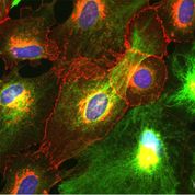
Inspired by cellular images captured by researchers in the lab of UBC scientist Christian Naus, a Peter Wall Distinguished Scholar in Residence, the project seeks to create artistic imagery based on the disease itself.

“My hope is that somehow through fashion, I more closely tap into what a woman might be feeling about her body as she undergoes the disease, but simultaneously reflect a strength, beauty, and resilience,” says Firkins, who will use the collection to raise money for cancer research, patients and survivors.

“This will be an opportunity for people to share their thoughts about the gowns,” says Firkins. “Are they too pretty to reflect something as destructive as cancer? Do they encourage you to tell your own story? Do they evoke any emotions related to your own experience?”

Before giving you where and when, here are two images (a cell and a dress based on the cell),

http://news.ubc.ca/2014/03/12/prof-challenges-cancer-fashion/

Cell7_brain_cells_in_a_dish; Astrocytes from the brain growing in a culture dish. Green colour indicates the cytoskeleton of these cells, red colour shows specific membrance [sic] channels (gap junctions), blue colour indicates the cell nuclei (DNA). The ability to grow cells in a dish has contributed to our understand of the changes these cells undergo when they become channels. Photo credit: John Bechberger, MSc., Christian Naus, PhD.

Cell7_Mercedes_de_la_Zerda: Dress modeled by BFA Acting student Mercedes de la Zerda.Black organza cap sleeve w/ sheer top and multicolour organza diagonal trim. Photo credit: Tim Matheson

Cell7_Mercedes_de_la_Zerda: Dress modeled by BFA Acting student Mercedes de la Zerda.Black organza cap sleeve w/ sheer top and multicolour organza diagonal trim. Photo credit: Tim Matheson

Details about the show (from the UBC event description webpage where you can also find a slide show more pictures),

  • Event: Fashioning Cancer: The Correlation between Destruction and Beauty
  • Date: Tue. March 25, 2014 | Time: 12-1pm
  • Location: UBC’s Frederic Wood Theatre, 6354 Crescent Rd.
  • MAP: http://bit.ly/1fZ4bC8

On a more or less related note, Aalto University (Finland) has announced a dress made of birch cellulose fibre, from a March 13, 2014 news item on ScienceDaily,

The first garment made out of birch cellulose fibre using the Ioncell method is displayed at a fashion show in Finland on 13 March [2014]. The Ioncell method, which was developed by researchers at Aalto University, is an environmentally friendly alternative to cotton in textile production. The dress produced for Marimekko is a significant step forward in the development of fibre for industrial production.

Researchers were looking for new alternatives to cotton, because demand for textile fibres is expected to nearly double by 2030. The raw material for the Ioncell fibre is a birch-based pulp from Finnish pulp mills. Growing birch wood does not require artificial irrigation in its native habitat, for instance.

The Aalto University March 12, 2014 news release, which originated the news item, describes the new Ioncell fibre and its relationship with Finnish clothing company Marimekko,

The production method for Ioncell has been developed by Professor Herbert Sixta’s research group. The method is based on a liquid salt (ionic liquid) developed under the guidance of Professor Ilkka Kilpeläinen which is a very efficient cellulose solvent. The fibres derived from it are carded and spun to yarns at the Textile University of Börås in Sweden.

‒ We made a breakthrough in the development of the method about a year ago. Progress has been rapid since then. [see my Oct. 3, 2013 posting for another Finnish team’s work with wood cellulose to create fabric]  Production of the fibre and the thread is still a cumbersome process, but we have managed to triple the amount of fibre that is produced in six months. The quality has also improved: the fibers are stronger and of more even quality, Professor Sixta says with satisfaction.

The surface of the ready textile has a dim glow and it is pleasing to the touch. According to Sixta, because of its strength, the strength properties of the Ioncell fibre are equal or even better than other pulp-based fibres on the market. The fibres are even stronger than cotton and viscose.

The Finnish textile and clothing design company Marimekko became inspired by the new fibre at an event organised by the Finnish Bioeconomy Cluster FIBIC, which coordinates bioeconomy research, and immediately got in touch with Professor Herbert Sixta at Aalto University.

‒ We monitor product development for materials closely in order to be able to offer our customers new and more ecological alternatives. It was a wonderful opportunity to be able to join this Aalto University development project at such an early stage. Fibre made from birch pulp seems to be a promising material by virtue of its durability and other characteristics, and we hope that we will soon be able to utilise this new material in our collections, says Noora Niinikoski, Head of Fashion at Marimekko.

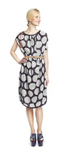
Here’s the birch cellulose dress,

Marimekko Birch Dress Courtesy: Aalto University

Let’s all have a fashionable day!