Tag Archives: CNRS

A race to find substitutes for graphene?

I have two items concerning research which seeks to replace graphene in one application or other.

Black phosporus and the École Polytechniqe de Montréal

A June 2, 2015 news item on Nanotechnology Now features work on developing a two-dimensional black phosphorus material, 2D phosphane,

A team of researchers from Universite de Montreal, Polytechnique Montreal and the Centre national de la recherche scientifique (CNRS) in France is the first to succeed in preventing two-dimensional layers of black phosphorus from oxidating. In so doing, they have opened the doors to exploiting their striking properties in a number of electronic and optoelectronic devices. …

Black phosphorus, a stable allotrope of phosphorus that presents a lamellar structure similar to that of graphite, has recently begun to capture the attention of physicists and materials researchers. It is possible to obtain single atomic layers from it, which researchers call 2D phosphane. A cousin of the widely publicized graphene, 2D phosphane brings together two very sought-after properties for device design.

A June 2, 2015 École Polytechniqe de Montréal news release, which originated the news item, expands on why 2D phosphane is an appealing material,

First, 2D phosphane is a semiconductor material that provides the necessary characteristics for making transistors and processors. With its high-mobility, it is estimated that 2D phosphane could form the basis for electronics that is both high-performance and low-cost.

Furthermore, this new material features a second, even more distinctive, characteristic: its interaction with light depends on the number of atomic layers used. One monolayer will emit red light, whereas a thicker sample will emit into the infrared. This variation makes it possible to manufacture a wide range of optoelectronic devices, such as lasers or detectors, in a strategic fraction of the electromagnetic spectrum.

The news release goes on to describe an important issue with phosphane and how the scientists addressed it,

Until now, the study of 2D phosphane’s properties was slowed by a major problem: in ambient  conditions, very thin layers of the material would degrade, to the point of compromising its future in the industry despite its promising potential.

As such, the research team has made a major step forward by succeeding in determining the physical mechanisms at play in this degradation, and in identifying the key elements that lead to the layers’ oxidation.

“We have demonstrated that 2D phosphane undergoes oxidation under ambient conditions, caused jointly by the presence of oxygen, water and light. We have also characterized the phenomenon’s evolution over time by using electron beam spectroscopy and Raman spectroscopy,” reports Professor Richard Martel of Université de Montréal’s Department of Chemistry.

Next, the researchers developed an efficient procedure for producing these very fragile single-atom layers and keeping them intact.

“We were able to study the vibration modes of the atoms in this new material. Since earlier studies had been carried out on heavily degraded materials, we revealed the as-yet-unsuspected effects of quantum confinement on atoms’ vibration modes,” notes Professor Sébastien Francoeur of Polytechnique’s Department of Engineering Physics.

The study’s results will help the world scientific community develop 2D phosphane’s very special properties with the aim of developing new nanotechnologies that could give rise to high-performance microprocessors, lasers, solar cells and more.

Here’s a link to and a citation for the paper,

Photooxidation and quantum confinement effects in exfoliated black phosphorus by Alexandre Favron, Etienne Gaufrès, Frédéric Fossard, Anne-Laurence Phaneuf-L’Heureux, Nathalie Y-W. Tang, Pierre L. Lévesque, Annick Loiseau, Richard Leonelli, Sébastien Francoeur, & Richard Martel. Nature Materials (2015)  doi:10.1038/nmat4299 Published online 25 May 2015

This paper is behind a paywall.

Now. for the second item about replacing graphene.

China’s new aerogel, a rival to graphene aerogels?

A June 2, 2015 American Institute of Physics news release (also on EurekAlert) describes research into an alternative to expensive graphene aerogels,

The electromagnetic radiation discharged by electronic equipment and devices is known to hinder their smooth operation. Conventional materials used today to shield from incoming electromagnetic waves tend to be sheets of metal or composites, which rely on reflection as a shielding mechanism.

But now, materials such as graphene aerogels are gaining traction as more desirable alternatives because they act as electromagnetic absorbers. They’re widely expected to improve energy storage, sensors, nanoelectronics, catalysis and separations, but graphene aerogels are prohibitively expensive and difficult to produce for large-scale applications because of the complicated purification and functionalization steps involved in their fabrication.

So a team of researchers in China set out to design a cheaper material with properties similar to a graphene aerogel–in terms of its conductivity, as well as a lightweight, anticorrosive, porous structure. In the journal Applied Physics Letters, from AIP Publishing, the researchers describe the new material they created and its performance.

Aming Xie, an expert in organic chemistry, and Fan Wu, both affiliated with PLA University of Science and Technology, worked with colleagues at Nanjing University of Science and Technology to tap into organic chemistry and conducting polymers to fabricate a three-dimensional (3-D) polypyrrole (PPy) aerogel-based electromagnetic absorber.

They chose to concentrate on this method because it enables them to “regulate the density and dielectric property of conducting polymers through the formation of pores during the oxidation polymerization of the pyrrole monomer,” explained Wu.

And the fabrication process is a simple one. “It requires only four common chemical reagents: pyrrole, ferric chloride (FeCl3), ethanol and water — which makes it cheap enough and enables large-scale fabrication,” Wu said. “We’re also able to pour the FeCl3 solution directly into the pyrrole solution — not drop by drop — to force the pyrrole to polymerize into a 3-D aerogel rather than PPy particles.”

In short, the team’s 3-D PPy aerogel is designed to exhibit “desirable properties such as a porous structure and low density,” Wu noted.

Beyond that, its electromagnetic absorption performance — with low loss — shows great promise. “We believe a ‘wide’ absorption range is more useful than high absorption within one frequency,” Wu said. Compared with previous works, the team’s new aerogel has the lowest adjunction and widest effective bandwidth — with a reflection loss below -10 decibels.

In terms of applications, based on the combination of low adjunction and a “wide” effective bandwidth, the researchers expect to see their 3-D PPy aerogel used in surface coatings for aircraft.

Another potential application is as coatings within the realm of corrosion prevention and control. “Common anticorrosion coatings contain a large amount of zinc (70 to 80 percent by weight), and these particles not only serve as a cathode by corroding to protect the iron structure but also to maintain a suitable conductivity for the electrochemistry process,” Wu pointed out. “If our 3-D PPy aerogel could build a conductivity network in this type of coating, the loss of zinc particles could be rapidly reduced.”

The team is now taking their work a step further by pursuing a 3-D PPy/PEDOT-based (poly(3,4-ethylenedioxythiophene) electromagnetic absorber. “Our goal is to grow solid-state polymerized PEDOT particles in the holes of the 3-D PPy aerogel formed by PPy chains,” Wu added.

Here’s a link to and a citation for the paper,

Self-assembled ultralight three-dimensional polypyrrole aerogel for effective electromagnetic absorption by Aming Xie, Fan Wu, Mengxiao Sun, Xiaoqing Dai, Zhuanghu Xu, Yanyu Qiu, Yuan Wang, and Mingyang Wang. Appl. Phys. Lett. 106, 222902 (2015); http://dx.doi.org/10.1063/1.4921180

This paper is open access.

2015 Science & You, a science communication conference in France

Science communicators can choose to celebrate June 2015 in Nancy, France and acquaint themselves with the latest and greatest in communication at the Science & You conference being held from June 1 – 6, 2015. Here’s the conference teaser being offered by the organizers,

The 2015 conference home page (ETA May 5, 2015 1045 hours PDT: the home page features change) offers this sampling of the workshops on offer,

No less than 180 communicators will be lined up to hold workshop sessions, from the 2nd to the 5th June in Nancy’s Centre Prouvé. In the meantime, here is an exclusive peek at some of the main themes which will be covered:

– Science communication and journalism. Abdellatif Bensfia will focus on the state of science communication in a country where major social changes are playing out, Morocco, while Olivier Monod will be speaking about “Chercheurs d’actu” (News Researchers), a system linking science with the news. Finally, Matthieu Ravaud and Fabrice Impériali from the CNRS (Centre National de Recherche Scientifique) will be presenting “CNRS Le journal”, the new on-line media for the general public.

– Using animals in biomedical research. This round-table, chaired by Victor Demaria-Pesce, from the Groupement Interprofessionnel de Réflexion et de Communication sur la Recherche (Gircor) will provide an opportunity to spotlight one of society’s great debates: the use of animals in research. Different actors working in biomedical research will present their point of view on the subject, and the results of an analysis of public perception of animal experimentation will be presented. What are the norms in this field? What are the living conditions of the animals in laboratories? How is this research to be made legitimate? This session will centre on all these questions.

– Science communication and the arts. This session will cover questions such as the relational interfaces between art and science, with in particular the presentation of “Pulse Project” with Michelle Lewis-King, and the Semaine du Cerveau (Brain Week) in Grenoble (Isabelle Le Brun).
Music will also be there with the talk by Milla Karvonen from the University of Oulu, who will be speaking about the interaction between science and music, while Philippe Berthelot will talk about the art of telling the story of science as a communication tool.

– Science on television. This workshop will also be in the form of a round table, with representatives from TVV (Vigyan Prasar, Inde), and Irene Lapuente (La Mandarina de Newton), Mico Tatalovic and Elizabeth Vidal (University of Cordoba), discussing how the world of science is represented on a mass media like television. Many questions will be debated, as for example the changing image of science on television, its historical context, or again, the impact these programmes have on audiences’ perceptions of science.

To learn more, you will find the detailed list of all the workshops and plenaries in the provisional programme on-line.

Science & You seems to be an ‘umbrella brand’ for the “Journées Hubert Curien” conference with plenaries and workshops and the “Science and Culture” forum, which may explain the variety of dates (June 1 – 6, June 2 – 5, and June 2 – 6) on the Science & You home page.

Here’s information about the Science & You organizers and more conference dates (from the Patrons page),

At the invitation of the President of the Université de Lorraine, the professors Etienne Klein, Cédric Villani and Brigitte Kieffer accepted to endorse Science & You. It is an honour to be able to associate them with this major event in science communication, in which they are particularly involved.

Cédric Villani, Fields Medal 2010

Cédric Villani is a French mathematician, the Director of the Institut Henri Poincaré and a professor at the Université Claude Bernard Lyon 1.
His main research interests are in kinetic theory (Boltzmann and Vlasov equations and their variants), and optimal transport and its applications (Monge equation).
He has received several national and international awards for his research, in particular the Fields Medal, which he received from the hands of the President of India at the 2010 International Congress of Mathematicians in Hyderabad (India). Since then he has played the role of spokesperson for the French mathematical community in media and political circles.
Cédric Villani regularly invests in science communication aiming at various audiences: conferences in schools, public conferences in France and abroad, regular participation in broadcasts and current affairs programmes and in science festivals.


Etienne Klein, physicist and philosopher

Etienne Klein is a French physicist, Director of Research at the CEA (Commissariat à l’énergie atomique et aux énergies alternatives – Alternative Energies and Atomic Energy Commission) and has a Ph.D. in philosophy of science. He teaches at the Ecole Centrale in Paris and is head of the Laboratoire de Recherche sur les Sciences de la Matière (LARSIM) at the CEA.

He has taken part in several major projects, such as developing a method of isotope separation involving the use of lasers, and the study of a particle accelerator with superconducting cavities. He was involved in the design of the Large Hadron Collider (LHC) at CERN.
He taught quantum physics and particle physics at Ecole Centrale in Paris for several years and currently teaches philosophy of science. He is a specialist on time in physics and is the author of a number of essays.
He is also a member of the OPECST (Conseil de l’Office parlementaire d’évaluation des choix scientifiques et technologiques – Parliamentary Office for the Evaluation of Scientific and Technological Choices), of the French Academy of Technologies, and of the Conseil d’Orientation (Advisory Board) of the Institut Diderot.
Until June 2014, he presented a weekly radio chronicle, Le Monde selon Etienne Klein, on the French national radio France Culture.

Photo by Philippe Matsas © Flammarion


Brigitte Kieffer, Campaigner for women in science

B. L. Kieffer is Professor at McGill University and at the Université de Strasbourg France. She is also Visiting Professor at UCLA (Los Angeles, USA). She develops her research activity at IGBMC, one of the leading European centres of biomedical research. She is recipient of the Jules Martin (French Academy of Science, 2001) and the Lounsbery (French and US Academies of Science, 2004) Awards, and has become an EMBO Member in 2009.
In 2012 she received the Lamonica Award of Neurology (French Academy of Science) and was nominated Chevalier de la Légion d’honneur. In December 2013 she was elected as a member of the French Academy of Sciences.
In March 2014, she received the International L’OREAL-UNESCO Award for Women in Science (European Laureate). She started as the Scientific Director of the Douglas Hospital Research Centre, affiliated to McGill University in January 2014, and remains Professor at the University of Strasbourg, France.

Photo by Julian Dufort

Here’s more about the conference at the heart of Science & You (from The Journées Hubert Curien International Conference webpage),

Following on the 2012 conference, this project will bring together all those interested in science communication: researchers, PhD students, science communicators, journalists, professionals from associations and museums, business leaders, politicians… A high-level scientific committee has been set up for this international conference, chaired by Professor Joëlle Le Marec, University of Paris 7, and counting among its members leading figures in science communication such as Bernard Schiele (Canada) or Hester du Plessis (South Africa).

The JHC Conference will take place from June 2nd to 6th at the Centre Prouvé, Nancy. These four days will be dedicated to a various programme of plenary conferences and workshops on the theme of science communication today and tomorrow.

You can find the Registration webpage here where you can get more information about the process and access the registration form.

Carbon nanotubes: OCSiAl’s deal in Korea and their effect on the body after one year

I have two news items related only by their focus on carbon nanotubes. First, there’s a July 3, 2014 news item on Azonano featuring OCSiAl’s deal with a Korean company announced at NANO KOREA 2014,

At NANO KOREA 2014 OCSiAl announced an unprecedentedly large-scale deal with Korean company Applied Carbon Nano Technology [ACN] Co., Ltd. – one of the key industry players.

OCSiAl, the dominating graphene tubes manufacturer, that successfully presented its products and technology in Europe and USA, now to enter Asian nanotech markets. At NANO KOREA 2014 the company introduced TUBALL, the universal nanomodifier of materials featuring >75% of single wall carbon nanotubes, and announced signing of supply agreement with Applied Carbon Nano Technology Co., Ltd. (hereinafter referred to as ACN), a recognized future-oriented innovative company.

A July 3, 2014 OCSiAl news release, which originated the news item, describes the memorandum of understanding (MOU) in greater detail,

Under this MoU ACN would buy 100 kg of TUBALL. The upcoming deal is the first of OCSiAl’s Korean contracts to be performed in 2015 and it turns up the largest throughout SWCNT market, which annual turnover recently hardly reached 500 kg. The agreement is exceptionally significant as it opens fundamental opportunities for manufacturing of new nanomaterial-based product with the unique properties that were not even possible before.

“OCSiAl’s entry to Korean market required thorough preparation. We invested time and efforts to prove that our company, our technology and our products worth credibility, – says Viktor Kim, OCSiAl Vice President, – we urged major playmakers to take TUBALL for testing to verify the quality and effectiveness. We believe that ACN is more than an appropriate partner to start – they are experts at the market and they understand its future perspectives very clearly. We believe that mutually beneficial partnership with ACN will path the way for future contracts, since it will become indicative to other companies in Asia and all over the world”.

“It comes as no surprise that OCSiAl’s products here in Korea will be in a great demand soon. The country strives to become world’s leader in advanced technology, and we do realize the benefits of nanomaterial’s exploitation. TUBALL is a truly versatile additive which may be used across many market sectors, where adoption of new materials with top-class performance is essential”, – says Mr. Dae-Yeol Lee, CEO of ACN.

OCSiAl’s entering to Korean market will undoubtedly have a high-reaching impact on the industry. The recent merger with American Zyvex Technologies made OCSiAl the not only the world’s largest nanomaterial producer but a first-rate developer of modifiers of different materials based on carbon nanotubes. To its Korean partners OCSiAl offers TUBALL, the raw ‘as produced’ SWCNT material and masterbatches, which can be either custom-made or ready-to-use mixtures for different applications, including li-ion batteries, car tires, transparent conductive coatings and many others. “Since Korea is increasingly dynamic, our success here will build on continuous development of our product, – adds Viktor Kim, – And we are constantly working on new applications of graphene tubes to meet sophisticated demands of nanotech-savvy Korean consumers”.

OCSiAl’s Zyvex acquisition was mentioned in a June 23, 2014 posting here.

My second tidbit concerns a July 4, 2014 news item on Nanowerk about carbon nanotubes and their effect on the body (Note: A link has been removed),

Having perfected an isotope labeling method allowing extremely sensitive detection of carbon nanotubes in living organisms, CEA and CNRS researchers have looked at what happens to nanotubes after one year inside an animal. Studies in mice revealed that a very small percentage (0.75%) of the initial quantity of nanotubes inhaled crossed the pulmonary epithelial barrier and translocated to the liver, spleen, and bone marrow. Although these results cannot be extrapolated to humans, this work highlights the importance of developing ultrasensitive methods for assessing the behavior of nanoparticles in animals. It has been published in the journal ACS Nano (“Carbon Nanotube Translocation to Distant Organs after Pulmonary Exposure: Insights from in Situ 14C-Radiolabeling and Tissue Radioimaging”).

A July 1, 2014 CNRS [France Centre national de la recherche scientifique] press release, which originated the news item, describes both applications for carbon nanotubes and the experiment in greater detail,

Carbon nanotubes are highly specific nanoparticles with outstanding mechanical and electronic properties that make them suitable for use in a wide range of applications, from structural materials to certain electronic components. Their many present and future uses explain why research teams around the world are now focusing on their impact on human health and the environment.

Researchers from CEA and the CNRS joined forces to study the distribution over time of these nanoparticles in mice, following contamination by inhalation. They combined radiolabeling with radio imaging tools for optimum detection sensitivity. When making the carbon nanotubes, stable carbon (12C) atoms were replaced directly by radioactive carbon (14C) atoms in the very structure of the tubes. This method allows the use of carbon nanotubes similar to those produced in industry, but labeled with 14C. Radio imaging tools make it possible to detect up to twenty or so carbon nanotubes on an animal tissue sample.

A single dose of 20 µg [micrograms] of labeled nanotubes was administered at the start of the protocol, then monitored for one year. The carbon nanotubes were observed to translocate from the lungs to other organs, especially the liver, spleen, and bone marrow. The study demonstrates that these nanoparticles are capable of crossing the pulmonary epithelial barrier, or air-blood barrier. It was also observed that the quantity of carbon nanotubes in these organs rose steadily over time, thus demonstrating that these particles are not eliminated on this timescale. Further studies will have to determine whether this observation remains true beyond a year.

The CEA [French Alternative Energies and Atomic Energy Commission {Commissariat à l’énergie atomique et aux énergies alternatives}] and CNRS teams have developed highly specific skills that enable them to study the health and environmental impact of nanoparticles from various angles. Nanotoxicology and nanoecotoxicology research such as this is both a priority for society and a scientific challenge, involving experimental approaches and still emerging concepts.

This work is conducted as part of CEA’s interdisciplinary Toxicology and Nanosciences programs. These are management, coordination and support structures set up to promote multidisciplinary approaches for studying the potential impact on living organisms of various components of industrial interest, including heavy metals, radionuclides, and new products.

At the CNRS, these concerns are reflected in particular in major initiatives such as the International Consortium for the Environmental Implications of Nano Technology (i-CEINT), a CNRS-led international initiative focusing on the ecotoxicology of nanoparticles. CNRS teams also have a long tradition of close involvement in matters relating to standards and regulations. Examples of this include the ANR NanoNORMA program, led by the CNRS, or ongoing work within the French C’Nano network.

For those who would either prefer or like to check out  the French language version of the July 1, 2014 CNRS press release (La biodistribution des nanotubes de carbone dans l’organisme), it can be found here.

Here’s a link to and a citation for the paper,

Carbon Nanotube Translocation to Distant Organs after Pulmonary Exposure: Insights from in Situ 14C-Radiolabeling and Tissue Radioimaging by Bertrand Czarny, Dominique Georgin, Fannely Berthon, Gael Plastow, Mathieu Pinault, Gilles Patriarche, Aurélie Thuleau, Martine Mayne L’Hermite, Frédéric Taran, and Vincent Dive. ACS Nano, 2014, 8 (6), pp 5715–5724 DOI: 10.1021/nn500475u Publication Date (Web): May 22, 2014

Copyright © 2014 American Chemical Society

This paper is behind a paywall.

Feel the vibe on Nanophonics Day

Officially, Nanophonics Day was held on May 26, 2014 but it’s never too late to appreciate good vibrations. Here’s more about the ‘day’ and nanophonics from a May 27, 2014 news item on Azonano (Note: A link has been removed),

The Nanophononics Day, collocated with the European Materials Research Society Spring Meeting (Lille, 26-30 May), aims to raise awareness about this emergent research area and the EUPHONON Project. ICREA Prof Dr Clivia Sotomayor, Group Leader at ICN2, coordinates this initiative.

A phonon is a collective excitation of atoms or molecules, a vibration of matter which plays a major role in physical properties of solids and liquids. Nanophononics is the science and engineering of these vibrations at the nanometre scale. Applications of the knowledge generated in the field might include novel devices aiming to decrease the power consumption for a low-power information society. It also includes phonon lasers and phenomena involving ultra-fast acoustic processes, or exceeding the limits of mass and pressure detections in membranes which might have an impact in safety and technology standards. Nanophononics links classical and quantum physics and translates this knowledge into everyday applications.

A May 26, 2014 Institut Català de Nanociència i Nanotecnologia (ICN2) news release, which originated the news item, provides more details about European research into nanophonics,

The EUPHONON project aims to amalgamate the activities on phonon science and technology in Europe to establish a strong community in this emerging research field. It started in November 2013, coordinated by Prof. Sebastian Volz from CNRS – École Central Paris. ICREA Prof Dr Clivia M Sotomayor Torres, Phononic and Photonic Nanostructures (P2N) Group Leader at the Institut Català de Nanociència i Nanotecnologia (ICN2), is among the 7 members of the consortium. She is the coordinator of the Nanophononics Day, intended to raise awareness about this emergent research area and the EUPHONON Project.

The Nanophononics Day is celebrated in May 26th 2014, collocated with Symposium D of the European Materials Research Society (E-MRS) Spring Meeting 2014 in Lille, entitled “Phonons and Fluctuation in Low Dimensional Structures” and with ICREA Prof Dr Clivia M Sotomayor Torres again among its organizers. It is probably the largest nanophononic event in Europe and a perfect context for a lively discussion about the most recent theoretical and experimental findings.

The Nanophononics Day includes conferences by leading scientists about recent breakthroughs in nano-scale thermal transport and how the recent achievements constitute solid base for nanophononics. Prof Gang Chen (MIT, USA) and Prof Olivier Bourgeois (CNRS Inst. Neel) will cover phonons in solid materials while phonons in biological matter will be addressed by Prof Thomas Dehoux (University of Bordeaux). Experimental methods using scanning probes will be illustrated by Prof Oleg Kolosov (Lancaster University) and Prof Severine Gomez (University of Lyon).

I wish you a belated Happy Nanophonics Day!

Are science cities London, Paris, New York and Tokyo losing prominence?

I am more accustomed to thinking about great art cities than great science cities but it appears I lack imagination if a Dec. 13, 2013 news item on Nanowerk is to be believed (Note: A link has been removed),

The world’s largest scientific centers are losing some of their prominence due to geographical decentralization at the global scale, according to a team of researchers from the LISST (Laboratoire Interdisciplinaire Solidarités, Sociétés, Territoires, CNRS / Université de Toulouse II-Le Mirail / EHESS) who conducted a systematic statistical analysis of millions of articles and papers published in thousands of scientific reviews between 1987 and 2007. Their project, whose results were recently published on the Urban Studies website (“Cities and the geographical deconcentration of scientific activity: A multilevel analysis of publications (1987–2007)”), was the first to focus on the geography of science in all the world’s cities.

Here’s an image illustrating the researchers’ work,

Courtesy o CNRS [downloaded from http://www2.cnrs.fr/presse/communique/3353.htm]

Courtesy o CNRS [downloaded from http://www2.cnrs.fr/presse/communique/3353.htm]

The Dec. 10, 2013 CNRS (France’s Centre national de la recherche scientifique) news release, [English language version] [en français]), which originated the news item, provides more details,

Geographic encoding, city by city, of all of the articles listed in the Science Citation Index (SCI) (1) between 1987 and 2007 shows that traditional scientific centers are not as prominent as they used to be: the combined share of the world’s top 10 science cities dropped from 20% in 1987 to 13% in 2007. Researchers at the LISST (Laboratoire Interdisciplinaire Solidarités, Sociétés, Territoires, CNRS /Université de Toulouse II-Le Mirail / EHESS), aided by two collaborators at the CIRST (Centre Interuniversitaire de Recherche sur la Science et la Technologie) in Montreal, concluded that this phenomenon is accompanied by a general trend toward decentralization worldwide, especially in emerging nations. China offers a good illustration: the main provincial capitals are playing a much stronger role than they did in the past, and the skyrocketing development of science in China goes alongside with a geographical realignment. Whereas Beijing and Shanghai together accounted for 52.8% of the articles published by Chinese researchers in the Science Citation Index in 1987, this percentage dropped to 31.9% in 2007. Turkey is another striking example of an emerging nation whose scientific system has seen very rapid growth. In terms of the number of articles published, the country rose from 44th to 16th place worldwide between 1987 and 2007. Over the same period, its two main scientific hubs, Ankara and Istanbul, lost some of their pre-eminence within the country. While these two cities represented more than 60% of Turkey’s scientific production in 1987, they now produce slightly less than half of the articles published by Turkish researchers. And, as in China, growth in scientific activity is accompanied by geographical decentralization: Turkey has more science hubs now than it did two decades ago, and its two traditional scientific capitals play a lesser role.

The US, which remains the world leader in terms of scientific production, is an exceptional case: the number of articles published by American researchers continues to rise steadily, but at a slower pace than in the emerging nations. Consequently, the country’s share of worldwide scientific production is lower than it used to be: in 1987, the US represented 34% of the SCI, but by 2007 this figure had fallen to 25%. Nonetheless, the American scientific scene remains quite stable geographically: the role of its main research centers has not evolved significantly because the US scientific establishment has always been one of the least centralized in the world, with research activities scattered across hundreds of cities of all sizes.

Does this development herald the decline of the great scientific centers? The fact that scientific activity is becoming more geographically decentralized on a worldwide scale does not imply that it is declining in large cities with a strong research tradition. The number of articles published in London, Paris, New York and Tokyo continues to rise every year. But the pace of growth in those traditional centers is slower than in others in the global scientific system. As more research is conducted in an increasing number of cities, the main scientific centers contribute a lesser share to the total.

The findings of this project, funded as part of an ANR program (2010-2013), challenge the assumption that scientific production inevitably tends to be concentrated in a few large urban areas, which therefore should be given priority in the allocation of resources.

(1) The Science Citation Index (or SCI) is a bibliographical database created in the US in 1964 for the purpose of documenting all scientific production worldwide. In its current version (SCI-Expanded), which is part of the Thomson Reuters Web of Science database (WoS), it registers more than one million scientific articles every year, encompassing the experimental sciences and sciences of the universe, medicine, the engineering sciences, etc., but not the humanities and social sciences, which are included in the SSCI. The SCI-Expanded records contain information on the content of each article (title, name of publication, summary, keywords), its author or authors (name, institution, city, country), and the list of references cited in the article.

This is especially fascinating in light of a recently published book claiming that the major city centres for art in the 21st century will shifting to some unexpected places. From Phaidon Press’ Art Cities of the Future webpage,

The volume profiles 12 global cities to watch for exciting contemporary art: Beirut, Bogotá, Cluj, Delhi, Istanbul, Johannesburg, Lagos, San Juan, São Paulo, Seoul, Singapore and Vancouver.

Thankfully, in both the old world and the new, commentators appear to agree. “It’s great to have a look around and discover truly interesting new work,” said Simon Armstrong, book buyer at Tate Modern and Tate Britain, in The Bookseller, “and there are some great examples of emergent artists here in this huge presentation of contemporary art from 12 cities on the fringes of the art map.”

Hannah Clugston, writing in Aesthetica concurred, describing the title as “brilliantly executed” with “stunning images,” and possessing an awareness “of the wider concerns behind the work.”

It appears that the geography of creative endeavours in the arts and the sciences is shifting. For those curious about the science end of things, here’s a link to and a citation for the paper about geography and scientific activity,

Cities and the geographical deconcentration of scientific activity: A multilevel analysis of publications (1987–2007) by Michel Grossetti, Denis Eckert, Yves Gingras, Laurent Jégou, Vincent Larivière, and Béatrice Milard. Urban Studies, 0042098013506047, November 20, 2013, doi:10.1177/0042098013506047

This paper is behind a paywall.

Should October 2013 be called ‘the month of graphene’?

Since the Oct. 10-11, 2013 Graphene Flagship (1B Euros investment) launch, mentioned in my preview Oct. 7, 2013 posting, there’ve been a flurry of graphene-themed news items both on this blog and elsewhere and I’ve decided to offer a brief roundup what I’ve found elsewhere.

Dexter Johnson offers a commentary in the pithily titled, Europe Invests €1 Billion to Become “Graphene Valley,” an Oct. 15, 2013 posting on his Nanoclast blog (on the IEEE [Institute of Electrical and Electronics Engineers] website) Note: Links have been removed,

The initiative has been dubbed “The Graphene Flagship,” and apparently it is the first in a number of €1 billion, 10-year plans the EC is planning to launch. The graphene version will bring together 76 academic institutions and industrial groups from 17 European countries, with an initial 30-month-budget of €54M ($73 million).

Graphene research is still struggling to find any kind of applications that will really take hold, and many don’t expect it will have a commercial impact until 2020. What’s more, manufacturing methods are still undeveloped. So it would appear that a 10-year plan is aimed at the academic institutions that form the backbone of this initiative rather than commercial enterprises.

Just from a political standpoint the choice of Chalmers University in Sweden as the base of operations for the Graphene Flagship is an intriguing choice. …

I have to agree with Dexter that choosing Chalmers University over the University of Manchester where graphene was first isolated is unexpected. As a companion piece to reading Dexter’s posting in its entirety and which features a video from the flagship launch, you might want to try this Oct. 15, 2013 article by Koen Mortelmans for Youris (h/t Oct. 15, 2013 news item on Nanowerk),

Andre Konstantin Geim is the only person who ever received both a Nobel and an Ig Nobel. He was born in 1958 in Russia, and is a Dutch-British physicist with German, Polish, Jewish and Ukrainian roots. “Having lived and worked in several European countries, I consider myself European. I don’t believe that any further taxonomy is necessary,” he says. He is now a physics professor at the University of Manchester. …

He shared the Noble [Nobel] Prize in 2010 with Konstantin Novoselov for their work on graphene. It was following on their isolation of microscope visible grapheme flakes that the worldwide research towards practical applications of graphene took off.  “We did not invent graphene,” Geim says, “we only saw what was laid up for five hundred year under our noses.”

Geim and Novoselov are often thought to have succeeded in separating graphene from graphite by peeling it off with ordinary duct tape until there only remained a layer. Graphene could then be observed with a microscope, because of the partial transparency of the material. That is, after dissolving the duct tape material in acetone, of course. That is also the story Geim himself likes to tell.

However, he did not use – as the urban myth goes – graphite from a common pencil. Instead, he used a carbon sample of extreme purity, specially imported. He also used ultrasound techniques. But, probably the urban legend will survive, as did Archimedes’ bath and Newtons apple. “It is nice to keep some of the magic,” is the expression Geim often uses when he does not want a nice story to be drowned in hard facts or when he wants to remain discrete about still incomplete, but promising research results.

Mortelmans’ article fills in some gaps for those not familiar with the graphene ‘origins’ story while Tim Harper’s July 22, 2012 posting on Cientifica’s (an emerging technologies consultancy where Harper is the CEO and founder) TNT blog offers an insight into Geim’s perspective on the race to commercialize graphene with a paraphrased quote for the title of Harper’s posting, “It’s a bit silly for society to throw a little bit of money at (graphene) and expect it to change the world.” (Note: Within this context, mention is made of the company’s graphene opportunities report.)

With all this excitement about graphene (and carbon generally), the magazine titled Carbon has just published a suggested nomenclature for 2D carbon forms such as graphene, graphane, etc., according to an Oct. 16, 2013 news item on Nanowerk (Note: A link has been removed),

There has been an intense research interest in all two-dimensional (2D) forms of carbon since Geim and Novoselov’s discovery of graphene in 2004. But as the number of such publications rise, so does the level of inconsistency in naming the material of interest. The isolated, single-atom-thick sheet universally referred to as “graphene” may have a clear definition, but when referring to related 2D sheet-like or flake-like carbon forms, many authors have simply defined their own terms to describe their product.

This has led to confusion within the literature, where terms are multiply-defined, or incorrectly used. The Editorial Board of Carbon has therefore published the first recommended nomenclature for 2D carbon forms (“All in the graphene family – A recommended nomenclature for two-dimensional carbon materials”).

This proposed nomenclature comes in the form of an editorial, from Carbon (Volume 65, December 2013, Pages 1–6),

All in the graphene family – A recommended nomenclature for two-dimensional carbon materials

  • Alberto Bianco
    CNRS, Institut de Biologie Moléculaire et Cellulaire, Immunopathologie et Chimie Thérapeutique, Strasbourg, France
  • Hui-Ming Cheng
    Shenyang National Laboratory for Materials Science, Institute of Metal Research, Chinese Academy of Sciences, 72 Wenhua Road, Shenyang 110016, China
  • Toshiaki Enoki
    Department of Chemistry, Graduate School of Science and Engineering, Tokyo Institute of Technology, Tokyo, Japan
  • Yury Gogotsi
    Materials Science and Engineering Department, A.J. Drexel Nanotechnology Institute, Drexel University, 3141 Chestnut Street, Philadelphia, PA 19104, USA
  • Robert H. Hurt
    Institute for Molecular and Nanoscale Innovation, School of Engineering, Brown University, Providence, RI 02912, USA
  • Nikhil Koratkar
    Department of Mechanical, Aerospace and Nuclear Engineering, The Department of Materials Science and Engineering, Rensselaer Polytechnic Institute, 110 8th Street, Troy, NY 12180, USA
  • Takashi Kyotani
    Institute of Multidisciplinary Research for Advanced Materials, Tohoku University, 2-1-1 Katahira, Aoba-ku, Sendai 980-8577, Japan
  • Marc Monthioux
    Centre d’Elaboration des Matériaux et d’Etudes Structurales (CEMES), UPR-8011 CNRS, Université de Toulouse, 29 Rue Jeanne Marvig, F-31055 Toulouse, France
  • Chong Rae Park
    Carbon Nanomaterials Design Laboratory, Global Research Laboratory, Research Institute of Advanced Materials, Department of Materials Science and Engineering, Seoul National University, Seoul 151-744, Republic of Korea
  • Juan M.D. Tascon
    Instituto Nacional del Carbón, INCAR-CSIC, Apartado 73, 33080 Oviedo, Spain
  • Jin Zhang
    Center for Nanochemistry, College of Chemistry and Molecular Engineering, Peking University, Beijing 100871, China

This editorial is behind a paywall.

INFERNOS: realizing Maxwell’s Demon

Before getting to the INFERNOS project and its relationship to Maxwell’s demon, I want to share a pretty good example of this ‘demon’ thought experiment which, as recently as Feb. 4, 2013, I featured in a piece about quantum dots,

James Clerk Maxwell, physicist,  has entered the history books for any number reasons but my personal favourite is Maxwell’s demon, a thought experiment he proposed in the 1800s to violate the 2nd law of thermodynamics. Lisa Zyga in her Feb. 1, 2013 article for phys.org provides an explanation,

When you open your door on a cold winter day, the warm air from your home and the cold air from outside begin to mix and evolve toward thermal equilibrium, a state of complete entropy where the temperatures outside and inside are the same. This situation is a rough example of the second law of thermodynamics, which says that entropy in a closed system never decreases. If you could control the air flow in a way that uses a sufficiently small amount of energy, so that the entropy of the system actually decreases overall, you would have a hypothetical mechanism called Maxwell’s demon.

An Oct. 9, 2013 news item on Nanowerk ties together INFERNOS and the ‘demon’,

Maxwell’s Demon is an imaginary creature that the mathematician James Clerk Maxwell created in 1897. The creature could turn heat into work without causing any other change, which violates the second law of thermodynamics. The primary goal of the European project INFERNOS (Information, fluctuations, and energy control in small systems) is to realize experimentally Maxwell’s Demon; in other words, to develop the electronic and biomolecular nanodevices that support this principle.

The Universitat de Barcelona (University of Barcelona) Oct. 7, 2013 news release, which originated the news item, provides more details about the project,

Although Maxwell’s Demon is one of the cornerstones of theoretical statistical mechanisms, little has been done about its definite experimental realization. Marco Ribezzi, researcher from the Department of Fundamental Physics, explains that “the principal novelty of INFERNOS is to bring a robust and rigorous experimental base for this field of knowledge. We aim at creating a device that can use information to supply/extract energy to/from a system”. In this sense, the UB group, in which researcher Fèlix Ritort from the former department also participates, focuses their activity on understanding how information and temperature changes are used in individual molecules manipulation.

From the theory side, researchers will work in order to develop a theory of the fluctuation processes in small systems, which would then facilitate efficient algorithms for the Maxwell’s Demon operation.

INFERNOS is a three-year European project of the programme Future and Emerging Technologies (FET). Besides the University of Barcelona, INFERNOS partners are: Aalto University (Finland), project coordinator, Lund University (Sweden), the University of Oslo (Norway), Delf University of Technology (Netherlands), the National Center for Scientific Research (France) and the Research Foundation of State University of New York.

I like the INFERNOS logo, demon and all,

Logo of the European project INFERNOS (Information, fluctuations, and energy control in small systems).

Logo of the European project INFERNOS (Information, fluctuations, and energy control in small systems).

The INFERNOS project website can be found here.

And for anyone who finds that music is the best way to learn, here are Flanders & Swann* performing ‘First and Second Law’ from a 1964 show,

Enjoy!

* ‘Swan’ corrected to ‘Swann’ on April 1, 2014.

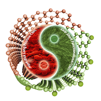
The yin and the yang of carbon nanotubes and toxicity


Illustration courtesy of the University College of London (UCL). Downloaded from http://www.ucl.ac.uk/news/news-articles/0113/130115-chemistry-resolves-toxic-concerns-about-carbon-nanotubes

Illustration courtesy of the University College of London (UCL). Downloaded from http://www.ucl.ac.uk/news/news-articles/0113/130115-chemistry-resolves-toxic-concerns-about-carbon-nanotubes

Researchers at the University College of London (UCL), France’s Centre national de la recherche scientifique (CNRS), and Italy’s University of Trieste have determined that carbon nanotube toxicity issues can be addressed be reducing their length and treating them chemically. From the Jan. 15,2013 news item on ScienceDaily,

In a new study, published January 15 [2013] in the journal Angewandte Chemie, evidence is provided that the asbestos-like reactivity and pathogenicity reported for long, pristine nanotubes can be completely alleviated if their surface is modified and their effective length is reduced as a result of chemical treatment.

First atomically described in the 1990s, carbon nanotubes are sheets of carbon atoms rolled up into hollow tubes just a few nanometres in diameter. Engineered carbon nanotubes can be chemically modified, with the addition of chemotherapeutic drugs, fluorescent tags or nucleic acids — opening up applications in cancer and gene therapy.

Furthermore, these chemically modified carbon nanotubes can pierce the cell membrane, acting as a kind of ‘nano-needle’, allowing the possibility of efficient transport of therapeutic and diagnostic agents directly into the cytoplasm of cells.

Among their downsides however, have been concerns about their safety profile. One of the most serious concerns, highlighted in 2008, involves the carcinogenic risk from the exposure and persistence of such fibres in the body. Some studies indicate that when long untreated carbon nanotubes are injected to the abdominal cavity of mice they can induce unwanted responses resembling those associated with exposure to certain asbestos fibres.

In this paper, the authors describe two different reactions which ask if any chemical modification can render the nanotubes non-toxic. They conclude that not all chemical treatments alleviate the toxicity risks associated with the material. Only those reactions that are able to render carbon nanotubes short and stably suspended in biological fluids without aggregation are able to result in safe, risk-free material.

Here’s a citation and link for this latest  research, from the ScienceDaily news item where you can also read the lead researcher’s comments about carbon nanotubes, safety, and unreasonable proposals to halt production,

Hanene Ali-Boucetta, Antonio Nunes, Raquel Sainz, M. Antonia Herrero, Bowen Tian, Maurizio Prato, Alberto Bianco, Kostas Kostarelos. Asbestos-like Pathogenicity of Long Carbon Nanotubes Alleviated by Chemical Functionalization. Angewandte Chemie International Edition, 2013; DOI: 10.1002/anie.201207664

The article is behind a paywall. I have mentioned long carbon nanotubes and their resemblance to asbestos fibres in several posts. The  Oct. 26, 2009 posting [scroll down about 1/3 of the way] highlights research which took place after the study where mice had carbon nanotubes injected into their bellies; in this second piece of research they inhaled the nanotubes.

ETA Jan. 21, 2013: Dexter Johnson gives context and commentary about this latest research into long multiwalled nanotubes (MWNTs) which he sums up as the answer to this question “What if you kept the MWNTs short?”  in a Jan. 18, 2013 posting on his Nanoclast blog (on the IEEE [Institute of Electrical and Electronics Engineers] website)

Where do buckyballs come from?

I’ve always wondered where buckyballs come from (as have scientists for the last 25 years) and now there’s an answer of sorts  (from the July 31, 2012 Florida State University news release Note: I have removed some links),

“We started with a paste of pre-existing fullerene molecules mixed with carbon and helium, shot it with a laser, and instead of destroying the fullerenes we were surprised to find they’d actually grown,” they wrote. The fullerenes were able to absorb and incorporate carbon from the surrounding gas.

By using fullenes  that contained heavy metal atoms in their centers, the scientists showed that the carbon cages remained closed throughout the process.

“If the cages grew by splitting open, we would have lost the metal atoms, but they always stayed locked inside,” Dunk [Paul Dunk, a doctoral student in chemistry and biochemistry at Florida State and lead author of the study published in Nature Communications] noted.

The researchers worked with a team of MagLab chemists using the lab’s 9.4-tesla Fourier transform ion cyclotron resonance mass spectrometer to analyze the dozens of molecular species produced when they shot the fullerene paste with the laser. The instrument works by separating molecules according to their masses, allowing the researchers to identify the types and numbers of atoms in each molecule. The process is used for applications as diverse as identifying oil spills, biomarkers and protein structures.

Dexter Johnson in his Aug. 6, 2012 posting on the Nanoclast blog on the IEEE (Institute of Electrical and Electronics Engineers) provides some context and commentary (Note: I have removed a link),

When Richard Smalley, Robert Curl, James Heath, Sean O’Brien, and Harold Kroto prepared the first buckminsterfullerene (C60) (or buckyball), they kicked off the next 25 years of nanomaterial science.

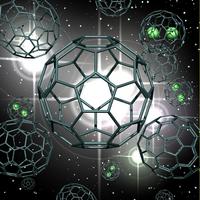
Here’s an artist’s illustration of  what these scientists have achieved, fullerene cage growth,

An artist’s representation of fullerene cage growth via carbon absorption from surrounding hot gases. Some of the cages contain lanthanum metal atoms. (Image courtesy National Science Foundation) [downloaded from Florida State University website]

 As I noted earlier I’m not alone in my fascination (from the news release),

Many people know the buckyball, also known by scientists as buckminsterfullerene, carbon 60 or C60, from the covers of their school chemistry textbooks. Indeed, the molecule represents the iconic image of “chemistry.” But how these often highly symmetrical, beautiful molecules with  fascinating properties form in the first place has been a mystery for a quarter-century. Despite worldwide investigation since the 1985 discovery of C60, buckminsterfullerene and other, non-spherical C60 molecules — known collectively as fullerenes — have kept their secrets. How? They’re born under highly energetic conditions and grow ultra-fast, making them difficult to analyze.

“The difficulty with fullerene formation is that the process is literally over in a flash — it’s next to impossible to see how the magic trick of their growth was performed,” said Paul Dunk, a doctoral student in chemistry and biochemistry at Florida State and lead author of the work.

There’s more than just idle curiosity at work (from the news release),

The buckyball research results will be important for understanding fullerene formation in extraterrestrial environments. Recent reports by NASA showed that crystals of C60 are in orbit around distant suns. This suggests that fullerenes may be more common in the universe than previously thought.

“The results of our study will surely be extremely valuable in deciphering fullerene formation in extraterrestrial environments,” said Florida State’s Harry Kroto, a Nobel Prize winner for the discovery of C60 and co-author of the current study.

The results also provide fundamental insight into self-assembly of other technologically important carbon nanomaterials such as nanotubes and the new wunderkind of the carbon family, graphene.

H/T to Nanowerk’s July 31, 2012 news item titled, Decades-old mystery how buckyballs form has been solved. In addition to Florida State University, National High Magnetic Field Laboratory (or MagLab), the CNRS  (Centre National de la Recherche Scientifique)Institute of Materials in France and Nagoya University in Japan were also involved in the research.

French scientists focus optical microscopes down to 30 nm

In fact, the French scientists are using two different imaging techniques to arrive at a resolution of 30 nm for their optical microscopes, according to the May 18, 2012 news item on Nanowerk.

Researchers from the Institut Pasteur and CNRS [Centre national de la recherche scientifique] have set up a new optical microscopy approach that combines two recent imaging techniques in order to visualize molecular assemblies without affecting their biological functions, at a resolution 10 times better than that of traditional microscopes. Using this approach, they were able to observe the AIDS virus and its capsids (containing the HIV genome) within cells at a scale of 30 nanometres, for the first time with light.

More specifically,

A study coordinated by Dr Christophe Zimmer (Institut Pasteur/CNRS), in collaboration with Dr Nathalie Arhel within the lab headed by Pr Pierre Charneau (Institut Pasteur/CNRS), shows that the association of two recent imaging techniques helps obtain unique images of molecular assemblies of HIV-1 capsids, with a resolution around 10 times better than that of traditional microscopes. This new approach, which uses super-resolution imaging and FlAsH labeling, does not affect the virus’ ability to self-replicate. It represents a major step forward in molecular biology studies, enabling the visualisation of microbial complexes at a scale of 30 nm without affecting their function.

The newly developed approach combines super-resolution PALM imaging and fluorescent FlAsH labeling. PALM imaging relies on the acquisition of thousands of low-resolution images, each of which showing only a few fluorescent molecules. The molecular positions are then calculated with high accuracy by computer programs and compiled into a single high-resolution image. FlAsH labeling involves the insertion of a 6-amino-acid peptide into the protein of interest. The binding of the FlAsH fluorophore to the peptide generates a fluorescent signal, thereby enabling the visualization of the protein. For the first time, researchers have combined these two methods in order to obtain high-resolution images of molecular structures in either fixed or living cells.

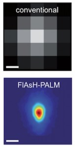
The researchers have supplied an image illustrating the difference between the conventional and new techniques will allow them to view (from the May 16, 2012 press release  [communiqué de presses] on the CNRS website),

© Institut Pasteur Reconstruction optique super-résolutive de la morphologie du VIH. L'image du dessous montre la distribution moyenne de l'enzyme intégrase observée par FlAsH-PALM. La résolution de cette technique (~30 nm) permet de retrouver la taille et la forme conique de la capside. Pour comparaison, la résolution de la microscopie conventionnelle (~200-300 nm), illustrée par l'image du dessus, ne permet pas une description détaillée de cette structure.

The conventional 200 – 300 nm resolution is shown at the top while the new 30 nm resolution achieved by combining the new techniques is shown below. This new technique has already allowed scientists to disprove a popular theory about the AIDS virus, from the May 18, 2012 news item on Nanowerk,

This new method has helped researchers visualise the AIDS Virus and localise its capsids in human cells, at a scale of 30 nm. Capsids are conical structures which contain the HIV genome. These structures must dismantle in order for the viral genome to integrate itself into the host cell’s genome. However, the timing of this disassembly has long been debated. According to a prevailing view, capsids disassemble right after infection of the host cell and, therefore, do not play an important role in the intracellular transport of the virus to the host cell’s nucleus. However, the results obtained by the researchers of the Institut Pasteur and CNRS indicate that numerous capsids remain unaltered until entry of the virus into the nucleus, confirming and strengthening earlier studies based on electron microscopy. Hence, capsids could play a more important role than commonly assumed in the replication cycle of HIV.

I gather excitement about this development is high as the scientists are suggesting that ‘microscopy’ could be known as ‘nanoscopy’ in the future.