Tag Archives: CNTs

Robust reverse osmosis membranes made of carbon nanotubes

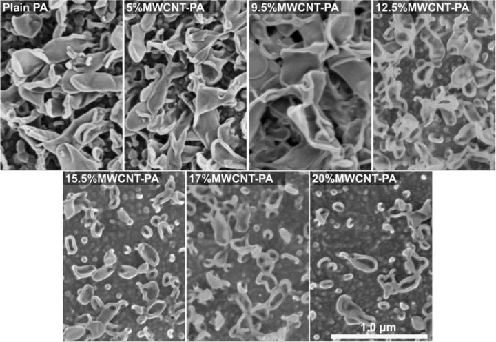
Caption: SEM images of MWCNT-PA (Multi-Walled Carbon Nanotube-Polyamide) nanocomposite membranes, for plain PA, and PA with 5, 9.5, 12.5, 15.5, 17 and 20 wt.% of MWCNT, where the typical lobe-like structures appear at the surface. Note the tendency towards a flatter membrane surface as the content of MWCNT increases. Scale bar corresponds to 1.0?μm for all the micrographs. Credit: Copyright 2018, Springer Nature, Licensed under CC BY 4.0

It seems unlikely that the image’s resemblance to a Japanese kimono on display is accidental. Either way, nicely done!

An April 12, 2018 news item on phys.org describes a technique that would allow large-scale water desalination,

A research team of Shinshu University, Japan, has developed robust reverse osmosis membranes that can endure large-scale water desalination. The team published their results in early February [2018] in Scientific Reports.

“Since more than 97 percent of the water in the world is saline water, reverse osmosis desalination plants for producing fresh water are increasingly important for providing a safe and consistent supply,” said Morinobu Endo, Ph.D., corresponding author on the paper. Endo is a distinguished professor of Shinshu University and the Honorary Director of the Institute of Carbon Science and Technology. “Even though reverse osmosis membrane technology has been under development for several decades, new threats like global warming and increasing clean water demand in populated urban centers challenge the conventional water supply systems.”

Reverse osmosis membranes typically consist of thin film composite systems, with an active layer of polymer film that restricts undesired substances, such as salt, from passing through a permeable porous substrate. Such membranes can turn seawater into drinkable water, as well as aid in agricultural and landscape irrigation, but they can be costly to operate and spend a large amount of energy.

To meet the demand of potable water at low cost, Endo says more robust membranes capable of withstanding harsh conditions, while remaining chemically stable to tolerate cleaning treatments, are necessary. The key lays in carbon nanotechnology.

An April 11, 2018 Shinshu University press release, which originated the news item, provides more details about the work,

Endo is a pioneer of carbon nanotubes [sic] synthesis by catalytic chemical vapor deposition. In this research, Endo and his team developed a multi-walled carbon nanotube-polyamide nanocomposite membrane, which is resistant to chlorine–one of the main cause of degradation or failure cases in reverse osmosis membranes. The added carbon nanotubes create a protective effect that stabilized the linked molecules of the polyamide against chlorine.

“Carbon nanotechnology has been expected to bring benefits, and this is one promising example of the contribution of carbon nanotubes to a very critical application: water purification,” Endo said. “Carbon nanotubes and fibers are already superb reinforcements for other applications in materials science and engineering, and this is yet another field where their exceptional properties can be used for improving conventional technologies.”

The researchers are working to stabilize and expand the production and processing of multi-walled carbon nanotube-polyamide nanocomposite membranes.

“We are currently working on scaling up our method of synthesis, which, in principle, is based on the same method used to prepare current polyamide membranes,” Endo said. He also noted that his team is planning a collaboration to produce commercial membranes.

Here’s a link to and a citation for the paper,

Robust water desalination membranes against degradation using high loads of carbon nanotubes by J. Ortiz-Medina, S. Inukai, T. Araki, A. Morelos-Gomez, R. Cruz-Silva, K. Takeuchi, T. Noguchi, T. Kawaguchi, M. Terrones, & M. Endo. Scientific Reports volume 8, Article number: 2748 (2018) doi:10.1038/s41598-018-21192-5 Published online: 09 February 2018

This paper is open access.

The new knitting: electronics and batteries

Researchers from China have developed a new type of yarn for flexible electronics. A March 28, 2018 news item on Nanowerk announces the work, (Note: A link has been removed),

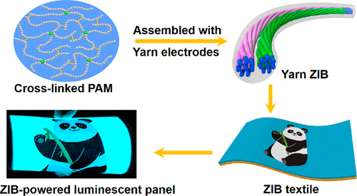
When someone thinks about knitting, they usually don’t conjure up an image of sweaters and scarves made of yarn that can power watches and lights. But that’s just what one group is reporting in ACS Nano (“Waterproof and Tailorable Elastic Rechargeable Yarn Zinc Ion Batteries by a Cross-Linked Polyacrylamide Electrolyte”). They have developed a rechargeable yarn battery that is waterproof and flexible. It also can be cut into pieces and still work.

A March 28, 2018 2018 American Chemical Society (ACS) news release (also on EurekAlert), which originated the news item, expands on the theme (Note: Links have been removed),

Most people are familiar with smartwatches, but for wearable electronics to progress, scientists will need to overcome the challenge of creating a device that is deformable, durable, versatile and wearable while still holding and maintaining a charge. One dimensional fiber or yarn has shown promise, since it is tiny, flexible and lightweight. Previous studies have had some success combining one-dimensional fibers with flexible Zn-MnO2 batteries, but many of these lose charge capacity and are not rechargeable. So, Chunyi Zhi and colleagues wanted to develop a rechargeable yarn zinc-ion battery that would maintain its charge capacity, while being waterproof and flexible.

The group twisted carbon nanotube fibers into a yarn, then coated one piece of yarn with zinc to form an anode, and another with magnesium oxide to form a cathode. These two pieces were then twisted like a double helix and coated with a polyacrylamide electrolyte and encased in silicone. Upon testing, the yarn zinc-ion battery was stable, had a high charge capacity and was rechargeable and waterproof. In addition, the material could be knitted and stretched. It also could be cut into several pieces, each of which could power a watch. In a proof-of-concept demonstration, eight pieces of the cut yarn battery were woven into a long piece that could power a belt containing 100 light emitting diodes (known as LEDs) and an electroluminescent panel.

The authors acknowledge funding from the National Natural Science Foundation of China and the Research Grants Council of Hong Kong Joint Research Scheme, City University of Hong Kong and the Sichuan Provincial Department of Science & Technology.

Here’s an image the researchers have used to illustrate their work,


Courtesy: American Chemical Society

Here’s a link to and a citation for the paper,

Waterproof and Tailorable Elastic Rechargeable Yarn Zinc Ion Batteries by a Cross-Linked Polyacrylamide Electrolyte by Hongfei Li, Zhuoxin Liu, Guojin Liang, Yang Huang, Yan Huang, Minshen Zhu, Zengxia Pe, Qi Xue, Zijie Tang, Yukun Wang, Baohua Li, and Chunyi Zhi. ACS Nano, Article ASAP DOI: 10.1021/acsnano.7b09003 Publication Date (Web): March 28, 2018

Copyright © 2018 American Chemical Society

This paper is behind a paywall.

Stronger than steel and spider silk: artificial, biodegradable, cellulose nanofibres

This is an artificial and biodegradable are two adjectives you don’t usually see united by the conjunction, and. However, it is worth noting that the artificial material is initially derived from a natural material, cellulose. Here’s more from a May 16, 2018 news item on ScienceDaily,

At DESY’s [Deutsches Elektronen-Synchrotron] X-ray light source PETRA III, a team led by Swedish researchers has produced the strongest bio-material that has ever been made. The artifical, but bio-degradable cellulose fibres are stronger than steel and even than dragline spider silk, which is usually considered the strongest bio-based material. The team headed by Daniel Söderberg from the KTH Royal Institute of Technology in Stockholm reports the work in the journal ACS Nano of the American Chemical Society.

A May 16, 2018 DESY press release (also on EurekAlert), which originated the news item, provides more detail,

The ultrastrong material is made of cellulose nanofibres (CNF), the essential building blocks of wood and other plant life. Using a novel production method, the researchers have successfully transferred the unique mechanical properties of these nanofibres to a macroscopic, lightweight material that could be used as an eco-friendly alternative for plastic in airplanes, cars, furniture and other products. “Our new material even has potential for biomedicine since cellulose is not rejected by your body”, explains Söderberg.

The scientists started with commercially available cellulose nanofibres that are just 2 to 5 nanometres in diameter and up to 700 nanometres long. A nanometre (nm) is a millionth of a millimetre. The nanofibres were suspended in water and fed into a small channel, just one millimetre wide and milled in steel. Through two pairs of perpendicular inflows additional deionized water and water with a low pH-value entered the channel from the sides, squeezing the stream of nanofibres together and accelerating it.

This process, called hydrodynamic focussing, helped to align the nanofibres in the right direction as well as their self-organisation into a well-packed macroscopic thread. No glue or any other component is needed, the nanofibres assemble into a tight thread held together by supramolecular forces between the nanofibres, for example electrostatic and Van der Waals forces.

With the bright X-rays from PETRA III the scientists could follow and optimise the process. “The X-rays allow us to analyse the detailed structure of the thread as it forms as well as the material structure and hierarchical order in the super strong fibres,” explains co-author Stephan Roth from DESY, head of the Micro- and Nanofocus X-ray Scattering Beamline P03 where the threads were spun. “We made threads up to 15 micrometres thick and several metres in length.”

Measurements showed a tensile stiffness of 86 gigapascals (GPa) for the material and a tensile strength of 1.57 GPa. “The bio-based nanocellulose fibres fabricated here are 8 times stiffer and have strengths higher than natural dragline spider silk fibres,” says Söderberg. “If you are looking for a bio-based material, there is nothing quite like it. And it is also stronger than steel and any other metal or alloy as well as glass fibres and most other synthetic materials.” The artificial cellulose fibres can be woven into a fabric to create materials for various applications. The researchers estimate that the production costs of the new material can compete with those of strong synthetic fabrics. “The new material can in principle be used to create bio-degradable components,” adds Roth.

The study describes a new method that mimics nature’s ability to accumulate cellulose nanofibres into almost perfect macroscale arrangements, like in wood. It opens the way for developing nanofibre material that can be used for larger structures while retaining the nanofibres’ tensile strength and ability to withstand mechanical load. “We can now transform the super performance from the nanoscale to the macroscale,” Söderberg underlines. “This discovery is made possible by understanding and controlling the key fundamental parameters essential for perfect nanostructuring, such as particle size, interactions, alignment, diffusion, network formation and assembly.” The process can also be used to control nanoscale assembly of carbon tubes and other nano-sized fibres.

(There are some terminology and spelling issues, which are described at the end of this post.)

Let’s get back to a material that rivals spider silk and steel for strength (for some reason that reminded me of an old carnival game where you’d test your strength by swinging a mallet down on a ‘teeter-totter-like’ board and sending a metal piece up a post to make a bell ring). From a May 16, 2018 DESY press release (also on EurekAlert), which originated the news item,

The ultrastrong material is made of cellulose nanofibres (CNF), the essential building blocks of wood and other plant life. Using a novel production method, the researchers have successfully transferred the unique mechanical properties of these nanofibres to a macroscopic, lightweight material that could be used as an eco-friendly alternative for plastic in airplanes, cars, furniture and other products. “Our new material even has potential for biomedicine since cellulose is not rejected by your body”, explains Söderberg.

The scientists started with commercially available cellulose nanofibres that are just 2 to 5 nanometres in diameter and up to 700 nanometres long. A nanometre (nm) is a millionth of a millimetre. The nanofibres were suspended in water and fed into a small channel, just one millimetre wide and milled in steel. Through two pairs of perpendicular inflows additional deionized water and water with a low pH-value entered the channel from the sides, squeezing the stream of nanofibres together and accelerating it.

This process, called hydrodynamic focussing, helped to align the nanofibres in the right direction as well as their self-organisation into a well-packed macroscopic thread. No glue or any other component is needed, the nanofibres assemble into a tight thread held together by supramolecular forces between the nanofibres, for example electrostatic and Van der Waals forces.

With the bright X-rays from PETRA III the scientists could follow and optimise the process. “The X-rays allow us to analyse the detailed structure of the thread as it forms as well as the material structure and hierarchical order in the super strong fibres,” explains co-author Stephan Roth from DESY, head of the Micro- and Nanofocus X-ray Scattering Beamline P03 where the threads were spun. “We made threads up to 15 micrometres thick and several metres in length.”

Measurements showed a tensile stiffness of 86 gigapascals (GPa) for the material and a tensile strength of 1.57 GPa. “The bio-based nanocellulose fibres fabricated here are 8 times stiffer and have strengths higher than natural dragline spider silk fibres,” says Söderberg. “If you are looking for a bio-based material, there is nothing quite like it. And it is also stronger than steel and any other metal or alloy as well as glass fibres and most other synthetic materials.” The artificial cellulose fibres can be woven into a fabric to create materials for various applications. The researchers estimate that the production costs of the new material can compete with those of strong synthetic fabrics. “The new material can in principle be used to create bio-degradable components,” adds Roth.

The study describes a new method that mimics nature’s ability to accumulate cellulose nanofibres into almost perfect macroscale arrangements, like in wood. It opens the way for developing nanofibre material that can be used for larger structures while retaining the nanofibres’ tensile strength and ability to withstand mechanical load. “We can now transform the super performance from the nanoscale to the macroscale,” Söderberg underlines. “This discovery is made possible by understanding and controlling the key fundamental parameters essential for perfect nanostructuring, such as particle size, interactions, alignment, diffusion, network formation and assembly.” The process can also be used to control nanoscale assembly of carbon tubes and other nano-sized fibres.

Here’s a link to and a citation for the paper,

Multiscale Control of Nanocellulose Assembly: Transferring Remarkable Nanoscale Fibril Mechanics to Macroscale Fibers by Nitesh Mittal, Farhan Ansari, Krishne Gowda V, Christophe Brouzet, Pan Chen, Per Tomas Larsson, Stephan V. Roth, Fredrik Lundell, Lars Wågberg, Nicholas A. Kotov, and L. Daniel Söderberg. ACS Nano, Article ASAP DOI: 10.1021/acsnano.8b01084 Publication Date (Web): May 9, 2018

Copyright © 2018 American Chemical Society

This paper is open access and accompanied by this image illustrating the work,
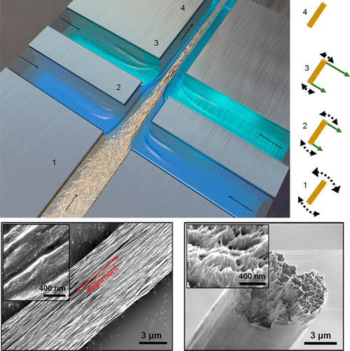
Courtesy: American Chemical Society and the researchers [Note: The bottom two images of cellulose nanofibres, which are constittuents of an artificial cellulose fibre, appear to be from a scanning tunneling microsscope. Credit: Nitesh Mittal, KTH Stockholm

This news has excited interest at General Electric (GE) (its Wikipedia entry), which has highlighted the work in a May 25, 2018 posting (The 5 Coolest Things On Earth This Week) by Tomas Kellner on the GE Reports blog.

Terminology and spelling

I’ll start with spelling since that’s the easier of the two. In some parts of the world it’s spelled ‘fibres’ and in other parts of the world it’s spelled ‘fibers’. When I write the text in my post, it tends to reflect the spelling used in the news/press releases. In other words, I swing in whichever direction the wind is blowing.

For diehards only

As i understand the terminology situation, nanocellulose and cellulose nanomaterials are interchangeable generic terms. Further, cellulose nanofibres (CNF) seems to be another generic term and it encompasses both cellulose nanocrystals (CNC) and cellulose nanofibrils (CNF). Yes, there appear to be two CNFs. Making matters more interesting is the fact that cellulose nanocrystals were originally christened nanocrystalline cellulose (NCC). For anyone who follows the science and technology scene, it becomes obvious that competing terminologies are the order of the day. Eventually the dust settles and naming conventions are resolved. More or less.

Ordinarily I would reference the Nanocellulose Wikipedia entry in my attempts to clarify the issues but it seems that the writers for the entry have not caught up to the current naming convention for cellulose nanocrystals, still referring to the material as nanocrystalline cellulose. This means, I can’t trust the rest of the entry, which has only one CNF (cellulose nanofibres).

I have paid more attention to the NCC/CNC situation and am not as familiar with the CNF situation. Using, NCC/CNC as an example of a terminology issue, I believe it was first developed in Canada and it was Canadian researchers who were pushing their NCC terminology while the international community pushed back with CNC.

In the end, NCC became a brand name, which was trademarked by CelluForce, a Canadian company in the CNC market. From the CelluForce Products page on Cellulose Nanocrystals,

CNC are not all made equal. The CNC produced by CelluForce is called CelluForce NCCTM and has specific properties and are especially easy to disperse. CelluForce NCCTM is the base material that CelluForce uses in all its products. This base material can be modified and tailored to suit the specific needs in various applications.

These, days CNC is almost universally used but NCC (not as a trademark) is a term still employed on occasion (and, oddly, the researchers are not necessarily Canadian).

Should anyone have better information about terminology issues, please feel free to comment.
I’ve got my eye on you (tissue paper sensors from the University of Washington [state])

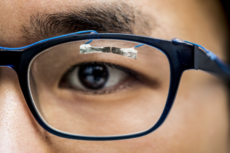
University of Washington graduate student, Jinyuan Zhang, demonstrates how wearable sensors can track eye movement. Dennis R. Wise/University of Washington

A February 13, 2018 news item on phys.org announces a technology that can transform tissue paper into a sensor,

University of Washington engineers have turned tissue paper – similar to toilet tissue – into a new kind of wearable sensor that can detect a pulse, a blink of an eye and other human movement. The sensor is light, flexible and inexpensive, with potential applications in health care, entertainment and robotics.

A February 12, 2018 University of Washington news release (also on EurekAlert) by Jackson Holtz, which originated the news item, provides more detail (Note: Links have been removed),

The technology, described in a paper published in January in the journal Advanced Materials Technologies, shows that by tearing tissue paper that’s loaded with nanocomposites and breaking the paper’s fibers, the paper acts as a sensor. It can detect a heartbeat, finger force, finger movement, eyeball movement and more, said Jae-Hyun Chung, a UW associate professor of mechanical engineering and senior author of the research.

Finger sensor

University of Washington graduate student, Jinyuan Zhang, demonstrates how a wearable sensor can measure finger pressure.Dennis R. Wise/University of Washington

“The major innovation is a disposable wearable sensor made with cheap tissue paper,” said Chung. “When we break the specimen, it will work as a sensor.”

These small, Band Aid-sized sensors could have a variety of applications in various fields. For example, monitoring a person’s gait or the movement of their eyes can be used to inspect brain function or a game player’s actions. The sensor could track how a special-needs child walks in a home test, sparing the child the need for hospital visits. Or the sensors could be used in occupational therapy for seniors.

“They can use these sensors and after one-time use, they can be thrown away,” said Chung.

In their research, the scientists used paper similar to toilet tissue. The paper – nothing more than conventional paper towels – is then doused with carbon nanotube-laced water. Carbon nanotubes are tiny materials that create electrical conductivity. Each piece of tissue paper has both horizontal and vertical fibers, so when the paper is torn, the direction of the tear informs the sensor of what’s happened. To trace eye movement, they’re attached to a person’s reading glasses.

For now, the work has been contained to a laboratory, and researchers are hoping to find a suitable commercial use. A provisional patent was filed in December 2017.

The study was funded partially by Samsung Research America through the Think Tank Team Award.

Foot sensor

University of Washington mechanical engineering undergraduate, Yared Shella, demonstrates how foot pressure is measured with a wearable sensor.Dennis R. Wise/University of Washington

Here’s a link to and a citation for the paper,

Fracture-Induced Mechanoelectrical Sensitivities of Paper-Based Nanocomposites by Jinyuan Zhang, Gil-Yong Lee, Chiew Cerwyn, Jinkyu Yang, Fabrice Fondjo, Jong-Hoon Kim, Minoru Taya, Dayong Gao, and Jae-Hyun Chung. Advanced Materials Technologies Vol. 3 Issue 1 DOI: 10.1002/admt.201700266 Version of Record online: 26 JAN 2018

© 2018 WILEY-VCH Verlag GmbH & Co. KGaA, Weinheim

This paper is behind a paywall.

How small can a carbon nanotube get before it stops being ‘electrical’?

Research, which began as an attempt to get reproducible electronics (?) measurements, yielded some unexpected results according ta January 3, 2018 news item on phys.org,

Carbon nanotubes bound for electronics not only need to be as clean as possible to maximize their utility in next-generation nanoscale devices, but contact effects may limit how small a nano device can be, according to researchers at the Energy Safety Research Institute (ESRI) at Swansea University [UK] in collaboration with researchers at Rice University [US].

ESRI Director Andrew Barron, also a professor at Rice University in the USA, and his team have figured out how to get nanotubes clean enough to obtain reproducible electronic measurements and in the process not only explained why the electrical properties of nanotubes have historically been so difficult to measure consistently, but have shown that there may be a limit to how “nano” future electronic devices can be using carbon nanotubes.

Swansea University Issued a January 3, 2018 press release (also on EurekAlert), which originated the news item, explains the work in more detail,

Like any normal wire, semiconducting nanotubes are progressively more resistant to current along their length. But conductivity measurements of nanotubes over the years have been anything but consistent. The ESRI team wanted to know why.

“We are interested in the creation of nanotube based conductors, and while people have been able to make wires their conduction has not met expectations. We were interested in determining the basic sconce behind the variability observed by other researchers.”

They discovered that hard-to-remove contaminants — leftover iron catalyst, carbon and water — could easily skew the results of conductivity tests. Burning them away, Barron said, creates new possibilities for carbon nanotubes in nanoscale electronics.

The new study appears in the American Chemical Society journal Nano Letters.

The researchers first made multiwalled carbon nanotubes between 40 and 200 nanometers in diameter and up to 30 microns long. They then either heated the nanotubes in a vacuum or bombarded them with argon ions to clean their surfaces.

They tested individual nanotubes the same way one would test any electrical conductor: By touching them with two probes to see how much current passes through the material from one tip to the other. In this case, their tungsten probes were attached to a scanning tunneling microscope.

In clean nanotubes, resistance got progressively stronger as the distance increased, as it should. But the results were skewed when the probes encountered surface contaminants, which increased the electric field strength at the tip. And when measurements were taken within 4 microns of each other, regions of depleted conductivity caused by contaminants overlapped, further scrambling the results.

“We think this is why there’s such inconsistency in the literature,” Barron said.

“If nanotubes are to be the next generation lightweight conductor, then consistent results, batch-to-batch, and sample-to-sample, is needed for devices such as motors and generators as well as power systems.”

Annealing the nanotubes in a vacuum above 200 degrees Celsius (392 degrees Fahrenheit) reduced surface contamination, but not enough to eliminate inconsistent results, they found. Argon ion bombardment also cleaned the tubes, but led to an increase in defects that degrade conductivity.

Ultimately they discovered vacuum annealing nanotubes at 500 degrees Celsius (932 Fahrenheit) reduced contamination enough to accurately measure resistance, they reported.

To now, Barron said, engineers who use nanotube fibers or films in devices modify the material through doping or other means to get the conductive properties they require. But if the source nanotubes are sufficiently decontaminated, they should be able to get the right conductivity by simply putting their contacts in the right spot.

“A key result of our work was that if contacts on a nanotube are less than 1 micron apart, the electronic properties of the nanotube changes from conductor to semiconductor, due to the presence of overlapping depletion zones” said Barron, “this has a potential limiting factor on the size of nanotube based electronic devices – this would limit the application of Moore’s law to nanotube devices.”

Chris Barnett of Swansea is lead author of the paper. Co-authors are Cathren Gowenlock and Kathryn Welsby, and Rice alumnus Alvin Orbaek White of Swansea. Barron is the Sêr Cymru Chair of Low Carbon Energy and Environment at Swansea and the Charles W. Duncan Jr.–Welch Professor of Chemistry and a professor of materials science and nanoengineering at Rice.

The Welsh Government Sêr Cymru National Research Network in Advanced Engineering and Materials, the Sêr Cymru Chair Program, the Office of Naval Research and the Robert A. Welch Foundation supported the research.

Rice University has published a January 4, 2018 Rice University news release (also on EurekAlert), which is almost (95%) identical to the press release from Swansea. That’s a bit unusual as collaborating institutions usually like to focus on their unique contributions to the research, hence, multiple news/press releases.

Dexter Johnson, in a January 11, 2018 post on his Nanoclast blog (on the IEEE [Institute of Electrical and Electronics Engineers] website,  adds a detail or two while writing in an accessible style.

Here’s a link to and a citation for the paper,

Spatial and Contamination-Dependent Electrical Properties of Carbon Nanotubes by Chris J. Barnett, Cathren E. Gowenlock, Kathryn Welsby, Alvin Orbaek White, and Andrew R. Barron. Nano Lett., Article ASAP DOI: 10.1021/acs.nanolett.7b03390 Publication Date (Web): December 19, 2017

Copyright © 2017 American Chemical Society

This paper is behind a paywall.

Carbon nanotubes for enhanced wheat growth?

It’s been a long time (Oct. 22, 2009 posting; scroll down about 20% of the way) since I’ve written about carbon nanotubes and their possible use in agriculture but now a December 6, 2017 news item on ScienceDaily raises the topic again,

The introduction of purified carbon nanotubes appears to have a beneficial effect on the early growth of wheatgrass, according to Rice University scientists. But in the presence of contaminants, those same nanotubes could do great harm.

The Rice lab of chemist Andrew Barron grew wheatgrass in a hydroponic garden to test the potential toxicity of nanoparticles on the plant. To their surprise, they found one type of particle dispersed in water helped the plant grow bigger and faster.

They suspect the results spring from nanotubes’ natural hydrophobic (water-avoiding) nature that in one experiment apparently facilitated the plants’ enhanced uptake of water.

The research appears in the Royal Society of Chemistry journal Environmental Science: Nano.

A December 6, 2017 Rice University news release (also on EurekAlert), which originated the news item, expands on the theme,

The lab mounted the small-scale study with the knowledge that the industrial production of nanotubes will inevitably lead to their wider dispersal in the environment. The study cited rapid growth in the market for nanoparticles in drugs, cosmetic, fabrics, water filters and military weapons, with thousands of tons produced annually.

Despite their widespread use, Barron said few researchers have looked at the impact of environmental nanoparticles — whether natural or man-made — on plant growth.

The researchers planted wheatgrass seeds in multiple replicates in cotton wool and fed them with dispersions that contained raw single-walled or multi-walled nanotubes, purified single-walled nanotubes or iron oxide nanoparticles that mimicked leftover catalyst often attached to nanotubes. The solutions were either water or tetrahydrofuran (THF), an industrial solvent. Some of the seeds were fed pure water or THF as a control.

Rice University researchers tested the effects of carbon nanotubes on the growth of wheatgrass. While some showed no effect, purified single-walled nanotubes in water (5) enhanced the plants' growth, while the same nanotubes in a solvent (6) retarded their development. The photos at left show the plants after four days and at right after eight days, with odd-numbered plants growing in water and evens in a solvent. Numbers 1 and 2 are controls without nanotubes; 3-4 contain raw single-walled tubes; 5-6 purified single-walled tubes; 7-8 raw multi-walled tubes; 9-10 low-concentration iron-oxide nanoparticles and 11-12 high-concentration iron-oxide nanoparticles.

Rice University researchers tested the effects of carbon nanotubes on the growth of wheatgrass. While some showed no effect, purified single-walled nanotubes in water (5) enhanced the plants’ growth, while the same nanotubes in a solvent (6) retarded their development. The photos at left show the plants after four days and at right after eight days, with odd-numbered plants growing in water and evens in a solvent. Numbers 1 and 2 are controls without nanotubes; 3-4 contain raw single-walled tubes; 5-6 purified single-walled tubes; 7-8 raw multi-walled tubes; 9-10 low-concentration iron-oxide nanoparticles and 11-12 high-concentration iron-oxide nanoparticles. Click on the image for a larger version. Photos by Seung Mook Lee

After eight days, the plantings showed that purified single-walled nanotubes in water enhanced the germination rate and shoot growth of wheatgrass, which grew an average of 13 percent larger than plants in plain water. Raw single- and multi-walled nanotubes and particles in either solution had little effect on the plants’ growth, they found.

However, purified single-walled nanotubes in THF retarded plant development by 45 percent compared to single-walled nanotubes in water, suggesting the nanotubes act as a carrier for the toxic substance.

The concern, Barron said, is that if single-walled nanotubes combine with organic pollutants like pesticides, industrial chemicals or solvents in the environment, they may concentrate and immobilize the toxins and enhance their uptake by plants.

Nothing seen in the limited study indicated whether carbon nanotubes in the environment, and potentially in plants, will rise up the food chain and be harmful to humans, he said.

On the other hand, the researchers said it may be worth looking at whether hydrophobic substrates that mimic the positive effects observed in single-walled nanotubes could be used for high-efficiency channeling of water to seeds.

“Our work confirms the importance of thinking of nanomaterials as part of a system rather in isolation,” Barron said. “It is the combination with other compounds that is important to understand.”

Seung Mook Lee, a former visiting student research assistant from Memorial High School in Houston and now an undergraduate student at the University of California, Berkeley, is lead author of the paper. Co-authors are Rice research scientist Pavan Raja and graduate student Gibran Esquenazi. Barron is the Charles W. Duncan Jr.–Welch Professor of Chemistry and a professor of materials science and nanoengineering at Rice and the Sêr Cymru Chair of Low Carbon Energy and Environment at Swansea University, Wales (UK).

The Welsh Government Sêr Cymru Program and the Robert A. Welch Foundation supported the research.

Here’s a link to and a citation for the paper,

Effect of raw and purified carbon nanotubes and iron oxide nanoparticles on the growth of wheatgrass prepared from the cotyledons of common wheat (triticum aestivum) by Seung Mook Lee, Pavan M. V. Raja, Gibran L. Esquenazi, and Andrew R. Barron. Environ. Sci.: Nano, 2018, Advance Article DOI: 10.1039/C7EN00680B First published on 09 Nov 2017

This paper appears to be behind a paywall.

Felted carbon nanotubes

Parachute (sculpted felt lantern). Artist and artisan felter: Chantal Cardinal. Studio: FELT à la main with LOVE

Scientists from Kiel University (Christian-Albrechts-Universität zu Kiel; Germany) and the University of Trento (Italy) claim to have developed a new method for integrating carbon nanotubes (CNTs) into new materials in a technique they describe as similar to felting according to a November 21, 2017 news item on Nanowerk,

Extremely lightweight, electrically highly conductive, and more stable than steel: due to their unique properties, carbon nanotubes would be ideal for numerous applications, from ultra-lightweight batteries to high-performance plastics, right through to medical implants. However, to date it has been difficult for science and industry to transfer the extraordinary characteristics at the nanoscale into a functional industrial application. The carbon nanotubes either cannot be combined adequately with other materials, or if they can be combined, they then lose their beneficial properties.

Scientists from the Functional Nanomaterials working group at Kiel University (CAU) and the University of Trento have now developed an alternative method, with which the tiny tubes can be combined with other materials, so that they retain their characteristic properties. As such, they “felt” the thread-like tubes into a stable 3D network that is able to withstand extreme forces.

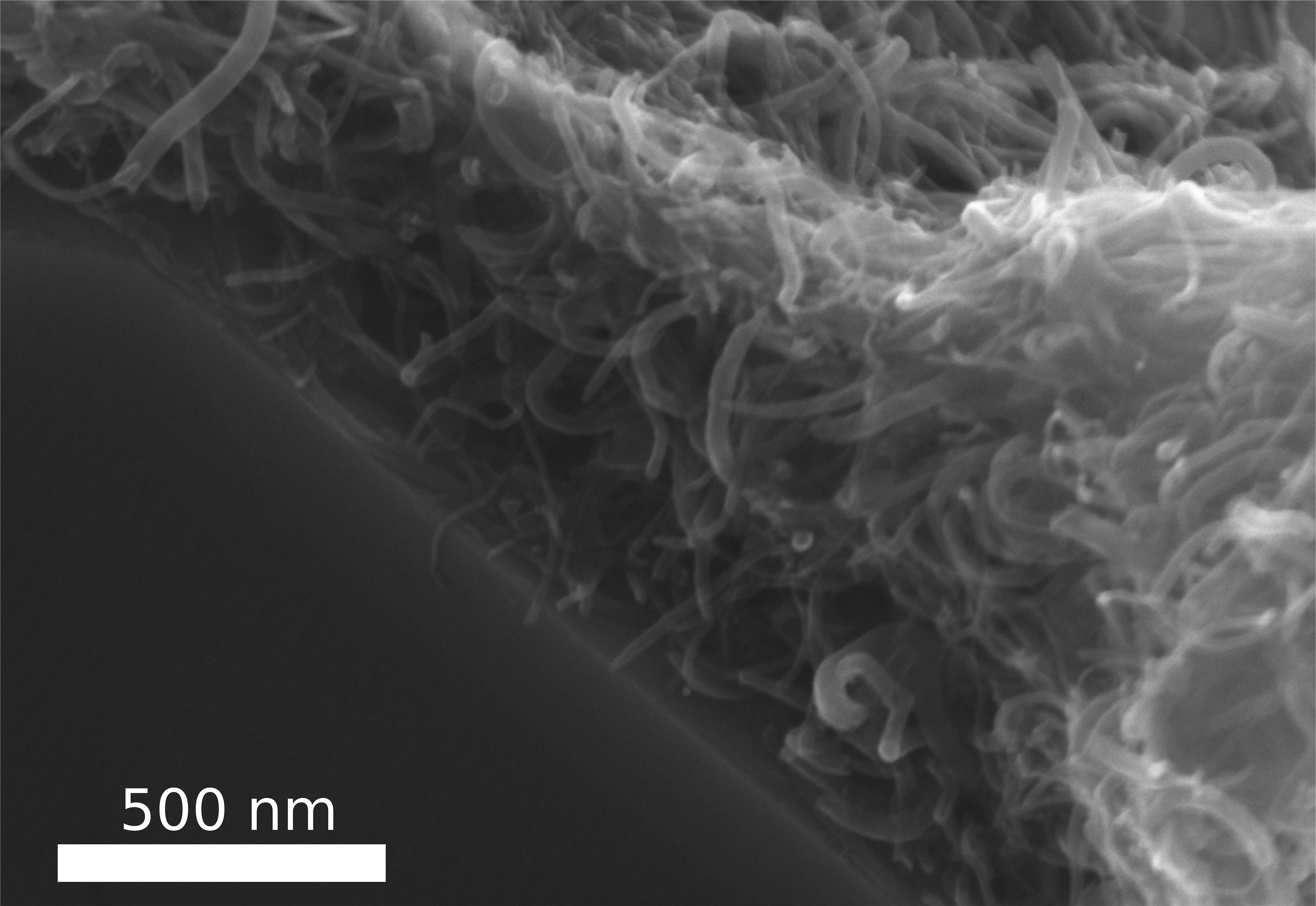
In contrast to the ‘felted’ image which opened this posting, here’s an image of the ‘felted’ carbon nanotubes,

In this new process, the tiny, thread-like carbon nanotubes (CNTs) arrange themselves – almost like felting – to form a stable, tear-resistant layer. Photo/Copyright: Fabian Schütt Courtesy: Kiel University

A November 21, 2017 Kiel University press release (also on EurekAlert), which originated the news item, expands on the theme and adds another analogy,

Industry and science have been intensively researching the significantly less than one hundred nanometre wide carbon tubes (carbon nanotubes, CNTs), in order to make use of the extraordinary properties of rolled graphene. Yet much still remains just theory. “Although carbon nanotubes are flexible like fibre strands, they are also very sensitive to changes,” explained Professor Rainer Adelung, head of the Functional Nanomaterials working group at the CAU. “With previous attempts to chemically connect them with other materials, their molecular structure also changed. This, however, made their properties deteriorate – mostly drastically.”

In contrast, the approach of the research team from Kiel and Trento is based on a simple wet chemical infiltration process. The CNTs are mixed with water and dripped into an extremely porous ceramic material made of zinc oxide, which absorbs the liquid like a sponge. The dripped thread-like CNTs attach themselves to the ceramic scaffolding, and automatically form a stable layer together, similar to a felt. The ceramic scaffolding is coated with nanotubes, so to speak. This has fascinating effects, both for the scaffolding as well as for the coating of nanotubes.

On the one hand, the stability of the ceramic scaffold increases so massively that it can bear 100,000 times its own weight. “With the CNT coating, the ceramic material can hold around 7.5kg, and without it just 50g – as if we had fitted it with a close-fitting pullover made of carbon nanotubes, which provide mechanical support,” summarised first author Fabian Schütt. “The pressure on the material is absorbed by the tensile strength of the CNT felt. Compressive forces are transformed into tensile forces.”

The principle behind this is comparable with bamboo buildings [emphasis mine], such as those widespread in Asia. Here, bamboo stems are bound so tightly with a simple rope that the lightweight material can form extremely stable scaffolding, and even entire buildings. “We do the same at the nano-scale with the CNT threads, which wrap themselves around the ceramic material – only much, much smaller,” said Helge Krüger, co-author of the publication.

The materials scientists were able to demonstrate another major advantage of their process. In a second step, they dissolved the ceramic scaffolding by using a chemical etching process. All that remains is a fine 3D network of tubes, each of which consists of a layer of tiny CNT tubes. In this way, the researchers were able to greatly increase the felt surface, and thus create more opportunities for reactions. “We basically pack the surface of an entire beach volleyball field into a one centimetre cube,” explained Schütt. The huge hollow spaces inside the three-dimensional structure can then be filled with a polymer. As such, CNTs can be connected mechanically with plastics, without their molecular structure – and thus their properties – being modified. “We can specifically arrange the CNTs and manufacture an electrically conductive composite material. To do so only requires a fraction of the usual quantity of CNTs, in order to achieve the same conductivity,” said Schütt.

Applications for use range from battery and filter technology as a filling material for conductive plastics, implants for regenerative medicine, right through to sensors and electronic components at the nano-scale. The good electrical conductivity of the tear-resistant material could in future also be interesting for flexible electronics applications, in functional clothing or in the field of medical technology, for example. “Creating a plastic which, for example, stimulates bone or heart cells to grow is conceivable,” said Adelung. Due to its simplicity, the scientists agree that the process could also be transferred to network structures made of other nanomaterials – which will further expand the range of possible applications.

So, we have ‘felting’ and bamboo buildings. I can appreciate the temptation to use multiple analogies especially since I’ve given into it, on occasion.  But, it’s never considered good style, not even when I do it.

Getting back to the work at hand, here’s a link to and a citation for the paper,

Hierarchical self-entangled carbon nanotube tube networks by Fabian Schütt, Stefano Signetti, Helge Krüger, Sarah Röder, Daria Smazna, Sören Kaps, Stanislav N. Gorb, Yogendra Kumar Mishra, Nicola M. Pugno, & Rainer Adelung. Nature Communications 8, Article number: 1215 (2017) doi:10.1038/s41467-017-01324-7 Published online: 31 October 2017

This is an open access paper.

One final comment, I notice that one of the authors is Nicola Pugno who was last mentioned here in an August 30, 2017 posting titled: Making spider silk stronger by feeding graphene and carbon nanotubes to spiders.

Calligraphy ink and cancer treatment

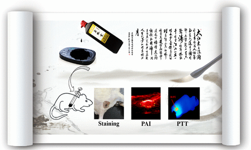
Courtesy of ACS Omega and the researchers

Nice illustration! I wish I could credit the artist. For anyone who needs a little text to make sense of it, there’s a Sept. 27, 2017 news item on Nanowerk (Note: A link has been removed),

For hundreds of years, Chinese calligraphers have used a plant-based ink to create beautiful messages and art. Now, one group reports in ACS Omega (“New Application of Old Material: Chinese Traditional Ink for Photothermal Therapy of Metastatic Lymph Nodes”) that this ink could noninvasively and effectively treat cancer cells that spread, or metastasize, to lymph nodes.

A Sept. 27, 2017 American Chemical Society (ACS) news release, which originated the news item, reveals more about the research,

As cancer cells leave a tumor, they frequently make their way to lymph nodes, which are part of the immune system. In this case, the main treatment option is surgery, but this can result in complications. Photothermal therapy (PTT) is an emerging noninvasive treatment option in which nanomaterials are injected and accumulate in cancer cells. A laser heats up the nanomaterials, and this heat kills the cells. Many of these nanomaterials are expensive, difficult-to-make and toxic. However, a traditional Chinese ink called Hu-Kaiwen ink (Hu-ink) has similar properties to the nanomaterials used in PTT. For example, they are the same color, and are both carbon-based and stable in water. So Wuli Yang and colleagues wanted to see if Hu-ink could be a good alternative material for PTT.

The researchers analyzed Hu-ink and found that it consists of nanoparticles and thin layers of carbon. When Hu-ink was heated with a laser, its temperature rose by 131 degrees Fahrenheit, much higher than current nanomaterials. Under PPT conditions, the Hu-ink killed cancer cells in a laboratory dish, but under normal conditions, the ink was non-toxic. This was also the scenario observed in mice with tumors. The researchers also noted that Hu-ink could act as a probe to locate tumors and metastases because it absorbs near-infrared light, which goes through skin.

Being a little curious about Hu-ink’s similarity to nanomaterial, I looked for more detail in the the paper (Note: Links have been removed), From the: Introduction,

Photothermal therapy (PTT) is an emerging tumor treatment strategy, which utilizes hyperthermia generated from absorbed near-infrared (NIR) light energy by photoabsorbing agents to kill tumor cells.(7-13) Different from chemotherapy, surgical treatment, and radiotherapy, PTT is noninvasive and more efficient.(7, 14, 15) In the past decade, PTT with diverse nanomaterials to eliminate cancer metastases lymph nodes has attracted extensive attention by several groups, including our group.(3, 16-20) For instance, Liu and his co-workers developed a treatment method based on PEGylated single-walled carbon nanotubes for PTT of tumor sentinel lymph nodes and achieved remarkably improved treatment effect in an animal tumor model.(21) To meet the clinical practice, the potential metastasis of deeper lymph nodes was further ablated in our previous work, using magnetic graphene oxide as a theranostic agent.(22) However, preparation of these artificial nanomaterials usually requires high cost, complicated synthetic process, and unavoidably toxic catalyst or chemicals,(23, 24) which impede their future clinical application. For the clinical application, exploring an environment-friendly material with simple preparation procedure, good biocompatibility, and excellent therapeutic efficiency is still highly desired. [emphases mine]

From the: Preparation and Characterization of Hu-Ink

To obtain an applicable sample, the condensed Hu-ink was first diluted into aqueous dispersion with a lower concentration. The obtained Hu-ink dispersion without any further treatment was black in color and stable in physiological environment, including water, phosphate-buffered saline (PBS), and Roswell Park Memorial Institute (RPMI) 1640; furthermore, no aggregation was observed even after keeping undisturbed for 3 days (Figure 2a). The nanoscaled morphology of Hu-ink was examined by transmission electron microscopy (TEM) (Figure 2b), which demonstrates that Hu-ink mainly exist in the form of small aggregates. These small aggregates consist of a few nanoparticles with diameter of about 20–50 nm. Dynamic light scattering (DLS) measurement (Figure 2c) further shows that Hu-ink aqueous dispersion possesses a hydrodynamic diameter of about 186 nm (polydispersity index: 0.18), which was a crucial prerequisite for biomedical applications.(29) In the X-ray diffraction (XRD) pattern, no other characteristic peaks are found except carbon peak (Figure S1, Supporting Information), which confirms that the main component of Hu-ink is carbon.(25) Raman spectroscopy was a common tool to characterize graphene-related materials.(30) D band (∼1300 cm–1, corresponding to the defects) and G band (∼1600 cm–1, related to the sp2 carbon sites) peaks could be observed in Figure 2d with the ratio ID/IG = 0.96, which confirms the existence of graphene sheetlike structure in Hu-ink.(31) The UV–vis–NIR spectra (Figure 2e) also revealed that Hu-ink has high absorption in the NIR region around 650–900 nm, in which hemoglobin and water, the major absorbers of biological tissue, have their lowest absorption coefficient.(32) The high NIR absorption capability of Hu-ink encouraged us to investigate its photothermal properties.(33-35) Hu-ink dispersions with different concentrations were irradiated under an 808 nm laser (the commercial and widely used wavelength in photothermal therapy).(8-13) [emphases mine]

Curiosity satisfied! For those who’d like to investigate even further, here’s a link to and a citation for the paper,

New Application of Old Material: Chinese Traditional Ink for Photothermal Therapy of Metastatic Lymph Nodes by Sheng Wang, Yongbin Cao, Qin Zhang, Haibao Peng, Lei Liang, Qingguo Li, Shun Shen, Aimaier Tuerdi, Ye Xu, Sanjun Cai, and Wuli Yang. ACS Omega, 2017, 2 (8), pp 5170–5178 DOI: 10.1021/acsomega.7b00993 Publication Date (Web): August 30, 2017

Copyright © 2017 American Chemical Society

This paper appears to be open access.

Liquid circuitry, shape-shifting fluids and more

I’d have to see it to believe it but researchers at the US Dept. of Energy (DOE) Lawrence Berkeley National Laboratory (LBNL) have developed a new kind of ‘bijel’ which would allow for some pretty nifty robotics. From a Sept. 25, 2017 news item on ScienceDaily,

A new two-dimensional film, made of polymers and nanoparticles and developed by researchers at the Department of Energy’s Lawrence Berkeley National Laboratory (Berkeley Lab), can direct two different non-mixing liquids into a variety of exotic architectures. This finding could lead to soft robotics, liquid circuitry, shape-shifting fluids, and a host of new materials that use soft, rather than solid, substances.

The study, reported today in the journal Nature Nanotechnology, presents the newest entry in a class of substances known as bicontinuous jammed emulsion gels, or bijels, which hold promise as a malleable liquid that can support catalytic reactions, electrical conductivity, and energy conversion.

A Sept. 25, 2017 LBNL news release (also on EurekAlert), which originated the news item, expands on the theme,

Bijels are typically made of immiscible, or non-mixing, liquids. People who shake their bottle of vinaigrette before pouring the dressing on their salad are familiar with such liquids. As soon as the shaking stops, the liquids start to separate again, with the lower density liquid – often oil – rising to the top.

Trapping, or jamming, particles where these immiscible liquids meet can prevent the liquids from completely separating, stabilizing the substance into a bijel. What makes bijels remarkable is that, rather than just making the spherical droplets that we normally see when we try to mix oil and water, the particles at the interface shape the liquids into complex networks of interconnected fluid channels.

Bijels are notoriously difficult to make, however, involving exact temperatures at precisely timed stages. In addition, the liquid channels are normally more than 5 micrometers across, making them too large to be useful in energy conversion and catalysis.

“Bijels have long been of interest as next-generation materials for energy applications and chemical synthesis,” said study lead author Caili Huang. “The problem has been making enough of them, and with features of the right size. In this work, we crack that problem.”

Huang started the work as a graduate student with Thomas Russell, the study’s principal investigator, at Berkeley Lab’s Materials Sciences Division, and he continued the project as a postdoctoral researcher at DOE’s Oak Ridge National Laboratory.

Creating a new bijel recipe

The method described in this new study simplifies the bijel process by first using specially coated particles about 10-20 nanometers in diameter. The smaller-sized particles line the liquid interfaces much more quickly than the ones used in traditional bijels, making the smaller channels that are highly valued for applications.

Illustration shows key stages of bijel formation. Clockwise from top left, two non-mixing liquids are shown. Ligands (shown in yellow) with amine groups are dispersed throughout the oil or solvent, and nanoparticles coated with carboxylic acids (shown as blue dots) are scattered in the water. With vigorous shaking, the nanoparticles and ligands form a “supersoap” that gets trapped at the interface of the two liquids. The bottom panel is a magnified view of the jammed nanoparticle supersoap. (Credit: Caili Huang/ORNL)

“We’ve basically taken liquids like oil and water and given them a structure, and it’s a structure that can be changed,” said Russell, a visiting faculty scientist at Berkeley Lab. “If the nanoparticles are responsive to electrical, magnetic, or mechanical stimuli, the bijels can become reconfigurable and re-shaped on demand by an external field.”

The researchers were able to prepare new bijels from a variety of common organic, water-insoluble solvents, such as toluene, that had ligands dissolved in it, and deionized water, which contained the nanoparticles. To ensure thorough mixing of the liquids, they subjected the emulsion to a vortex spinning at 3,200 revolutions per minute.

“This extreme shaking creates a whole bunch of new places where these particles and polymers can meet each other,” said study co-author Joe Forth, a postdoctoral fellow at Berkeley Lab’s Materials Sciences Division. “You’re synthesizing a lot of this material, which is in effect a thin, 2-D coating of the liquid surfaces in the system.”

The liquids remained a bijel even after one week, a sign of the system’s stability.

Russell, who is also a professor of polymer science and engineering at the University of Massachusetts-Amherst, added that these shape-shifting characteristics would be valuable in microreactors, microfluidic devices, and soft actuators.

Nanoparticle supersoap

Nanoparticles had not been seriously considered in bijels before because their small size made them hard to trap in the liquid interface. To resolve that problem, the researchers coated nano-sized particles with carboxylic acids and put them in water. They then took polymers with an added amine group – a derivative of ammonia – and dissolved them in the toluene.

At left is a vial of bijel stabilized with nanoparticle surfactants. On the right is the same vial after a week of inversion, showing that the nanoparticle kept the liquids from moving. (Credit: Caili Huang/ORNL)

This configuration took advantage of the amine group’s affinity to water, a characteristic that is comparable to surfactants, like soap. Their nanoparticle “supersoap” was designed so that the nanoparticles join ligands, forming an octopus-like shape with a polar head and nonpolar legs that get jammed at the interface, the researchers said.

“Bijels are really a new material, and also excitingly weird in that they are kinetically arrested in these unusual configurations,” said study co-author Brett Helms, a staff scientist at Berkeley Lab’s Molecular Foundry. “The discovery that you can make these bijels with simple ingredients is a surprise. We all have access to oils and water and nanocrystals, allowing broad tunability in bijel properties. This platform also allows us to experiment with new ways to control their shape and function since they are both responsive and reconfigurable.”

The nanoparticles were made of silica, but the researchers noted that in previous studies they used graphene and carbon nanotubes to form nanoparticle surfactants.

“The key is that the nanoparticles can be made of many materials,” said Russell.  “The most important thing is what’s on the surface.”

This is an animation of the bijel

3-D rendering of the nanoparticle bijel taken by confocal microscope. (Credit: Caili Huang/ORNL [Oak Ridge National Laboratory] and Joe Forth/Berkeley Lab)

Here’s a link to and a citation for the paper,

Bicontinuous structured liquids with sub-micrometre domains using nanoparticle surfactants by Caili Huang, Joe Forth, Weiyu Wang, Kunlun Hong, Gregory S. Smith, Brett A. Helms & Thomas P. Russell. Nature Nanotechnology (2017) doi:10.1038/nnano.2017.182 25 September 2017

This paper is behind a paywall.

Vampire nanogenerators: 2017

Researchers have been working on ways to harvest energy from bloodstreams. I last wrote about this type of research in an April 3, 2009 posting about ‘vampire batteries ‘(for use in pacemakers). The latest work according to a Sept. 8, 2017 news item on Nanowerk comes from China,

Men build dams and huge turbines to turn the energy of waterfalls and tides into electricity. To produce hydropower on a much smaller scale, Chinese scientists have now developed a lightweight power generator based on carbon nanotube fibers suitable to convert even the energy of flowing blood in blood vessels into electricity. They describe their innovation in the journal Angewandte Chemie (“A One-Dimensional Fluidic Nanogenerator with a High Power Conversion Efficiency”)

A Sept. 8, 2017 Wiley Publishing news release (also on EurekAlert), which originated the news item, expands on the theme,

For thousands of years, people have used the energy of flowing or falling water for their purposes, first to power mechanical engines such as watermills, then to generate electricity by exploiting height differences in the landscape or sea tides. Using naturally flowing water as a sustainable power source has the advantage that there are (almost) no dependencies on weather or daylight. Even flexible, minute power generators that make use of the flow of biological fluids are conceivable. How such a system could work is explained by a research team from Fudan University in Shanghai, China. Huisheng Peng and his co-workers have developed a fiber with a thickness of less than a millimeter that generates electrical power when surrounded by flowing saline solution—in a thin tube or even in a blood vessel.

The construction principle of the fiber is quite simple. An ordered array of carbon nanotubes was continuously wrapped around a polymeric core. Carbon nanotubes are well known to be electroactive and mechanically stable; they can be spun and aligned in sheets. In the as-prepared electroactive threads, the carbon nanotube sheets coated the fiber core with a thickness of less than half a micron. For power generation, the thread or “fiber-shaped fluidic nanogenerator” (FFNG), as the authors call it, was connected to electrodes and immersed into flowing water or simply repeatedly dipped into a saline solution. “The electricity was derived from the relative movement between the FFNG and the solution,” the scientists explained. According to the theory, an electrical double layer is created around the fiber, and then the flowing solution distorts the symmetrical charge distribution, generating an electricity gradient along the long axis.

The power output efficiency of this system was high. Compared with other types of miniature energy-harvesting devices, the FFNG was reported to show a superior power conversion efficiency of more than 20%. Other advantages are elasticity, tunability, lightweight, and one-dimensionality, thus offering prospects of exciting technological applications. The FFNG can be made stretchable just by spinning the sheets around an elastic fiber substrate. If woven into fabrics, wearable electronics become thus a very interesting option for FFNG application. Another exciting application is the harvesting of electrical energy from the bloodstream for medical applications. First tests with frog nerves proved to be successful.

Here’s a link to and a citation for the paper,

A One-Dimensional Fluidic Nanogenerator with a High Power Conversion Efficiency by Yifan Xu, Dr. Peining Chen, Jing Zhang, Songlin Xie, Dr. Fang Wan, Jue Deng, Dr. Xunliang Cheng, Yajie Hu, Meng Liao, Dr. Bingjie Wang, Dr. Xuemei Sun, and Prof. Dr. Huisheng Peng. Angewandte Chemie International Edition DOI: 10.1002/anie.201706620 Version of Record online: 7 SEP 2017

© 2017 Wiley-VCH Verlag GmbH & Co. KGaA, Weinheim

This paper is behind a paywall.