Tag Archives: cobalt

Rijksmuseum’s ‘live’ restoration of Rembrandt’s masterpiece: The Nightwatch: is it or isn’t it like watching paint dry?

Somewhere in my travels, I saw ‘like watching paint dry’ as a description for the experience of watching researchers examining Rembrandt’s Night Watch. Granted it’s probably not that exciting but there has to be something to be said for being present while experts undertake an extraordinary art restoration effort. The Night Watch is not only a masterpiece—it’s huge.

This posting was written closer to the time the ‘live’ restoration first began. I have an update at the end of this posting.

A July 8, 2019 news item on the British Broadcasting Corporation’s (BBC) news online sketches in some details,

The masterpiece, created in 1642, has been placed inside a specially designed glass chamber so that it can still be viewed while being restored.

Enthusiasts can follow the latest on the restoration work online.

The celebrated painting was last restored more than 40 years ago after it was slashed with a knife.

The Night Watch is considered Rembrandt’s most ambitious work. It was commissioned by the mayor and leader of the civic guard of Amsterdam, Frans Banninck Cocq, who wanted a group portrait of his militia company.

The painting is nearly 4m tall and 4.5m wide (12.5 x 15 ft) and weighs 337kg (743lb) [emphasis mine]. As well as being famous for its size, the painting is acclaimed for its use of dramatic lighting and movement.

But experts at Amsterdam’s Rijksmuseum are concerned that aspects of the masterpiece are changing, pointing as an example to the blanching of the figure of a small dog. The museum said the multi-million euro research and restoration project under way would help staff gain a better understanding of the painting’s condition.

An October 16, 2018 Rijksmuseum press release announced the restoration work months prior to the start (Note: Some of the information is repetitive;),

Before the restoration begins, The Night Watch will be the centrepiece of the Rijksmuseum’s display of their entire collection of more than 400 works by Rembrandt in an exhibition to mark the 350th anniversary of the artist’s death opening on 15 February 2019.

Commissioned in 1642 by the mayor and leader of the civic guard of Amsterdam, Frans Banninck Cocq, to create a group portrait of his shooting company, The Night Watch is recognised as one of the most important works of art in the world today and hangs in the specially designed “Gallery of Honour” at the Rijksmuseum. It is more than 40 years since The Night Watch underwent its last major restoration, following an attack on the painting in 1975.

The Night Watch will be encased in a state-of-the-art clear glass chamber designed by the French architect Jean Michel Wilmotte. This will ensure that the painting can remain on display for museum visitors. A digital platform will allow viewers from all over the world to follow the entire process online [emphasis mine] continuing the Rijksmuseum innovation in the digital field.

Taco Dibbits, General Director Rijksmuseum: The Night Watch is one of the most famous paintings in the world. It belongs to us all, and that is why we have decided to conduct the restoration within the museum itself – and everyone, wherever they are, will be able to follow the process online.

The Rijksmuseum continually monitors the condition of The Night Watch, and it has been discovered that changes are occurring, such as the blanching [emphasis mine] on the dog figure at the lower right of the painting. To gain a better understanding of its condition as a whole, the decision has been taken to conduct a thorough examination. This detailed study is necessary to determine the best treatment plan, and will involve imaging techniques, high-resolution photography and highly advanced computer analysis. Using these and other methods, we will be able to form a very detailed picture of the painting – not only of the painted surface, but of each and every layer, from varnish to canvas.

A great deal of experience has been gained in the Rijksmuseum relating to the restoration of Rembrandt’s paintings. Last year saw the completion of the restoration of Rembrandt’s spectacular portraits of Marten Soolmans and Oopjen Coppit. The research team working on The Night Watch is made up of researchers, conservators and restorers from the Rijksmuseum, which will conduct this research in close collaboration with museums and universities in the Netherlands and abroad.

The Night Watch

The group portrait of the officers and other members of the militia company of District II, under the command of Captain Frans Banninck Cocq and Lieutenant Willem van Ruytenburch, now known as The Night Watch, is Rembrandt’s most ambitious painting. This 1642 commission by members of Amsterdam’s civic guard is Rembrandt’s first and only painting of a militia group. It is celebrated particularly for its bold and energetic composition, with the musketeers being depicted ‘in motion’, rather than in static portrait poses. The Night Watch belongs to the city of Amsterdam, and it been the highlight of the Rijksmuseum collection since 1808. The architect of the Rijksmuseum building Pierre Cuypers (1827-1921) even created a dedicated gallery of honour for The Night Watch, and it is now admired there by more than 2.2 million people annually.

2019, The Year of Rembrandt

The Year of Rembrandt, 2019, marks the 350th anniversary of the artist’s death with two major exhibitions honouring the great master painter. All the Rembrandts of the Rijksmuseum (15 February to 10 June 2019) will bring together the Rijksmuseum’s entire collection of Rembrandt’s paintings, drawings and prints, for the first time in history. The second exhibition, Rembrandt-Velázquez (11 October 2019 to 19 January 2020), will put the master in international context by placing 17th-century Spanish and Dutch masterpieces in dialogue with each another.

First, the restoration work is not being livestreamed; the digital platform Operation Night Watch is a collection of resources, which are being updated constantly, For example, the first scan was placed online in Operation Night Watch on July 16, 2019.

Second, ‘blanching’ reminded me of a June 22, 2017 posting where I featured research into why masterpieces were turning into soap, (Note: The second paragraph should be indented to indicated that it’s an excerpt fro the news release. Unfortunately, the folks at WordPress appear to have removed the tools that would allow me to do that and more),

This piece of research has made a winding trek through the online science world. First it was featured in an April 20, 2017 American Chemical Society news release on EurekAlert

A good art dealer can really clean up in today’s market, but not when some weird chemistry wreaks havoc on masterpieces. Art conservators started to notice microscopic pockmarks forming on the surfaces of treasured oil paintings that cause the images to look hazy. It turns out the marks are eruptions of paint caused, weirdly, by soap that forms via chemical reactions. Since you have no time to watch paint dry, we explain how paintings from Rembrandts to O’Keefes are threatened by their own compositions — and we don’t mean the imagery.

….

Getting back to the Night Watch, there’s a July 8, 2019 Rijksmuseum press release which provides some technical details,

On 8 July 2019 the Rijksmuseum starts Operation Night Watch. It will be the biggest and most wide-ranging research and conservation project in the history of Rembrandt’s masterpiece. The goal of Operation Night Watch is the long-term preservation of the painting. The entire operation will take place in a specially designed glass chamber so the visiting public can watch.

Never before has such a wide-ranging and thorough investigation been made of the condition of The Night Watch. The latest and most advanced research techniques will be used, ranging from digital imaging and scientific and technical research, to computer science and artificial intelligence. The research will lead to a better understanding of the painting’s original appearance and current state, and provide insight into the many changes that The Night Watch has undergone over the course of the last four centuries. The outcome of the research will be a treatment plan that will form the basis for the restoration of the painting.

Operation Night Watch can also be followed online from 8 July 2019 at rijksmuseum.nl/nightwatch

From art historical research to artificial intelligence

Operation Night Watch will look at questions regarding the original commission, Rembrandt’s materials and painting technique, the impact of previous treatments and later interventions, as well as the ageing, degradation and future of the painting. This will involve the newest and most advanced research methods and technologies, including art historical and archival research, scientific and technical research, computer science and artificial intelligence.

During the research phase The Night Watch will be unframed and placed on a specially designed easel. Two platform lifts will make it possible to study the entire canvas, which measures 379.5 cm in height and 454.5 cm in width.

Advanced imaging techniques

Researchers will make use of high resolution photography, as well as a variety of advanced imaging techniques, such as macro X-ray fluorescence scanning (macro-XRF) and hyperspectral imaging, also called infrared reflectance imaging spectroscopy (RIS), to accurately determine the condition of the painting.

56 macro-XRF scans

The Night Watch will be scanned millimetre by millimetre using a macro X-ray fluorescence scanner (macro-XRF scanner). This instrument uses X-rays to analyse the different chemical elements in the paint, such as calcium, iron, potassium and cobalt. From the resulting distribution maps of the various chemical elements in the paint it is possible to determine which pigments were used. The macro-XRF scans can also reveal underlying changes in the composition, offering insights into Rembrandt’s painting process. To scan the entire surface of the The Night Watch it will be necesary to make 56 scans, each one of which will take 24 hours.

12,500 high-resolution photographs

A total of some 12,500 photographs will be taken at extremely high resolution, from 180 to 5 micrometres, or a thousandth of a millimetre. Never before has such a large painting been photographed at such high resolution. In this way it will be possible to see details such as pigment particles that normally would be invisible to the naked eye. The cameras and lamps will be attached to a dynamic imaging frame designed specifically for this purpose.

Glass chamber

Operation Night Watch is for everyone to follow and will take place in full view of the visiting public in an ultra-transparent glass chamber designed by the French architect Jean Michel Wilmotte.

Research team

The Rijksmuseum has extensive experience and expertise in the investigation and treatment of paintings by Rembrandt. The conservation treatment of Rembrandt’s portraits of Marten Soolmans and Oopjen Coppit was completed in 2018. The research team working on The Night Watch is made up of more than 20 Rijksmuseum scientists, conservators, curators and photographers. For this research, the Rijksmuseum is also collaborating with museums and universities in the Netherlands and abroad, including the Dutch Cultural Heritage Agency (RCE), Delft University of Technology (TU Delft), the University of Amsterdam (UvA), Amsterdam University Medical Centre (AUMC), University of Antwerp (UA) and National Gallery of Art, Washington DC.

The Night Watch

Rembrandt’s Night Watch is one of the world’s most famous works of art. The painting is the property of the City of Amsterdam, and it is the heart of Amsterdam’s Rijksmuseum, where it is admired by more than two million visitors each year. The Night Watch is the Netherland’s foremost national artistic showpiece, and a must-see for tourists.

Rembrandt’s group portrait of officers and other civic guardsmen of District 2 in Amsterdam under the command of Captain Frans Banninck Cocq and Lieutenant Willem van Ruytenburch has been known since the 18th century as simply The Night Watch. It is the artist’s most ambitious painting. One of Amsterdam’s 20 civic guard companies commissioned the painting for its headquarters, the Kloveniersdoelen, and Rembrandt completed it in 1642. It is Rembrandt’s only civic guard piece, and it is famed for the lively and daring composition that portrays the troop in active poses rather than the traditional static ones.

Donors and partners

AkzoNobel is main partner of Operation Night Watch.

Operation Night Watch is made possible by The Bennink Foundation, PACCAR Foundation, Piet van der Slikke & Sandra Swelheim, American Express Foundation, Familie De Rooij, Het AutoBinck Fonds, Segula Technologies, Dina & Kjell Johnsen, Familie D. Ermia, Familie M. van Poecke, Henry M. Holterman Fonds, Irma Theodora Fonds, Luca Fonds, Piek-den Hartog Fonds, Stichting Zabawas, Cevat Fonds, Johanna Kast-Michel Fonds, Marjorie & Jeffrey A. Rosen, Stichting Thurkowfonds and the Night Watch Fund.

With the support of the Ministry of Education, Culture and Science, the City of Amsterdam, Founder Philips and main sponsors ING, BankGiro Loterij and KPN every year more than 2 million people visit the Rijksmuseum and The Night Watch.

Details:
Rembrandt van Rijn (1606-1669)
The Night Watch, 1642
oil on canvas
Rijksmuseum, on loan from the Municipality of Amsterdam

Update as of November 22, 2019

I just clicked on the Operation Night Watch link and found a collection of resources including videos of live updates from October 2019. As noted earlier, they’re not livestreaming the restoration. The October 29, 2019 ‘live update’ features a host speaking in Dutch (with English subtitles in the version I was viewing) and interviews with the scientists conducting the research necessary before they start actually restoring the painting.
Jiggly jell-o as a new hydrogen fuel catalyst

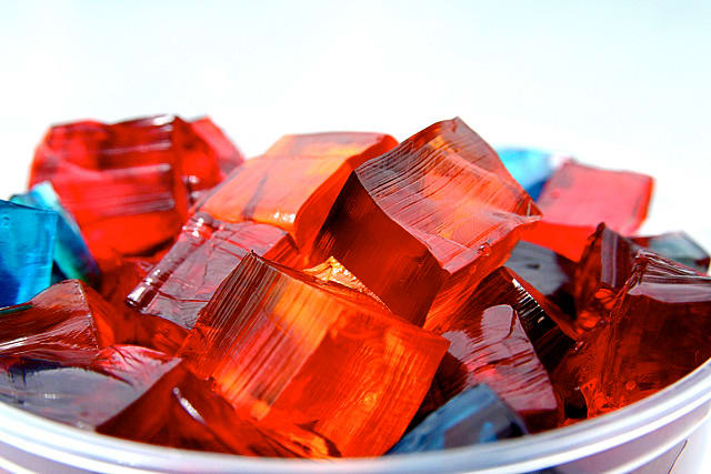
Jello [uploaded from https://www.organicauthority.com/eco-chic-table/new-jell-o-mold-jiggle-chic-holidays]

I’m quite intrigued by this ‘jell-o’ story. It’s hard to believe a childhood dessert might prove to have an application as a catalyst for producing hydrogen fuel. From a December 14, 2018 news item on Nanowerk,

A cheap and effective new catalyst developed by researchers at the University of California, Berkeley, can generate hydrogen fuel from water just as efficiently as platinum, currently the best — but also most expensive — water-splitting catalyst out there.

The catalyst, which is composed of nanometer-thin sheets of metal carbide, is manufactured using a self-assembly process that relies on a surprising ingredient: gelatin, the material that gives Jell-O its jiggle.

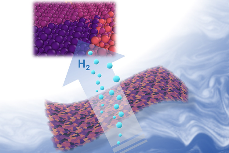
Two-dimensional metal carbides spark a reaction that splits water into oxygen and valuable hydrogen gas. Berkeley researchers have discovered an easy new recipe for cooking up these nanometer-thin sheets that is nearly as simple as making Jell-O from a box. (Xining Zang graphic, copyright Wiley)

A December 13, 2018 University of California at Berkeley (UC Berkeley) news release by Kara Manke (also on EurekAlert but published on Dec. 14, 2018), which originated the news item, provides more technical detail,

“Platinum is expensive, so it would be desirable to find other alternative materials to replace it,” said senior author Liwei Lin, professor of mechanical engineering at UC Berkeley. “We are actually using something similar to the Jell-O that you can eat as the foundation, and mixing it with some of the abundant earth elements to create an inexpensive new material for important catalytic reactions.”

The work appears in the Dec. 13 [2018] print edition of the journal Advanced Materials.

A zap of electricity can break apart the strong bonds that tie water molecules together, creating oxygen and hydrogen gas, the latter of which is an extremely valuable source of energy for powering hydrogen fuel cells. Hydrogen gas can also be used to help store energy from renewable yet intermittent energy sources like solar and wind power, which produce excess electricity when the sun shines or when the wind blows, but which go dormant on rainy or calm days.

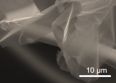
A black and white image of metal carbide under high magnification.

When magnified, the two-dimensional metal carbides resemble sheets of cell[o]phane. (Xining Zang photo, copyright Wiley)

But simply sticking an electrode in a glass of water is an extremely inefficient method of generating hydrogen gas. For the past 20 years, scientists have been searching for catalysts that can speed up this reaction, making it practical for large-scale use.

“The traditional way of using water gas to generate hydrogen still dominates in industry. However, this method produces carbon dioxide as byproduct,” said first author Xining Zang, who conducted the research as a graduate student in mechanical engineering at UC Berkeley. “Electrocatalytic hydrogen generation is growing in the past decade, following the global demand to lower emissions. Developing a highly efficient and low-cost catalyst for electrohydrolysis will bring profound technical, economical and societal benefit.”

To create the catalyst, the researchers followed a recipe nearly as simple as making Jell-O from a box. They mixed gelatin and a metal ion — either molybdenum, tungsten or cobalt — with water, and then let the mixture dry.

“We believe that as gelatin dries, it self-assembles layer by layer,” Lin said. “The metal ion is carried by the gelatin, so when the gelatin self-assembles, your metal ion is also arranged into these flat layers, and these flat sheets are what give Jell-O its characteristic mirror-like surface.”

Heating the mixture to 600 degrees Celsius triggers the metal ion to react with the carbon atoms in the gelatin, forming large, nanometer-thin sheets of metal carbide. The unreacted gelatin burns away.

The researchers tested the efficiency of the catalysts by placing them in water and running an electric current through them. When stacked up against each other, molybdenum carbide split water the most efficiently, followed by tungsten carbide and then cobalt carbide, which didn’t form thin layers as well as the other two. Mixing molybdenum ions with a small amount of cobalt boosted the performance even more.

“It is possible that other forms of carbide may provide even better performance,” Lin said.

On the left, an illustration of blue spheres, representing gelatin molecules, arranged in a lattice shape. On the right, an illustration of thin sheets of metal carbide.

Molecules in gelatin naturally self-assemble in flat sheets, carrying the metal ions with them (left). Heating the mixture to 600 degrees Celsius burns off the gelatin, leaving nanometer-thin sheets of metal carbide. (Xining Zang illustration, copyright Wiley)

The two-dimensional shape of the catalyst is one of the reasons why it is so successful. That is because the water has to be in contact with the surface of the catalyst in order to do its job, and the large surface area of the sheets mean that the metal carbides are extremely efficient for their weight.

Because the recipe is so simple, it could easily be scaled up to produce large quantities of the catalyst, the researchers say.

“We found that the performance is very close to the best catalyst made of platinum and carbon, which is the gold standard in this area,” Lin said. “This means that we can replace the very expensive platinum with our material, which is made in a very scalable manufacturing process.”

Co-authors on the study are Lujie Yang, Buxuan Li and Minsong Wei of UC Berkeley, J. Nathan Hohman and Chenhui Zhu of Lawrence Berkeley National Lab; Wenshu Chen and Jiajun Gu of Shanghai Jiao Tong University; Xiaolong Zou and Jiaming Liang of the Shenzhen Institute; and Mohan Sanghasadasa of the U.S. Army RDECOM AMRDEC.

Here’s a link to and a citation for the paper,

Self‐Assembly of Large‐Area 2D Polycrystalline Transition Metal Carbides for Hydrogen Electrocatalysis by Xining Zang, Wenshu Chen, Xiaolong Zou, J. Nathan Hohman, Lujie Yang
Buxuan Li, Minsong Wei, Chenhui Zhu, Jiaming Liang, Mohan Sanghadasa, Jiajun Gu, Liwei Lin. Advanced Materials Volume30, Issue 50 December 13, 2018 1805188 DOI: https://doi.org/10.1002/adma.201805188 First published [online]: 09 October 2018

This paper is behind a paywall.

Bristly hybrid materials

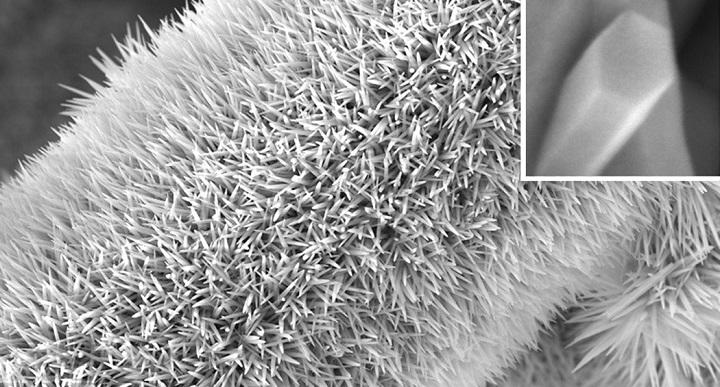
Caption: [Image 1] A carbon fiber covered with a spiky forest of NiCoHC nanowires. Credit: All images reproduced from reference 1 under a Creative Commons Attribution 4.0 International License© 2018 KAUST

It makes me think of small, cuddly things like cats and dogs but it’s not. From an August 7, 2018 King Abdullah University of Science and Technology (KAUST; Saudi Arabia) news release (also published on August 12, 2018 on EurekAlert),

By combining multiple nanomaterials into a single structure, scientists can create hybrid materials that incorporate the best properties of each component and outperform any single substance. A controlled method for making triple-layered hollow nanostructures has now been developed at KAUST. The hybrid structures consist of a conductive organic core sandwiched between layers of electrocatalytically active metals: their potential uses range from better battery electrodes to renewable fuel production.

Although several methods exist to create two-layer materials, making three-layered structures has proven much more difficult, says Peng Wang from the Water Desalination and Reuse Center who co-led the current research with Professor Yu Han, member of the Advanced Membranes and Porous Materials Center at KAUST. The researchers developed a new, dual-template approach, explains Sifei Zhuo, a postdoctoral member of Wang’s team.

The researchers grew their hybrid nanomaterial directly on carbon paper–a mat of electrically conductive carbon fibers. They first produced a bristling forest of nickel cobalt hydroxyl carbonate (NiCoHC) nanowires onto the surface of each carbon fiber (image 1). Each tiny inorganic bristle was coated with an organic layer called hydrogen substituted graphdiyne (HsGDY) (image 2 [not included here]).

Next was the key dual-template step. When the team added a chemical mixture that reacts with the inner NiCoHC, the HsGDY acted as a partial barrier. Some nickel and cobalt ions from the inner layer diffused outward, where they reacted with thiomolybdate from the surrounding solution to form the outer nickel-, cobalt-co-doped MoS2 (Ni,Co-MoS2) layer. Meanwhile, some sulfur ions from the added chemicals diffused inwards to react with the remaining nickel and cobalt. The resulting substance (image 3 [not included here]) had the structure Co9S8, Ni3S2@HsGDY@Ni,Co-MoS2, in which the conductive organic HsGDY layer is sandwiched between two inorganic layers (image 4 [not included here]).

The triple layer material showed good performance at electrocatalytically breaking up water molecules to generate hydrogen, a potential renewable fuel. The researchers also created other triple-layer materials using the dual-template approach

“These triple-layered nanostructures hold great potential in energy conversion and storage,” says Zhuo. “We believe it could be extended to serve as a promising electrode in many electrochemical applications, such as in supercapacitors and sodium-/lithium-ion batteries, and for use in water desalination.”

Here’s a link to and a citation for the paper,

Dual-template engineering of triple-layered nanoarray electrode of metal chalcogenides sandwiched with hydrogen-substituted graphdiyne by Sifei Zhuo, Yusuf Shi, Lingmei Liu, Renyuan Li, Le Shi, Dalaver H. Anjum, Yu Han, & Peng Wang. Nature Communicationsvolume 9, Article number: 3132 (2018) DOI: https://doi.org/10.1038/s41467-018-05474-0 Published 07 August 2018

This paper is open access.


Mixing the unmixable for all new nanoparticles

This news comes out of the University of Maryland and the discovery could led to nanoparticles that have never before been imagined. From a March 29, 2018 news item on ScienceDaily,

Making a giant leap in the ‘tiny’ field of nanoscience, a multi-institutional team of researchers is the first to create nanoscale particles composed of up to eight distinct elements generally known to be immiscible, or incapable of being mixed or blended together. The blending of multiple, unmixable elements into a unified, homogenous nanostructure, called a high entropy alloy nanoparticle, greatly expands the landscape of nanomaterials — and what we can do with them.

This research makes a significant advance on previous efforts that have typically produced nanoparticles limited to only three different elements and to structures that do not mix evenly. Essentially, it is extremely difficult to squeeze and blend different elements into individual particles at the nanoscale. The team, which includes lead researchers at University of Maryland, College Park (UMD)’s A. James Clark School of Engineering, published a peer-reviewed paper based on the research featured on the March 30 [2018] cover of Science.

A March 29, 2018 University of Maryland press release (also on EurekAlert), which originated the news item, delves further (Note: Links have been removed),

“Imagine the elements that combine to make nanoparticles as Lego building blocks. If you have only one to three colors and sizes, then you are limited by what combinations you can use and what structures you can assemble,” explains Liangbing Hu, associate professor of materials science and engineering at UMD and one of the corresponding authors of the paper. “What our team has done is essentially enlarged the toy chest in nanoparticle synthesis; now, we are able to build nanomaterials with nearly all metallic and semiconductor elements.”

The researchers say this advance in nanoscience opens vast opportunities for a wide range of applications that includes catalysis (the acceleration of a chemical reaction by a catalyst), energy storage (batteries or supercapacitors), and bio/plasmonic imaging, among others.

To create the high entropy alloy nanoparticles, the researchers employed a two-step method of flash heating followed by flash cooling. Metallic elements such as platinum, nickel, iron, cobalt, gold, copper, and others were exposed to a rapid thermal shock of approximately 3,000 degrees Fahrenheit, or about half the temperature of the sun, for 0.055 seconds. The extremely high temperature resulted in uniform mixtures of the multiple elements. The subsequent rapid cooling (more than 100,000 degrees Fahrenheit per second) stabilized the newly mixed elements into the uniform nanomaterial.

“Our method is simple, but one that nobody else has applied to the creation of nanoparticles. By using a physical science approach, rather than a traditional chemistry approach, we have achieved something unprecedented,” says Yonggang Yao, a Ph.D. student at UMD and one of the lead authors of the paper.

To demonstrate one potential use of the nanoparticles, the research team used them as advanced catalysts for ammonia oxidation, which is a key step in the production of nitric acid (a liquid acid that is used in the production of ammonium nitrate for fertilizers, making plastics, and in the manufacturing of dyes). They were able to achieve 100 percent oxidation of ammonia and 99 percent selectivity toward desired products with the high entropy alloy nanoparticles, proving their ability as highly efficient catalysts.

Yao says another potential use of the nanoparticles as catalysts could be the generation of chemicals or fuels from carbon dioxide.

“The potential applications for high entropy alloy nanoparticles are not limited to the field of catalysis. With cross-discipline curiosity, the demonstrated applications of these particles will become even more widespread,” says Steven D. Lacey, a Ph.D. student at UMD and also one of the lead authors of the paper.

This research was performed through a multi-institutional collaboration of Prof. Liangbing Hu’s group at the University of Maryland, College Park; Prof. Reza Shahbazian-Yassar’s group at University of Illinois at Chicago; Prof. Ju Li’s group at the Massachusetts Institute of Technology; Prof. Chao Wang’s group at Johns Hopkins University; and Prof. Michael Zachariah’s group at the University of Maryland, College Park.

What outside experts are saying about this research:

“This is quite amazing; Dr. Hu creatively came up with this powerful technique, carbo-thermal shock synthesis, to produce high entropy alloys of up to eight different elements in a single nanoparticle. This is indeed unthinkable for bulk materials synthesis. This is yet another beautiful example of nanoscience!,” says Peidong Yang, the S.K. and Angela Chan Distinguished Professor of Energy and professor of chemistry at the University of California, Berkeley and member of the American Academy of Arts and Sciences.

“This discovery opens many new directions. There are simulation opportunities to understand the electronic structure of the various compositions and phases that are important for the next generation of catalyst design. Also, finding correlations among synthesis routes, composition, and phase structure and performance enables a paradigm shift toward guided synthesis,” says George Crabtree, Argonne Distinguished Fellow and director of the Joint Center for Energy Storage Research at Argonne National Laboratory.

More from the research coauthors:

“Understanding the atomic order and crystalline structure in these multi-element nanoparticles reveals how the synthesis can be tuned to optimize their performance. It would be quite interesting to further explore the underlying atomistic mechanisms of the nucleation and growth of high entropy alloy nanoparticle,” says Reza Shahbazian-Yassar, associate professor at the University of Illinois at Chicago and a corresponding author of the paper.

“Carbon metabolism drives ‘living’ metal catalysts that frequently move around, split, or merge, resulting in a nanoparticle size distribution that’s far from the ordinary, and highly tunable,” says Ju Li, professor at the Massachusetts Institute of Technology and a corresponding author of the paper.

“This method enables new combinations of metals that do not exist in nature and do not otherwise go together. It enables robust tuning of the composition of catalytic materials to optimize the activity, selectivity, and stability, and the application will be very broad in energy conversions and chemical transformations,” says Chao Wang, assistant professor of chemical and biomolecular engineering at Johns Hopkins University and one of the study’s authors.

Here’s a link to and a citation for the paper,

Carbothermal shock synthesis of high-entropy-alloy nanoparticles by Yonggang Yao, Zhennan Huang, Pengfei Xie, Steven D. Lacey, Rohit Jiji Jacob, Hua Xie, Fengjuan Chen, Anmin Nie, Tiancheng Pu, Miles Rehwoldt, Daiwei Yu, Michael R. Zachariah, Chao Wang, Reza Shahbazian-Yassar, Ju Li, Liangbing Hu. Science 30 Mar 2018: Vol. 359, Issue 6383, pp. 1489-1494 DOI: 10.1126/science.aan5412

This paper is behind a paywall.

Nanoparticles from tattoo inks circulate through your body

English: Tattoo of Hand of Fatima,. Model: Casini. Date: 4 July 2017, 18:13:41. Source : Own work. Author: Stephencdickson.

For those who like their news in video format, there’s this Canadian Broadcasting Corporation (CBC) news item broadcast on Sep. 11, 2017 (after the commercials),

For those who like text and more detail, scientists at the European Synchrotron Radiation Facility (ESRF) have produced a study of the (at the nanoparticle scale) inks in tattoos. From a Sept. 12, 2017 news item on phys.org,

The elements that make up the ink in tattoos travel inside the body in micro and nanoparticle forms and reach the lymph nodes, according to a study published in Scientific Reports on 12 September [2017] by scientists from Germany and the ESRF, the European Synchrotron, Grenoble (France). It is the first time researchers have found analytical evidence of the transport of organic and inorganic pigments and toxic element impurities as well as in depth characterization of the pigments ex vivo in tattooed tissues. Two ESRF beamlines were crucial in this breakthrough.

A Sept. 12, 2017 ESRF press release (also on EurkeAlert), which originated the news item, explains further,

The reality is that little is known about the potential impurities in the colour mixture applied to the skin. Most tattoo inks contain organic pigments, but also include preservatives and contaminants like nickel, chromium, manganese or cobalt. Besides carbon black, the second most common ingredient used in tattoo inks is titanium dioxide (TiO2), a white pigment usually applied to create certain shades when mixed with colorants. Delayed healing, along with skin elevation and itching, are often associated with white tattoos, and by consequence with the use of TiO2. TiO2 is also commonly used in food additives, sun screens and paints. Scientists from the ESRF, the German Federal Institute for Risk Assessment, Ludwig-Maximilians University, and the Physikalisch-Technische Bundesanstalt have managed to get a very clear picture on the location of titanium dioxide once it gets in the tissue. This work was done on the ESRF beamlines ID21 and ID16B.

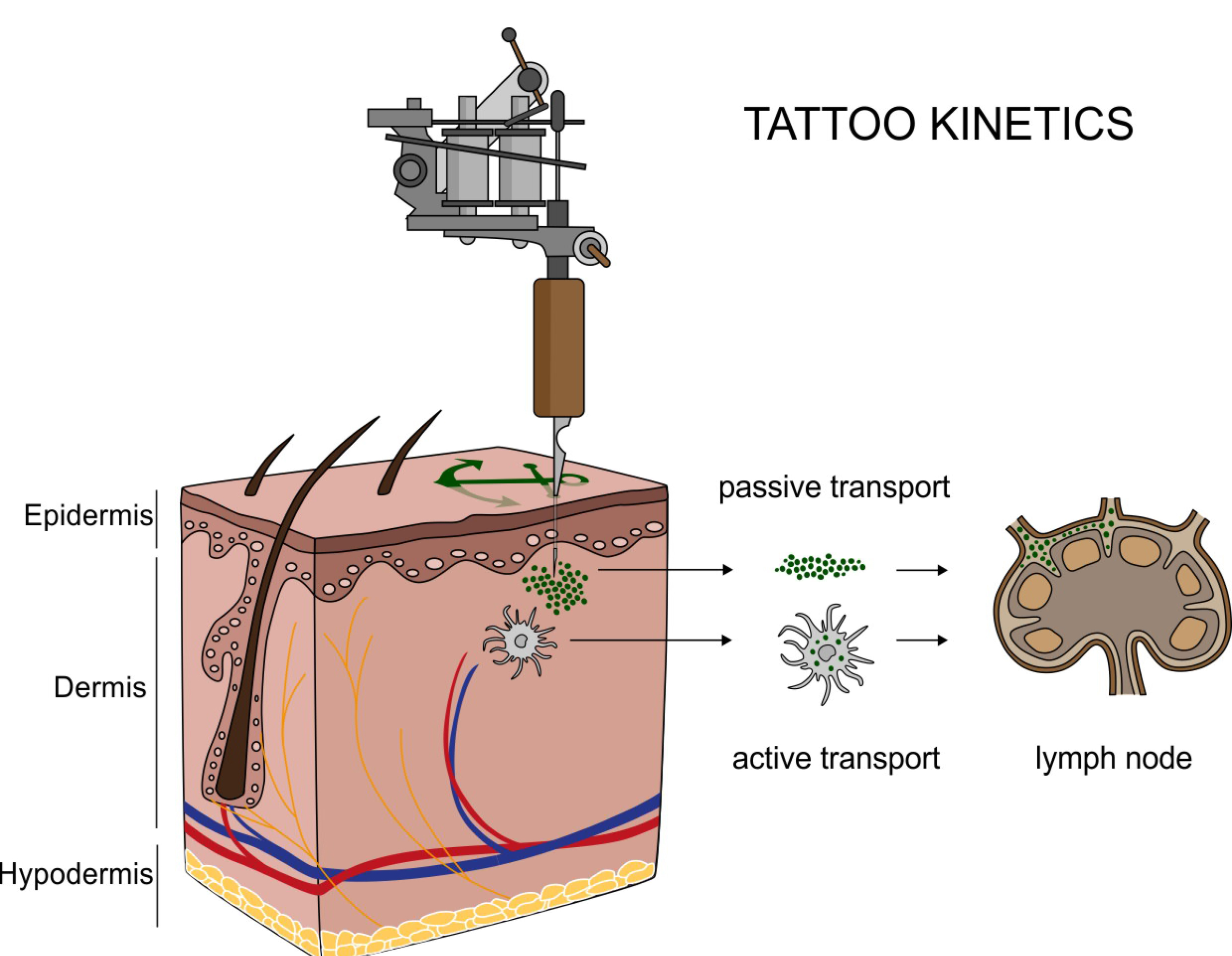
drawing tattookinetics.jpg

Translocation of tattoo particles from skin to lymph nodes. Upon injection of tattoo inks, particles can be either passively transported via blood and lymph fluids or phagocytized by immune cells and subsequently deposited in regional lymph nodes. After healing, particles are present in the dermis and in the sinusoids of the draining lymph nodes. Credits: C. Seim.

The hazards that potentially derive from tattoos were, until now, only investigated by chemical analysis of the inks and their degradation products in vitro. “We already knew that pigments from tattoos would travel to the lymph nodes because of visual evidence: the lymph nodes become tinted with the colour of the tattoo. It is the response of the body to clean the site of entrance of the tattoo. What we didn’t know is that they do it in a nano form, which implies that they may not have the same behaviour as the particles at a micro level. And that is the problem: we don’t know how nanoparticles react”, explains Bernhard Hesse, one of the two first authors of the study (together with Ines Schreiver, from the German Federal Institute for Risk Assessment) and ESRF visiting scientist.

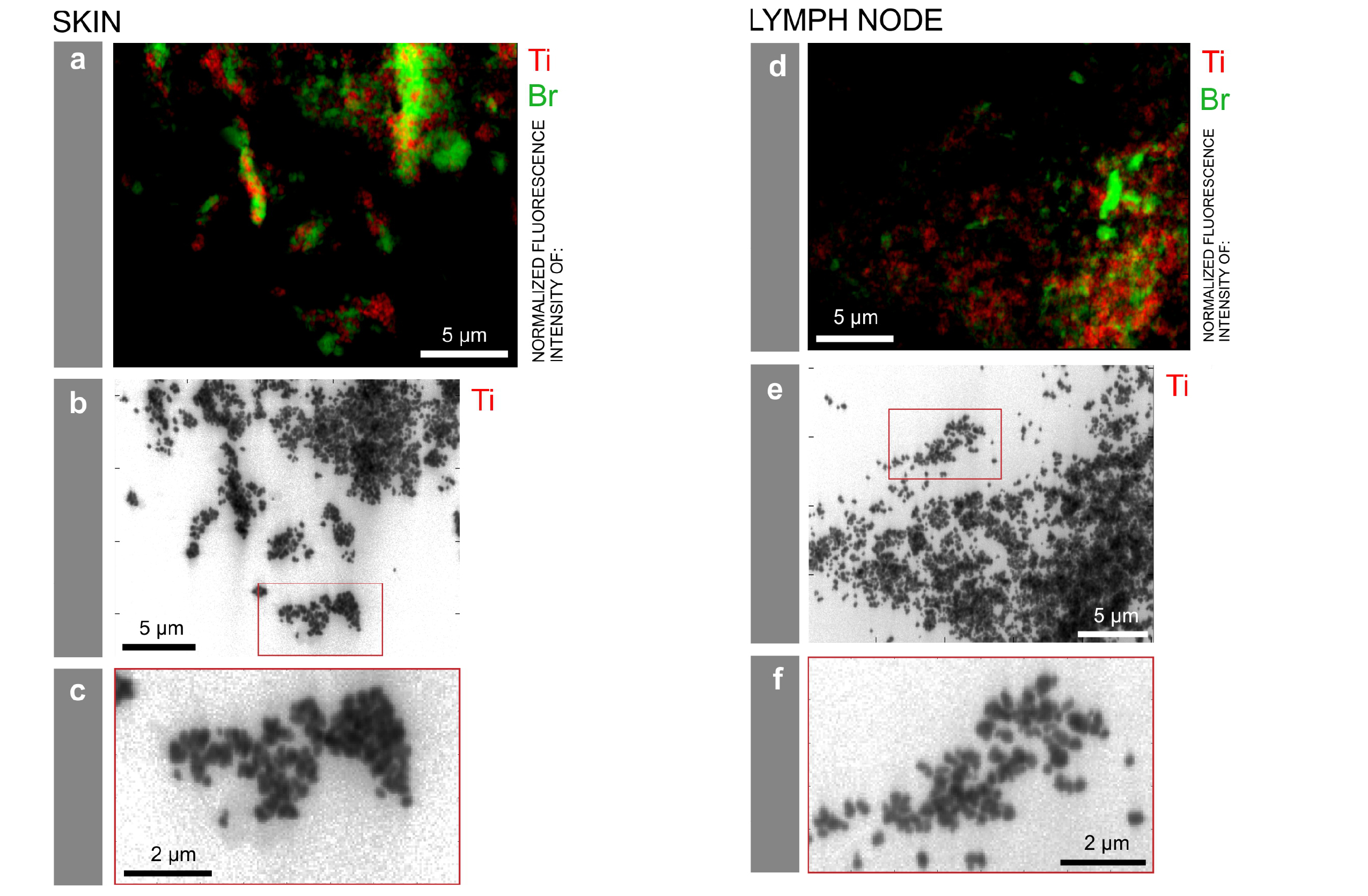
titaniumdistribution.jpg

Particle mapping and size distribution of different tattoo pigment elements.  a, d) Ti and the Br containing pigment phthalocyanine green 36 are located next to each other. b, e) Log scale mappings of Ti, Br and Fe in the same areas as displayed in a) and d) reveal primary particle sizes of different pigment species. c, f) Magnifications of the indicated areas in b) and e), respectively. Credits: C. Seim.

X-ray fluorescence measurements on ID21 allowed the team to locate titanium dioxide at the micro and nano range in the skin and the lymphatic environment. They found a broad range of particles with up to several micrometres in size in human skin, but only smaller (nano) particles transported to the lymph nodes. This can lead to the chronic enlargement of the lymph nodes and lifelong exposure. Scientists also used the technique of Fourier transform infrared spectroscopy to assess biomolecular changes in the tissues in the proximity of the tattoo particles.

ESRF16_Tattoo-1low.jpg

Ines Schreiver doing experiments on ID16B with Julie Villanova. Credits: B. Hesse.

Altogether the scientists report strong evidence for both migration and long-term deposition of toxic elements and tattoo pigments as well as for conformational alterations of biomolecules that are sometimes linked to cutaneous adversities upon tattooing.

Then next step for the team is to inspect further samples of patients with adverse effects in their tattoos in order to find links with chemical and structural properties of the pigments used to create these tattoos.

Here’s a link to and  a citation for the paper,

Synchrotron-based ν-XRF mapping and μ-FTIR microscopy enable to look into the fate and effects of tattoo pigments in human skin by Ines Schreiver, Bernhard Hesse, Christian Seim, Hiram Castillo-Michel, Julie Villanova, Peter Laux, Nadine Dreiack, Randolf Penning, Remi Tucoulou, Marine Cotte, & Andreas Luch. Scientific Reports 7, Article number: 11395 (2017) doi:10.1038/s41598-017-11721-z Published online: 12 September 2017

This paper is open access.

Refining metals more sustainably

We don’t just extract and refine metals from the earth, increasingly, we extract and refine them from consumer goods. Researchers from McGill University (Montréal, Québec, Canada) have devised a ‘greener’ technique to do this. From a June 7, 2017 McGill University news release (received via email and also on EurekAlert),

A team of chemists in Canada has developed a way to process metals without using toxic solvents and reagents.

The system, which also consumes far less energy than conventional techniques, could greatly shrink the environmental impact of producing metals from raw materials or from post-consumer electronics.

“At a time when natural deposits of metals are on the decline, there is a great deal of interest in improving the efficiency of metal refinement and recycling, but few disruptive technologies are being put forth,” says Jean-Philip Lumb, an associate professor in McGill University’s Department of Chemistry. “That’s what makes our advance so important.”

The discovery stems from a collaboration between Lumb and Tomislav Friscic at McGill in Montreal, and Kim Baines of Western University in London, Ont. In an article published recently in Science Advances, the researchers outline an approach that uses organic molecules, instead of chlorine and hydrochloric acid, to help purify germanium, a metal used widely in electronic devices. Laboratory experiments by the researchers have shown that the same technique can be used with other metals, including zinc, copper, manganese and cobalt.

The research could mark an important milestone for the “green chemistry” movement, which seeks to replace toxic reagents used in conventional industrial manufacturing with more environmentally friendly alternatives. Most advances in this area have involved organic chemistry – the synthesis of carbon-based compounds used in pharmaceuticals and plastics, for example.

“Applications of green chemistry lag far behind in the area of metals,” Lumb says. “Yet metals are just as important for sustainability as any organic compound. For example, electronic devices require numerous metals to function.”

Taking a page from biology

There is no single ore rich in germanium, so it is generally obtained from mining operations as a minor component in a mixture with many other materials. Through a series of processes, that blend of matter can be reduced to germanium and zinc.

“Currently, in order to isolate germanium from zinc, it’s a pretty nasty process,” Baines explains. The new approach developed by the McGill and Western chemists “enables you to get germanium from zinc, without those nasty processes.”

To accomplish this, the researchers took a page from biology. Lumb’s lab for years has conducted research into the chemistry of melanin, the molecule in human tissue that gives skin and hair their color. Melanin also has the ability to bind to metals. “We asked the question: ‘Here’s this biomaterial with exquisite function, would it be possible to use it as a blueprint for new, more efficient technologies?'”

The scientists teamed up to synthesize a molecule that mimics some of the qualities of melanin. In particular, this “organic co-factor” acts as a mediator that helps to extract germanium at room temperature, without using solvents.

Next step: industrial scale

The system also taps into Friscic’s expertise in mechanochemistry, an emerging branch of chemistry that relies on mechanical force – rather than solvents and heat – to promote chemical reactions. Milling jars containing stainless-steel balls are shaken at high speeds to help purify the metal.

“This shows how collaborations naturally can lead to sustainability-oriented innovation,” Friscic says. “Combining elegant new chemistry with solvent-free mechanochemical techniques led us to a process that is cleaner by virtue of circumventing chlorine-based processing, but also eliminates the generation of toxic solvent waste”

The next step in developing the technology will be to show that it can be deployed economically on industrial scales, for a range of metals.

“There’s a tremendous amount of work that needs to be done to get from where we are now to where we need to go,” Lumb says. “But the platform works on many different kinds of metals and metal oxides, and we think that it could become a technology adopted by industry. We are looking for stakeholders with whom we can partner to move this technology forward.”

Here’s a link to and a citation for the paper,

A chlorine-free protocol for processing germanium by Martin Glavinovic, Michael Krause, Linju Yang, John A. McLeod, Lijia Liu, Kim M. Baines, Tomislav Friščić, and Jean-Philip Lumb. Science Advances 05 May 2017: Vol. 3, no. 5, e1700149 DOI: 10.1126/sciadv.1700149

This paper is open access.

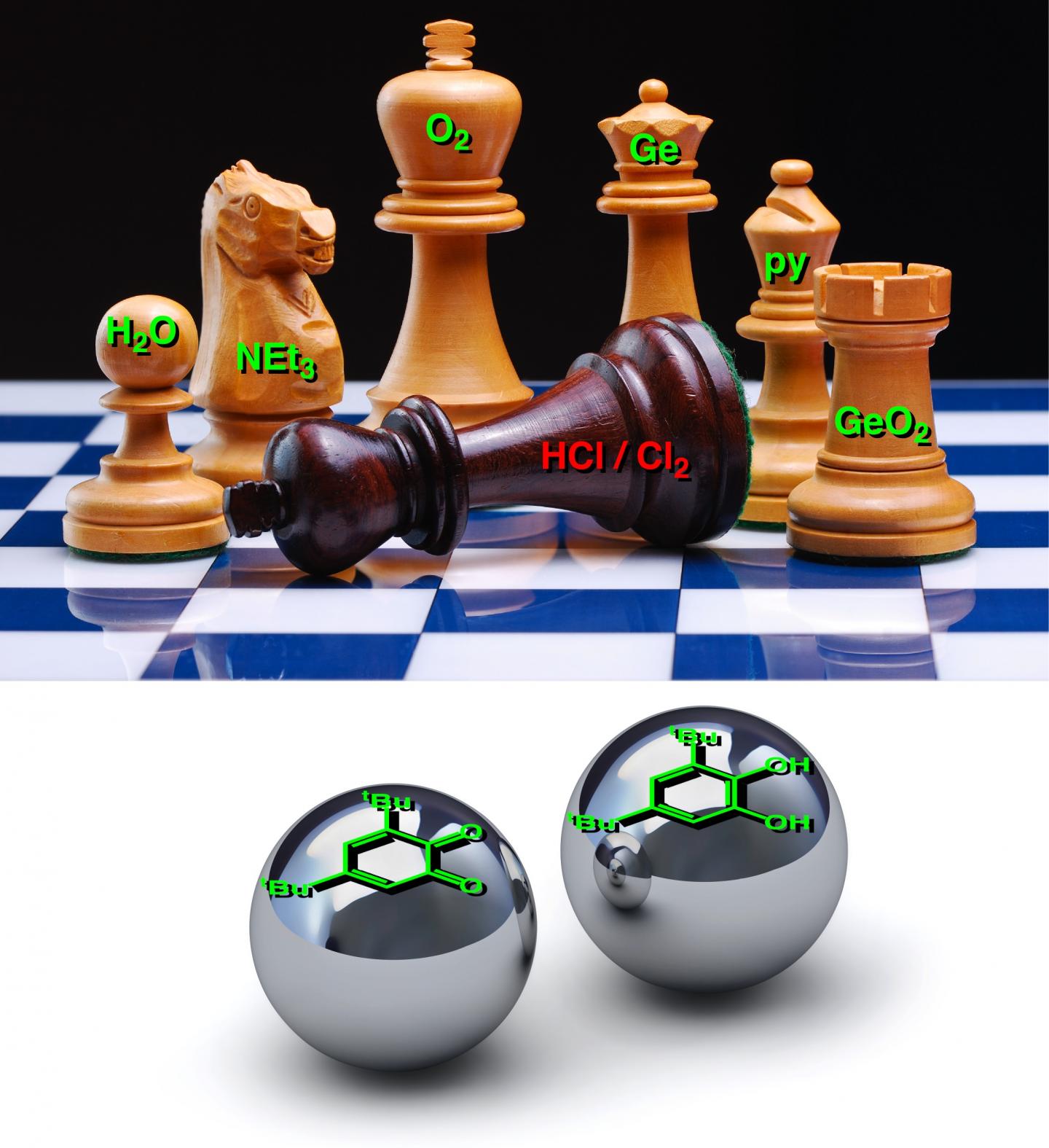
ETA June 9, 2017 at 1700 hours PDT: I have to give them marks for creativity. Here’s the image being used to illustrate the work,

Caption: Strategy for reducing the environmental impact of a refining process: replace hazardous chemicals with more benign and recyclable compounds. Credit: Michael J. Krause (Western University)

Vitamin-driven lithium-ion battery from the University of Toronto

It seems vitamins aren’t just good for health, they’re also good for batteries. My Aug. 2, 2016 post on vitamins and batteries focused on work from Harvard, this time the work is from the University of Toronto (Canada). From an Aug. 3, 2016 news item on ScienceDaily,

A team of University of Toronto chemists has created a battery that stores energy in a biologically derived unit, paving the way for cheaper consumer electronics that are easier on the environment.

The battery is similar to many commercially-available high-energy lithium-ion batteries with one important difference. It uses flavin from vitamin B2 as the cathode: the part that stores the electricity that is released when connected to a device.

“We’ve been looking to nature for a while to find complex molecules for use in a number of consumer electronics applications,” says Dwight Seferos, an associate professor in U of T’s Department of Chemistry and Canada Research Chair in Polymer Nanotechnology.

“When you take something made by nature that is already complex, you end up spending less time making new material,” says Seferos.

An Aug. 2, 2016 University of Toronto news release (also on EurekAlert) by Peter McMahon, which originated the news item, explains further,

To understand the discovery, it’s important to know that modern batteries contain three basic parts:

  • a positive terminal – the metal part that touches devices to power them – connected to a cathode inside the battery casing
  • a negative terminal connected to an anode inside the battery casing
  • an electrolyte solution, in which ions can travel between the cathode and anode electrodes

When a battery is connected to a phone, iPod, camera or other device that requires power, electrons flow from the anode – the negatively charged electrode of the device supplying current – out to the device, then into the cathode and ions migrate through the electrolyte solution to balance the charge. When connected to a charger, this process happens in reverse.

The reaction in the anode creates electrons and the reaction in the cathode absorbs them when discharging. The net product is electricity. The battery will continue to produce electricity until one or both of the electrodes run out of the substance necessary for the reactions to occur.

Organic chemistry is kind of like Lego

While bio-derived battery parts have been created previously, this is the first one that uses bio-derived polymers – long-chain molecules – for one of the electrodes, essentially allowing battery energy to be stored in a vitamin-created plastic, instead of costlier, harder to process, and more environmentally-harmful metals such as cobalt.

“Getting the right material evolved over time and definitely took some test reactions,” says paper co-author and doctoral student Tyler Schon. “In a lot of ways, it looked like this could have failed. It definitely took a lot of perseverance.”

Schon, Seferos and colleagues happened upon the material while testing a variety of long-chain polymers – specifically pendant group polymers: the molecules attached to a ‘backbone’ chain of a long molecule.

“Organic chemistry is kind of like Lego,” he says. “You put things together in a certain order, but some things that look like they’ll fit together on paper don’t in reality. We tried a few approaches and the fifth one worked,” says Seferos.

Building a better power pack

The team created the material from vitamin B2 that originates in genetically-modified fungi using a semi-synthetic process to prepare the polymer by linking two flavin units to a long-chain molecule backbone.

This allows for a green battery with high capacity and high voltage – something increasingly important as the ‘Internet of Things’ continues to link us together more and more through our battery-powered portable devices.

“It’s a pretty safe, natural compound,” Seferos adds. “If you wanted to, you could actually eat the source material it comes from.”

B2’s ability to be reduced and oxidized makes its well-suited for a lithium ion battery.

“B2 can accept up to two electrons at a time,” says Seferos. “This makes it easy to take multiple charges and have a high capacity compared to a lot of other available molecules.”

A step to greener electronics

“It’s been a lot of trial-and-error,” says Schon. “Now we’re looking to design new variants that can be recharged again and again.”

While the current prototype is on the scale of a hearing aid battery, the team hopes their breakthrough could lay the groundwork for powerful, thin, flexible, and even transparent metal-free batteries that could support the next wave of consumer electronics.

Here’s a link to and a citation for the paper,

Bio-Derived Polymers for Sustainable Lithium-Ion Batteries by Tyler B. Schon, Andrew J. Tilley, Colin R. Bridges, Mark B. Miltenburg, and Dwight S. Seferos. Advanced Functional Materials DOI: 10.1002/adfm.201602114 Version of Record online: 14 JUL 2016

© 2016 WILEY-VCH Verlag GmbH & Co. KGaA, Weinheim

This paper is behind a paywall.

Ultimate discovery tool?

For anyone familiar with the US nanomedicine scene, Chad Mirkin’s appearance in this announcement from Northwestern University isn’t much of a surprise.  From a June 23, 2016 news item on ScienceDaily,

The discovery power of the gene chip is coming to nanotechnology. A Northwestern University research team is developing a tool to rapidly test millions and perhaps even billions or more different nanoparticles at one time to zero in on the best particle for a specific use.

When materials are miniaturized, their properties—optical, structural, electrical, mechanical and chemical—change, offering new possibilities. But determining what nanoparticle size and composition are best for a given application, such as catalysts, biodiagnostic labels, pharmaceuticals and electronic devices, is a daunting task.

“As scientists, we’ve only just begun to investigate what materials can be made on the nanoscale,” said Northwestern’s Chad A. Mirkin, a world leader in nanotechnology research and its application, who led the study. “Screening a million potentially useful nanoparticles, for example, could take several lifetimes. Once optimized, our tool will enable researchers to pick the winner much faster than conventional methods. We have the ultimate discovery tool.”

A June 23, 2016 Northwestern University news release (also on EurekAlert), which originated the news item, describes the work in more detail,

Using a Northwestern technique that deposits materials on a surface, Mirkin and his team figured out how to make combinatorial libraries of nanoparticles in a very controlled way. (A combinatorial library is a collection of systematically varied structures encoded at specific sites on a surface.) Their study will be published June 24 by the journal Science.

The nanoparticle libraries are much like a gene chip, Mirkin says, where thousands of different spots of DNA are used to identify the presence of a disease or toxin. Thousands of reactions can be done simultaneously, providing results in just a few hours. Similarly, Mirkin and his team’s libraries will enable scientists to rapidly make and screen millions to billions of nanoparticles of different compositions and sizes for desirable physical and chemical properties.

“The ability to make libraries of nanoparticles will open a new field of nanocombinatorics, where size — on a scale that matters — and composition become tunable parameters,” Mirkin said. “This is a powerful approach to discovery science.”

“I liken our combinatorial nanopatterning approach to providing a broad palette of bold colors to an artist who previously had been working with a handful of dull and pale black, white and grey pastels,” said co-author Vinayak P. Dravid, the Abraham Harris Professor of Materials Science and Engineering in the McCormick School of Engineering.

Using five metallic elements — gold, silver, cobalt, copper and nickel — Mirkin and his team developed an array of unique structures by varying every elemental combination. In previous work, the researchers had shown that particle diameter also can be varied deliberately on the 1- to 100-nanometer length scale.

Some of the compositions can be found in nature, but more than half of them have never existed before on Earth. And when pictured using high-powered imaging techniques, the nanoparticles appear like an array of colorful Easter eggs, each compositional element contributing to the palette.

To build the combinatorial libraries, Mirkin and his team used Dip-Pen Nanolithography, a technique developed at Northwestern in 1999, to deposit onto a surface individual polymer “dots,” each loaded with different metal salts of interest. The researchers then heated the polymer dots, reducing the salts to metal atoms and forming a single nanoparticle. The size of the polymer dot can be varied to change the size of the final nanoparticle.

This control of both size and composition of nanoparticles is very important, Mirkin stressed. Having demonstrated control, the researchers used the tool to systematically generate a library of 31 nanostructures using the five different metals.

To help analyze the complex elemental compositions and size/shape of the nanoparticles down to the sub-nanometer scale, the team turned to Dravid, Mirkin’s longtime friend and collaborator. Dravid, founding director of Northwestern’s NUANCE Center, contributed his expertise and the advanced electron microscopes of NUANCE to spatially map the compositional trajectories of the combinatorial nanoparticles.

Now, scientists can begin to study these nanoparticles as well as build other useful combinatorial libraries consisting of billions of structures that subtly differ in size and composition. These structures may become the next materials that power fuel cells, efficiently harvest solar energy and convert it into useful fuels, and catalyze reactions that take low-value feedstocks from the petroleum industry and turn them into high-value products useful in the chemical and pharmaceutical industries.

Here’s a diagram illustrating the work,

 Caption: A combinatorial library of polyelemental nanoparticles was developed using Dip-Pen Nanolithography. This novel nanoparticle library opens up a new field of nanocombinatorics for rapid screening of nanomaterials for a multitude of properties. Credit: Peng-Cheng Chen/James Hedrick

Caption: A combinatorial library of polyelemental nanoparticles was developed using Dip-Pen Nanolithography. This novel nanoparticle library opens up a new field of nanocombinatorics for rapid screening of nanomaterials for a multitude of properties. Credit: Peng-Cheng Chen/James Hedrick

Here’s a link to and a citation for the paper,

Polyelemental nanoparticle libraries by Peng-Cheng Chen, Xiaolong Liu, James L. Hedrick, Zhuang Xie, Shunzhi Wang, Qing-Yuan Lin, Mark C. Hersam, Vinayak P. Dravid, Chad A. Mirkin. Science  24 Jun 2016: Vol. 352, Issue 6293, pp. 1565-1569 DOI: 10.1126/science.aaf8402

This paper is behind a paywall.

Split some water molecules and save solar and wind (energy) for a future day

Professor Ted Sargent’s research team at the University of Toronto has a developed a new technique for saving the energy harvested by sun and wind farms according to a March 28, 2016 news item on Nanotechnology Now,

We can’t control when the wind blows and when the sun shines, so finding efficient ways to store energy from alternative sources remains an urgent research problem. Now, a group of researchers led by Professor Ted Sargent at the University of Toronto’s Faculty of Applied Science & Engineering may have a solution inspired by nature.

The team has designed the most efficient catalyst for storing energy in chemical form, by splitting water into hydrogen and oxygen, just like plants do during photosynthesis. Oxygen is released harmlessly into the atmosphere, and hydrogen, as H2, can be converted back into energy using hydrogen fuel cells.

Discovering a better way of storing energy from solar and wind farms is “one of the grand challenges in this field,” Ted Sargent says (photo above by Megan Rosenbloom via flickr) Courtesy: University of Toronto

Discovering a better way of storing energy from solar and wind farms is “one of the grand challenges in this field,” Ted Sargent says (photo above by Megan Rosenbloom via flickr) Courtesy: University of Toronto

A March 24, 2016 University of Toronto news release by Marit Mitchell, which originated the news item, expands on the theme,

“Today on a solar farm or a wind farm, storage is typically provided with batteries. But batteries are expensive, and can typically only store a fixed amount of energy,” says Sargent. “That’s why discovering a more efficient and highly scalable means of storing energy generated by renewables is one of the grand challenges in this field.”

You may have seen the popular high-school science demonstration where the teacher splits water into its component elements, hydrogen and oxygen, by running electricity through it. Today this requires so much electrical input that it’s impractical to store energy this way — too great proportion of the energy generated is lost in the process of storing it.

This new catalyst facilitates the oxygen-evolution portion of the chemical reaction, making the conversion from H2O into O2 and H2 more energy-efficient than ever before. The intrinsic efficiency of the new catalyst material is over three times more efficient than the best state-of-the-art catalyst.

Details are offered in the news release,

The new catalyst is made of abundant and low-cost metals tungsten, iron and cobalt, which are much less expensive than state-of-the-art catalysts based on precious metals. It showed no signs of degradation over more than 500 hours of continuous activity, unlike other efficient but short-lived catalysts. …

“With the aid of theoretical predictions, we became convinced that including tungsten could lead to a better oxygen-evolving catalyst. Unfortunately, prior work did not show how to mix tungsten homogeneously with the active metals such as iron and cobalt,” says one of the study’s lead authors, Dr. Bo Zhang … .

“We invented a new way to distribute the catalyst homogenously in a gel, and as a result built a device that works incredibly efficiently and robustly.”

This research united engineers, chemists, materials scientists, mathematicians, physicists, and computer scientists across three countries. A chief partner in this joint theoretical-experimental studies was a leading team of theorists at Stanford University and SLAC National Accelerator Laboratory under the leadership of Dr. Aleksandra Vojvodic. The international collaboration included researchers at East China University of Science & Technology, Tianjin University, Brookhaven National Laboratory, Canadian Light Source and the Beijing Synchrotron Radiation Facility.

“The team developed a new materials synthesis strategy to mix multiple metals homogeneously — thereby overcoming the propensity of multi-metal mixtures to separate into distinct phases,” said Jeffrey C. Grossman, the Morton and Claire Goulder and Family Professor in Environmental Systems at Massachusetts Institute of Technology. “This work impressively highlights the power of tightly coupled computational materials science with advanced experimental techniques, and sets a high bar for such a combined approach. It opens new avenues to speed progress in efficient materials for energy conversion and storage.”

“This work demonstrates the utility of using theory to guide the development of improved water-oxidation catalysts for further advances in the field of solar fuels,” said Gary Brudvig, a professor in the Department of Chemistry at Yale University and director of the Yale Energy Sciences Institute.

“The intensive research by the Sargent group in the University of Toronto led to the discovery of oxy-hydroxide materials that exhibit electrochemically induced oxygen evolution at the lowest overpotential and show no degradation,” said University Professor Gabor A. Somorjai of the University of California, Berkeley, a leader in this field. “The authors should be complimented on the combined experimental and theoretical studies that led to this very important finding.”

Here’s a link to and a citation for the paper,

Homogeneously dispersed, multimetal oxygen-evolving catalysts by Bo Zhang, Xueli Zheng, Oleksandr Voznyy, Riccardo Comin, Michal Bajdich, Max García-Melchor, Lili Han, Jixian Xu, Min Liu, Lirong Zheng, F. Pelayo García de Arquer, Cao Thang Dinh, Fengjia Fan, Mingjian Yuan, Emre Yassitepe, Ning Chen, Tom Regier, Pengfei Liu, Yuhang Li, Phil De Luna, Alyf Janmohamed, Huolin L. Xin, Huagui Yang, Aleksandra Vojvodic, Edward H. Sargent. Science  24 Mar 2016: DOI: 10.1126/science.aaf1525

This paper is behind a paywall.

Carbon nanotubes sense spoiled food

CNT_FoodSpolage

Courtesy: MIT (Massachusetts Institute of Technology)

I love this .gif; it says a lot without a word. However for details, you need words and here’s what an April 15, 2015 news item on Nanowerk has to say about the research illustrated by the .gif,

MIT [Massachusetts Institute of Technology] chemists have devised an inexpensive, portable sensor that can detect gases emitted by rotting meat, allowing consumers to determine whether the meat in their grocery store or refrigerator is safe to eat.

The sensor, which consists of chemically modified carbon nanotubes, could be deployed in “smart packaging” that would offer much more accurate safety information than the expiration date on the package, says Timothy Swager, the John D. MacArthur Professor of Chemistry at MIT.

An April 14, 2015 MIT news release (also on EurekAlert), which originated the news item, offers more from Dr. Swager,

It could also cut down on food waste, he adds. “People are constantly throwing things out that probably aren’t bad,” says Swager, who is the senior author of a paper describing the new sensor this week in the journal Angewandte Chemie.

This latest study is builds on previous work at Swager’s lab (Note: Links have been removed),

The sensor is similar to other carbon nanotube devices that Swager’s lab has developed in recent years, including one that detects the ripeness of fruit. All of these devices work on the same principle: Carbon nanotubes can be chemically modified so that their ability to carry an electric current changes in the presence of a particular gas.

In this case, the researchers modified the carbon nanotubes with metal-containing compounds called metalloporphyrins, which contain a central metal atom bound to several nitrogen-containing rings. Hemoglobin, which carries oxygen in the blood, is a metalloporphyrin with iron as the central atom.

For this sensor, the researchers used a metalloporphyrin with cobalt at its center. Metalloporphyrins are very good at binding to nitrogen-containing compounds called amines. Of particular interest to the researchers were the so-called biogenic amines, such as putrescine and cadaverine, which are produced by decaying meat.

When the cobalt-containing porphyrin binds to any of these amines, it increases the electrical resistance of the carbon nanotube, which can be easily measured.

“We use these porphyrins to fabricate a very simple device where we apply a potential across the device and then monitor the current. When the device encounters amines, which are markers of decaying meat, the current of the device will become lower,” Liu says.

In this study, the researchers tested the sensor on four types of meat: pork, chicken, cod, and salmon. They found that when refrigerated, all four types stayed fresh over four days. Left unrefrigerated, the samples all decayed, but at varying rates.

There are other sensors that can detect the signs of decaying meat, but they are usually large and expensive instruments that require expertise to operate. “The advantage we have is these are the cheapest, smallest, easiest-to-manufacture sensors,” Swager says.

“There are several potential advantages in having an inexpensive sensor for measuring, in real time, the freshness of meat and fish products, including preventing foodborne illness, increasing overall customer satisfaction, and reducing food waste at grocery stores and in consumers’ homes,” says Roberto Forloni, a senior science fellow at Sealed Air, a major supplier of food packaging, who was not part of the research team.

The new device also requires very little power and could be incorporated into a wireless platform Swager’s lab recently developed that allows a regular smartphone to read output from carbon nanotube sensors such as this one.

The funding sources are interesting, as I am appreciating with increasing frequency these days (from the news release),

The researchers have filed for a patent on the technology and hope to license it for commercial development. The research was funded by the National Science Foundation and the Army Research Office through MIT’s Institute for Soldier Nanotechnologies.

Here’s a link to and a citation for the paper,

Single-Walled Carbon Nanotube/Metalloporphyrin Composites for the Chemiresistive Detection of Amines and Meat Spoilage by Sophie F. Liu, Alexander R. Petty, Dr. Graham T. Sazama, and Timothy M. Swager. Angewandte Chemie International Edition DOI: 10.1002/anie.201501434 Article first published online: 13 APR 2015

© 2015 WILEY-VCH Verlag GmbH & Co. KGaA, Weinheim

This article is behind a paywall.

There are other posts here about the quest to create food sensors including this Sept. 26, 2013 piece which features a critique (by another blogger) about trying to create food sensors that may be more expensive than the item they are protecting, a problem Swager claims to have overcome in an April 17, 2015 article by Ben Schiller for Fast Company (Note: Links have been removed),

Swager has set up a company to commercialize the technology and he expects to do the first demonstrations to interested clients this summer. The first applications are likely to be for food workers working with meat and fish, but there’s no reason why consumers shouldn’t get their own devices in due time.

There are efforts to create visual clues for food status. But Swager says his method is better because it doesn’t rely on perception: it produces hard data that can be logged and tracked. And it also has potential to be very cheap.

“The resistance method is a game-changer because it’s two to three orders of magnitude cheaper than other technology. It’s hard to imagine doing this cheaper,” he says.