A May 19, 2021 news item on phys.org sheds some light on a new approach to stroke treatments,
Blocked blood vessels in the brains of stroke patients prevent oxygen-rich blood from getting to cells, causing severe damage. Plants and some microbes produce oxygen through photosynthesis. What if there was a way to make photosynthesis happen in the brains of patients? Now, researchers reporting in ACS’ Nano Letters have done just that in cells and in mice, using blue-green algae and special nanoparticles, in a proof-of-concept demonstration.
Strokes result in the deaths of 5 million people worldwide every year, according to the World Health Organization. Millions more survive, but they often experience disabilities, such as difficulties with speech, swallowing or memory. The most common cause is a blood vessel blockage in the brain, and the best way to prevent permanent brain damage from this type of stroke is to dissolve or surgically remove the blockage as soon as possible. However, those options only work within a narrow time window after the stroke happens and can be risky. Blue-green algae, such as Synechococcus elongatus, have been studied previously to treat the lack of oxygen in heart tissue and tumors using photosynthesis. But the visible light needed to trigger the microbes can’t penetrate the skull, and although near-infrared light can pass through, it is insufficient to directly power photosynthesis. “Up-conversion” nanoparticles, often used for imaging, can absorb near-infrared photons and emit visible light. So, Lin Wang, Zheng Wang, Guobin Wang and colleagues at Huazhong University of Science and Technology wanted to see if they could develop a new approach that could someday be used for stroke patients by combining these parts — S. elongatus, nanoparticles and near-infrared light — in a new “nano-photosynthetic” system.
The researchers paired S. elongatus with neodymium up-conversion nanoparticles that transform tissue-penetrating near-infrared light to a visible wavelength that the microbes can use to photosynthesize. In a cell study, they found that the nano-photosynthesis approach reduced the number of neurons that died after oxygen and glucose deprivation. They then injected the microbes and nanoparticles into mice with blocked cerebral arteries and exposed the mice to near-infrared light. The therapy reduced the number of dying neurons, improved the animals’ motor function and even helped new blood vessels to start growing. Although this treatment is still in the animal testing stage, it has promise to advance someday toward human clinical trials, the researchers say.
The authors acknowledge funding from the National Key Basic Research Program of China, the National Natural Science Foundation of China, the Chinese Ministry of Education’s Science and Technology Program, the Major Scientific and Technological Innovation Projects in Hubei Province, and the Joint Fund of Ministry of Education for Equipment Pre-research.
Gold stars for everyone who recognized the loose paraphrasing of the title, Love in the Time of Cholera, for Gabrial Garcia Marquez’s 1985 novel.
I wrote my headline and first paragraph yesterday and found this in my email box this morning, from a March 25, 2020 University of British Columbia news release, which compares times, diseases, and scares of the past with today’s COVID-19 (Perhaps politicians and others could read this piece and stop using the word ‘unprecedented’ when discussing COVID-19?),
How globalization stoked fear of disease during the Romantic era
In the late 18th and early 19th centuries, the word “communication” had several meanings. People used it to talk about both media and the spread of disease, as we do today, but also to describe transport—via carriages, canals and shipping.
Miranda Burgess, an associate professor in UBC’s English department, is working on a book called Romantic Transport that covers these forms of communication in the Romantic era and invites some interesting comparisons to what the world is going through today.
We spoke with her about the project.
What is your book about?
It’s about global infrastructure at the dawn of globalization—in particular the extension of ocean navigation through man-made inland waterways like canals and ship’s canals. These canals of the late 18th and early 19th century were like today’s airline routes, in that they brought together places that were formerly understood as far apart, and shrunk time because they made it faster to get from one place to another.
This book is about that history, about the fears that ordinary people felt in response to these modernizations, and about the way early 19th-century poets and novelists expressed and responded to those fears.
What connections did those writers make between transportation and disease?
In the 1810s, they don’t have germ theory yet, so there’s all kinds of speculation about how disease happens. Works of tropical medicine, which is rising as a discipline, liken the human body to the surface of the earth. They talk about nerves as canals that convey information from the surface to the depths, and the idea that somehow disease spreads along those pathways.
When the canals were being built, some writers opposed them on the grounds that they could bring “strangers” through the heart of the city, and that standing water would become a breeding ground for disease. Now we worry about people bringing disease on airplanes. It’s very similar to that.
What was the COVID-19 of that time?
Probably epidemic cholera [emphasis mine], from about the 1820s onward. The Quarterly Review, a journal that novelist Walter Scott was involved in editing, ran long articles that sought to trace the map of cholera along rivers from South Asia, to Southeast Asia, across Europe and finally to Britain. And in the way that its spread is described, many of the same fears that people are evincing now about COVID-19 were visible then, like the fear of clothes. Is it in your clothes? Do we have to burn our clothes? People were concerned.
What other comparisons can be drawn between those times and what is going on now?
Now we worry about the internet and “fake news.” In the 19th century, they worried about what William Wordsworth called “the rapid communication of intelligence,” which was the daily newspaper. Not everybody had access to newspapers, but each newspaper was read by multiple families and newspapers were available in taverns and coffee shops. So if you were male and literate, you had access to a newspaper, and quite a lot of women did, too.
Paper was made out of rags—discarded underwear. Because of the French Revolution and Napoleonic Wars that followed, France blockaded Britain’s coast and there was a desperate shortage of rags to make paper, which had formerly come from Europe. And so Britain started to import rags from the Caribbean that had been worn by enslaved people.
Papers of the time are full of descriptions of the high cost of rags, how they’re getting their rags from prisons, from prisoners’ underwear, and fear about the kinds of sweat and germs that would have been harboured in those rags—and also discussions of scarcity, as people stole and hoarded those rags. It rings very well with what the internet is telling us now about a bunch of things around COVID-19.
Pietsch, who is also curator emeritus of fishes at the Burke Museum of Natural History and Culture, has published over 200 articles and a dozen books on the biology and behavior of marine fishes. He wrote this book with Rachel J. Arnold, a faculty member at Northwest Indian College in Bellingham and its Salish Sea Research Center.
These walking fishes have stepped into the spotlight lately, with interest growing in recent decades. And though these predatory fishes “will almost certainly devour anything else that moves in a home aquarium,” Pietsch writes, “a cadre of frogfish aficionados around the world has grown within the dive community and among aquarists.” In fact, Pietsch said, there are three frogfish public groups on Facebook, with more than 6,000 members.
…
First, what is a frogfish?
Ted Pietsch: A member of a family of bony fishes, containing 52 species, all of which are highly camouflaged and whose feeding strategy consists of mimicking the immobile, inert, and benign appearance of a sponge or an algae-encrusted rock, while wiggling a highly conspicuous lure to attract prey.
This is a fish that “walks” and “hops” across the sea bottom, and clambers about over rocks and coral like a four-legged terrestrial animal but, at the same time, can jet-propel itself through open water. Some lay their eggs encapsulated in a complex, floating, mucus mass, called an “egg raft,” while some employ elaborate forms of parental care, carrying their eggs around until they hatch.
They are among the most colorful of nature’s productions, existing in nearly every imaginable color and color pattern, with an ability to completely alter their color and pattern in a matter of days or seconds. All these attributes combined make them one of the most intriguing groups of aquatic vertebrates for the aquarist, diver, and underwater photographer as well as the professional zoologist.
…
I couldn’t resist the ‘frog’ reference and I’m glad since this is a good read with a number of fascinating photographs and illustrations.,
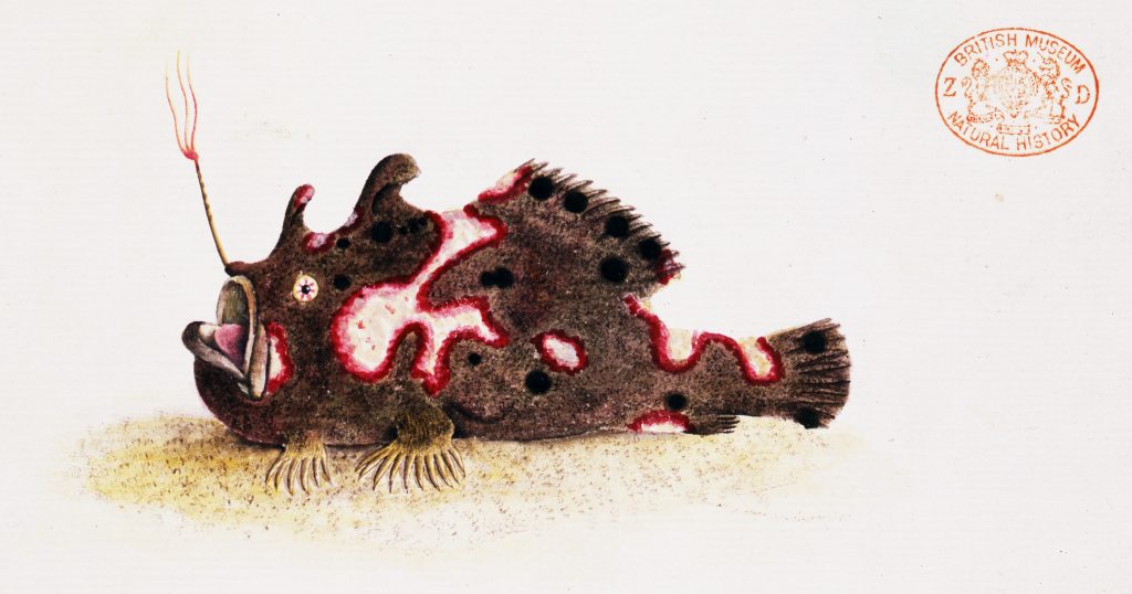
An illustration of the frogfish Antennarius pictus, published by George Shaw in 1794. From a new book by Ted Pietsch, UW professor of emeritus of aquatic and fishery sciences. Courtesy: University of Washington (state)
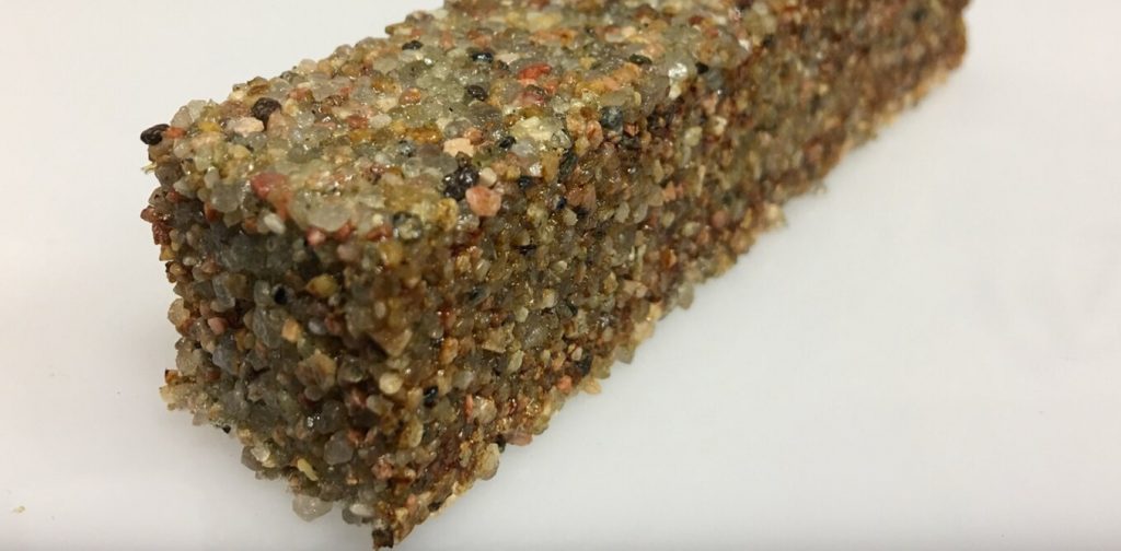
A block of sand particles held together by living cells. Credit: The University of Colorado Boulder College of Engineering and Applied Science
A March 24, 2020 news item on phys.org features the future of building construction as perceived by synthetic biologists,
Buildings are not unlike a human body. They have bones and skin; they breathe. Electrified, they consume energy, regulate temperature and generate waste. Buildings are organisms—albeit inanimate ones.
But what if buildings—walls, roofs, floors, windows—were actually alive—grown, maintained and healed by living materials? Imagine architects using genetic tools that encode the architecture of a building right into the DNA of organisms, which then grow buildings that self-repair, interact with their inhabitants and adapt to the environment.
…
A March 23, 2020 essay by Wil Srubar (Professor of Architectural Engineering and Materials Science, University of Colorado Boulder), which originated the news item, provides more insight,
Living architecture is moving from the realm of science fiction into the laboratory as interdisciplinary teams of researchers turn living cells into microscopic factories. At the University of Colorado Boulder, I lead the Living Materials Laboratory. Together with collaborators in biochemistry, microbiology, materials science and structural engineering, we use synthetic biology toolkits to engineer bacteria to create useful minerals and polymers and form them into living building blocks that could, one day, bring buildings to life.
In our most recent work, published in Matter, we used photosynthetic cyanobacteria to help us grow a structural building material – and we kept it alive. Similar to algae, cyanobacteria are green microorganisms found throughout the environment but best known for growing on the walls in your fish tank. Instead of emitting CO2, cyanobacteria use CO2 and sunlight to grow and, in the right conditions, create a biocement, which we used to help us bind sand particles together to make a living brick.
By keeping the cyanobacteria alive, we were able to manufacture building materials exponentially. We took one living brick, split it in half and grew two full bricks from the halves. The two full bricks grew into four, and four grew into eight. Instead of creating one brick at a time, we harnessed the exponential growth of bacteria to grow many bricks at once – demonstrating a brand new method of manufacturing materials.
Researchers have only scratched the surface of the potential of engineered living materials. Other organisms could impart other living functions to material building blocks. For example, different bacteria could produce materials that heal themselves, sense and respond to external stimuli like pressure and temperature, or even light up. If nature can do it, living materials can be engineered to do it, too.
It also take less energy to produce living buildings than standard ones. Making and transporting today’s building materials uses a lot of energy and emits a lot of CO2. For example, limestone is burned to make cement for concrete. Metals and sand are mined and melted to make steel and glass. The manufacture, transport and assembly of building materials account for 11% of global CO2 emissions. Cement production alone accounts for 8%. In contrast, some living materials, like our cyanobacteria bricks, could actually sequester CO2.
…
The field of engineered living materials is in its infancy, and further research and development is needed to bridge the gap between laboratory research and commercial availability. Challenges include cost, testing, certification and scaling up production. Consumer acceptance is another issue. For example, the construction industry has a negative perception of living organisms. Think mold, mildew, spiders, ants and termites. We’re hoping to shift that perception. Researchers working on living materials also need to address concerns about safety and biocontamination.
The [US] National Science Foundation recently named engineered living materials one of the country’s key research priorities. Synthetic biology and engineered living materials will play a critical role in tackling the challenges humans will face in the 2020s and beyond: climate change, disaster resilience, aging and overburdened infrastructure, and space exploration.
…
If you have time and interest, this is fascinating. Strubar is a little exuberant and, at this point, I welcome it.
Fitness
The Lithuanians are here for us. Scientists from the Kaunas University of Technology have just published a paper on better exercises for lower back pain in our increasingly sedentary times, from a March 23, 2020 Kaunas University of Technology press release (also on EurekAlert) Note: There are a few minor grammatical issues,
With the significant part of the global population forced to work from home, the occurrence of lower back pain may increase. Lithuanian scientists have devised a spinal stabilisation exercise programme for managing lower back pain for people who perform a sedentary job. After testing the programme with 70 volunteers, the researchers have found that the exercises are not only efficient in diminishing the non-specific lower back pain, but their effect lasts 3 times longer than that of a usual muscle strengthening exercise programme.
According to the World Health Organisation, lower back pain is among the top 10 diseases and injuries that are decreasing the quality of life across the global population. It is estimated that non-specific low back pain is experienced by 60% to 70% of people in industrialised societies. Moreover, it is the leading cause of activity limitation and work absence throughout much of the world. For example, in the United Kingdom, low back pain causes more than 100 million workdays lost per year, in the United States – an estimated 149 million.
Chronic lower back pain, which starts from long-term irritation or nerve injury affects the emotions of the afflicted. Anxiety, bad mood and even depression, also the malfunctioning of the other bodily systems – nausea, tachycardia, elevated arterial blood pressure – are among the conditions, which may be caused by lower back pain.
During the coronavirus disease (COVID-19) outbreak, with a significant part of the global population working from home and not always having a properly designed office space, the occurrence of lower back pain may increase.
“Lower back pain is reaching epidemic proportions. Although it is usually clear what is causing the pain and its chronic nature, people tend to ignore these circumstances and are not willing to change their lifestyle. Lower back pain usually comes away itself, however, the chances of the recurring pain are very high”, says Dr Irina Klizienė, a researcher at Kaunas University of Technology (KTU) Faculty of Social Sciences, Humanities and Arts.
Dr Klizienė, together with colleagues from KTU and from Lithuanian Sports University has designed a set of stabilisation exercises aimed at strengthening the muscles which support the spine at the lower back, i.e. lumbar area. The exercise programme is based on Pilates methodology.
According to Dr Klizienė, the stability of lumbar segments is an essential element of body biomechanics. Previous research evidence shows that in order to avoid the lower back pain it is crucial to strengthen the deep muscles, which are stabilising the lumbar area of the spine. One of these muscles is multifidus muscle.
“Human central nervous system is using several strategies, such as preparing for keeping the posture, preliminary adjustment to the posture, correcting the mistakes of the posture, which need to be rectified by specific stabilising exercises. Our aim was to design a set of exercises for this purpose”, explains Dr Klizienė.
The programme, designed by Dr Klizienė and her colleagues is comprised of static and dynamic exercises, which train the muscle strength and endurance. The static positions are to be held from 6 to 20 seconds; each exercise to be repeated 8 to 16 times.
Caption: The static positions are to be held from 6 to 20 seconds; each exercise to be repeated 8 to 16 times. Credit: KTU
The previous set is a little puzzling but perhaps you’ll find these ones below easier to follow,
Caption: The exercises are aimed at strengthening the muscles which support the spine at the lower back. Credit: KTU
I think more pictures of intervening moves would have been useful. Now. getting back to the press release,
In order to check the efficiency of the programme, 70 female volunteers were randomly enrolled either to the lumbar stabilisation exercise programme or to a usual muscle strengthening exercise programme. Both groups were exercising twice a week for 45 minutes for 20 weeks. During the experiment, ultrasound scanning of the muscles was carried out.
As soon as 4 weeks in lumbar stabilisation programme, it was observed that the cross-section area of the multifidus muscle of the subjects of the stabilisation group has increased; after completing the programme, this increase was statistically significant (p < 0,05). This change was not observed in the strengthening group.
Moreover, although both sets of exercises were efficient in eliminating lower back pain and strengthening the muscles of the lower back area, the effect of stabilisation exercises lasted 3 times longer – 12 weeks after the completion of the stabilisation programme against 4 weeks after the completion of the muscle strengthening programme.
“There are only a handful of studies, which have directly compared the efficiency of stabilisation exercises against other exercises in eliminating lower back pain”, says Dr Klizienė, “however, there are studies proving that after a year, lower back pain returned only to 30% of people who have completed a stabilisation exercise programme, and to 84% of people who haven’t taken these exercises. After three years these proportions are 35% and 75%.”
According to her, research shows that the spine stabilisation exercises are more efficient than medical intervention or usual physical activities in curing the lower back pain and avoiding the recurrence of the symptoms in the future.
The dream is to find sunscreens that don’t endanger humans or pollute the environment and it seems that Spanish scientists may have taken a step closer to making that dream a reality (from a Jan. 30, 2017 Wiley Publications press release (also on EurekAlert),
The ideal sunscreen should block UVB and UVA radiation while being safe and stable. In the journal Angewandte Chemie, Spanish scientists have introduced a new family of UVA and UVB filters based on natural sunscreen substances found in algae and cyanobacteria. They are highly stable and enhance the effectivity [sic] of commercial sunscreens.
Good news for sunseekers. Commercial [sic] available sunscreen lotions can very effectively protect from dangerous radiation in the ultraviolet [spectrum], but they need to be applied regularly and in high amounts to develop their full potential. One of the most critical issues is the limited stability of the UV filter molecules. Inspired by nature, Diego Sampedro and his colleagues from La Rioja University in Logrono and collaborators from Malaga University and Alcala University, Madrid, Spain, have screened a natural class of UV-protecting [blocking?] molecules for their possible use in skin protection. They adjusted the nature-given motif [sic] to the requirements of chemical synthesis and found that the molecules could indeed boost the sun protection factor of common formulations.
The natural sunscreen molecules are called microsporine-like amino acids (MAAs) and are widespread in the microbial world, most prominently in marine algae and cyanobacteria. MAAs are small molecules derived from amino acids, thermally stable, and they absorb light in the ultraviolet region, protecting the microbial DNA from radiation damage. Thus they are natural sunscreens, which inspired Sampedro and his colleagues to create [a] new class of organic sunscreen compounds.
Theoretical calculations revealed what is chemically needed for a successful design. “We performed a computer calculation of several basic scaffolds [..] to identify the simplest compound that fulfills the requisites for efficient sunscreens”, the authors write. The result of their search was a set of molecules which were readily synthesized, “avoiding the decorating substituents that come from the biosynthetic route.” Thus the small basic molecules can be tuned to give them more favorable properties.
The authors found that the synthesized compounds are characterized by excellent filter capacities in the relevant UV range. In addition they are photostable, much more than, for example, oxybenzene [sic] which is a widely used sunscreen in commercial formulations. They do not react chemically and dissipate radiation as heat (but not to such an extent that the skin temperature would rise as well). And, most importantly, when tested in real formulations, the sun protection factor (SPF) rose by a factor of more than two. Thus they could be promising targets for more stable, more efficient sunscreen lotions. Good news for your next summer vacation.
There’s some unusual phrasing so, I’m guessing that the writer it not accustomed to writing press releases in English. One other comment, it’s oxybenzone that’s often used as an ingredient in commercial sunscreens.
Here’s a link to and a citation for the paper,
Rational Design and Synthesis of Efficient Sunscreens To Boost the Solar Protection Factor by Raúl Losantos, Ignacio Funes-Ardoiz, Dr. José Aguilera, Prof. Enrique Herrera-Ceballos, Dr. Cristina García-Iriepa, Prof. Pedro J. Campos, and Diego Sampedro. Angewandte Chemie International Edition Volume 56, Issue 10, pages 2632–2635, March 1, 2017 DOI: 10.1002/anie.201611627 Version of Record online: 27 JAN 2017
I have previously featured work on another natural sunscreen. In that case it was to be derived from English ivy (July 22, 2010 posting); there was an update on the English ivy work in a May 30, 2016 posting but the researcher has moved in a different direction looking at wound healing and armour as possible applications for the research.
This story made me think of fractals where a pattern at one scale is repeated at a smaller scale. Here’s more about the earth’s rotation and the protein from a June 25, 2015 news item on ScienceDaily,
A collaborative group of Japanese researchers has demonstrated that the Earth’s daily rotation period (24 hours) is encoded in the KaiC protein at the atomic level, a small, 10 nm-diameter biomolecule expressed in cyanobacterial cells.
For anyone who’s unfamiliar (me) with cyanobacteria, here’s a definition from its Wikipedia entry (Note: Links have been removed),
Cyanobacteria /saɪˌænoʊbækˈtɪəriə/, also known as Cyanophyta, is a phylum of bacteria that obtain their energy through photosynthesis.[3] The name “cyanobacteria” comes from the color of the bacteria (Greek: κυανός (kyanós) = blue). They are often called blue-green algae (but some consider that name a misnomer, as cyanobacteria are prokaryotic and algae should be eukaryotic,[4] although other definitions of algae encompass prokaryotic organisms).[5]
By producing gaseous oxygen as a byproduct of photosynthesis, cyanobacteria are thought to have converted the early reducing atmosphere into an oxidizing one, causing the “rusting of the Earth”[6] and dramatically changing the composition of life forms on Earth by stimulating biodiversity and leading to the near-extinction of oxygen-intolerant organisms. According to endosymbiotic theory, the chloroplasts found in plants and eukaryotic algae evolved from cyanobacterial ancestors via endosymbiosis.
The idea that cyanobacteria may have changed the earth’s atmosphere into an oxidizing one and stimulating biodiversity is fascinating to me. Plus, cyanobacteria are pretty,
CC BY-SA 3.0 File:Tolypothrix (Cyanobacteria).JPG Uploaded by Matthewjparker Created: January 22, 2013 Location: 29° 38′ 58.2″ N, 82° 20′ 40.8″ W [downloaded from https://en.wikipedia.org/wiki/Cyanobacteria]
The results of this joint research will help elucidate a longstanding question in chronobiology: How is the circadian period of biological clocks determined? The results will also help understand the basic molecular mechanism of the biological clock. This knowledge might contribute to the development of therapies for disorders associated with abnormal circadian rhythms.
The results will be disclosed online on June 25, 2015 (North American Eastern Standard Time) in ScienceExpress, the electronic version of Science, published by the American Association for the Advancement of Science (AAAS). 1. Research Background
In accordance with diurnal changes in the environment (notably light intensity and temperature) resulting from the Earth’s daily rotation around its axis, many organisms regulate their biological activities to ensure optimal fitness and efficiency. The biological clock refers to the mechanism whereby organisms adjust the timing of their biological activities. The period of this clock is set to approximately 24 hours. A wide range of studies have investigated the biological clock in organisms ranging from bacteria to mammals. Consequently, the relationship between the biological clock and multiple diseases has been clarified. However, it remains unclear how 24-hour circadian rhythms are implemented.
The research group mentioned above addressed this question using cyanobacteria. The cyanobacterial circadian clock can be reconstructed by mixing three clock proteins (KaiA, KaiB, and KaiC) and ATP. A study published in 2007 showed that KaiC ATPase activity, which mediates the ATP hydrolysis reaction, is strongly associated with circadian periodicity. The results of that study indicated that the functional structure of KaiC could be responsible for determining the circadian rhythm.
Figure 1 Earth and the circadian clock protein KaiC 2. Research Results
KaiC ATPase activity exhibits a robust circadian oscillation in the presence of KaiA and KaiB proteins (Figure 2). In the study reported here, the temporal profile of KaiC ATPase activity exhibited an attenuating and oscillating component even in the absence of KaiA and KaiB. A close analysis revealed that this signal had a frequency of 0.91 day-1, which approximately coincided with the 24-hour period. Thus, KaiC is the source of a steady cycle that is in tune with the Earth’s daily rotation.
Figure 2 KaiC ATPase activity-time profile
To identify causal structural factors, the N-terminal domain of KaiC was analyzed using high-resolution crystallography. The resultant atomic structures revealed the underlying cause of KaiC’s slowness relative to other ATPases (Figure 3). “A water molecule is prevented from attacking into the ideal position (a black dot in Figure 3) for the ATP hydrolysis by a steric hindrance near ATP phosphoryl groups. In addition, this hindrance is surely anchored to a spring-like structure derived from polypeptide isomerization,” elaborates Dr. Jun Abe. “The ATP hydrolysis, which involves access of a water molecule to the bound ATP and reverse isomerization of the polypeptide, is expected to require a significantly larger amount of free energy than for typical ATP hydrolysis. Thus, the three-dimensional atomic structure discovered in this study explains why the ATPase activity of KaiC is so much lower (by 100- to 1,000,000-fold) than that of typical ATPase molecules.”
Figure 3 Structural basis for steady slowness. The steric barrier prevents access of a water molecule to the catalytic site (indicated by a black dot).
The circadian clock’s period is independent of ambient temperature, a phenomenon known as temperature compensation. One KaiC molecule is composed of six identical subunits, each containing duplicated domains with a series of ATPase motifs. The asymmetric atomic-scale regulation by the aforementioned mechanism dictates a feedback mechanism that maintains the ATPase activity at a constant low level. The authors of this study discovered that the Earth’s daily rotation period (24 hours) is implemented as the time constant of the feedback mechanism mediated in this protein structure.
3. Technological Implications
KaiC and other protein molecules are capable of moving on short time scales, on the order of 10-12 to 10-1 seconds. This study provides the first atomic-level demonstration that small protein molecules can generate 24-hour rhythms by regulating molecular structure and reactivity. Lab head and CIMoS Director Prof. Shuji Akiyama sees, “The fact that a water molecule, ATP, the polypeptide chain, and other universal biological components are involved in this regulation suggests that humans and other complex organisms may also share a similar molecular machinery. In the crowded intracellular environment that contains a myriad of molecular signals, KaiC demonstrates long-paced oscillations using a small amount of energy generated through ATP consumption. This clever mechanism for timekeeping in a noisy environment may inspire development of highly efficient and sustainable chemical reaction processes and molecular-system-based information processing.” 4. Glossary
1) Clock protein
A clock protein plays an essential role in the circadian pacemaker. Mutations and deficiencies in clock proteins can alter the intrinsic characteristics of circadian rhythm.
2) ATP
Adenosine triphosphate is a source of energy required for muscle contraction and many other biological activities. ATP, a nucleotide that mediates the storage and consumption of energy, is sometimes referred to as the “currency of biological energy” due to its universality and importance in metabolism. ATP consists of an adenosine molecule bound to three phosphate groups. Upon hydrolysis, the ATPase releases one phosphate molecule plus approximately 8 kcal/mol of energy.
3) Polypeptide isomerization
Protein polypeptide main chains undergo isomerization on a time scale of seconds or longer; therefore, protein isomerization is one of the slowest biological reactions. Most functional protein main chains have a trans conformation, and a few proteins have a functional cis conformation.
Here’s a link to and a citation for the paper,
Atomic-scale origins of slowness in the cyanobacterial circadian clock by Jun Abe, Takuya B. Hiyama, Atsushi Mukaiyama, Seyoung Son, Toshifumi Mori, Shinji Saito, Masato Osako, Julie Wolanin, Eiki Yamashita, Takao Kondo, & Shuji Akiyama. Science DOI: 10.1126/science.1261040 Published Online June 25 2015 (on Science Express)
This paper is behind a paywall.
Kudos to the person(s) who wrote the news release.
The American Chemical Society (ACS) is holding its 245th meeting April 7 – 11, 2013 and its first International Symposium on Bacterial Nanocellulose simultaneously. I have written about nanocellulose previously but it’s always been concerned with the type derived from plant matter; bacterial nanocellulose is new to me but not the scientific community as the Apr. 8, 2013 news item on Azonano notes,
In the 1800s, French scientist Louis Pasteur first discovered that vinegar-making [and Kombucha tea and nata de coco] bacteria make “a sort of moist skin, swollen, gelatinous and slippery” — a “skin” now known as bacterial nanocellulose. Nanocellulose made by bacteria has advantages, including ease of production and high purity that fostered the kind of scientific excitement reflected in the first international symposium on the topic, Brown [R. Malcolm Brown, Jr., Ph.D.] pointed out.
Before going on to this latest research, here’s a description of cellulose and nanocellulose as per its presence in plant material (from the news item),
Cellulose is the most abundant organic polymer on Earth, a material, like plastics, consisting of molecules linked together into long chains. Cellulose makes up tree trunks and branches, corn stalks and cotton fibers, and it is the main component of paper and cardboard. People eat cellulose in “dietary fiber,” the indigestible material in fruits and vegetables. Cows, horses and termites can digest the cellulose in grass, hay and wood.
Most cellulose consists of wood fibers and cell wall remains. Very few living organisms can actually synthesize and secrete cellulose in its native nanostructure form of microfibrils. At this level, nanometer-scale fibrils are very hydrophilic and look like jelly. A nanometer is one-millionth the thickness of a U.S. dime. Nevertheless, cellulose shares the unique properties of other nanometer-sized materials — properties much different from large quantities of the same material. Nanocellulose-based materials can be stronger than steel and stiffer than Kevlar. Great strength, light weight and other advantages has fostered interest in using it in everything from lightweight armor and ballistic glass to wound dressings and scaffolds for growing replacement organs for transplantation.
A new kind of bacteria actively entered the nanocellulose picture in 2001 (from the news item) allowing Brown to exploit research he had been pursuing since the 1970s (from the news item),
Brown recalled that in 2001, a discovery by David Nobles, Ph.D., a member of the research team at the University of Texas at Austin, refocused their research on nanocellulose, but with a different microbe. Nobles established that several kinds of blue-green algae, which are mainly photosynthetic bacteria much like the vinegar-making bacteria in basic structure; however, these blue-green algae, or cyanobacteria, as they are called, can produce nanocellulose. One of the largest problems with cyanobacterial nanocellulose is that it is not made in abundant amounts in nature. If it could be scaled up, Brown describes this as “one of the most important discoveries in plant biology.”
While I find the science interesting, it’s Brown’s comments about the policy and politics of commercializing nanocellulose-based fuels that intrigue me (from the news item),
In his report at the ACS meeting, Brown described how his team already has genetically engineered the cyanobacteria to produce one form of nanocellulose, the long-chain, or polymer, form of the material. And they are moving ahead with the next step, engineering the cyanobacteria to synthesize a more complete form of nanocellulose, one that is a polymer with a crystalline architecture. He also said that operations are being scaled up, with research moving from laboratory-sized tests to larger outdoor facilities.
Brown expressly pointed out that one of the major barriers to commercializing nanocellulose fuels involves national policy and politics, rather than science. Biofuels, he said, will face a difficult time for decades into the future in competing with the less-expensive natural gas now available with hydraulic fracturing, or “fracking.” [emphasis mine] In the long run, the United States will need sustainable biofuels, he said, citing the importance of national energy policies that foster parallel development and commercialization of biofuels.


![CC BY-SA 3.0 File:Tolypothrix (Cyanobacteria).JPG Uploaded by Matthewjparker Created: January 22, 2013 Location: 29° 38′ 58.2″ N, 82° 20′ 40.8″ W [downloaded from https://en.wikipedia.org/wiki/Cyanobacteria]](http://www.frogheart.ca/wp-content/uploads/2015/06/Cyanobacteria.jpg)
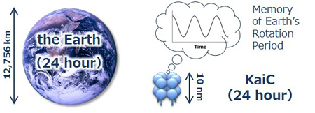
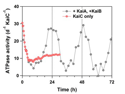
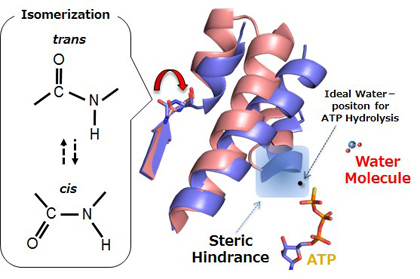 Figure 3 Structural basis for steady slowness. The steric barrier prevents access of a water molecule to the catalytic site (indicated by a black dot).