Tag Archives: cyborg tissue

Brain and machine as one (machine/flesh)

The essay on brains and machines becoming intertwined is making the rounds. First stop on my tour was its Oct. 4, 2016 appearance on the Mail & Guardian, then there was its Oct. 3, 2016 appearance on The Conversation, and finally (moving forward in time) there was its Oct. 4, 2016 appearance on the World Economic Forum website as part of their Final Frontier series.

The essay was written by Richard Jones of Sheffield University (mentioned here many times before but most recently in a Sept. 4, 2014 posting). His book ‘Soft Machines’ provided me with an important and eminently readable introduction to nanotechnology. He is a professor of physics at the University of Sheffield and here’s more from his essay (Oct. 3, 2016 on The Conversation) about brains and machines (Note: Links have been removed),

Imagine a condition that leaves you fully conscious, but unable to move or communicate, as some victims of severe strokes or other neurological damage experience. This is locked-in syndrome, when the outward connections from the brain to the rest of the world are severed. Technology is beginning to promise ways of remaking these connections, but is it our ingenuity or the brain’s that is making it happen?

Ever since an 18th-century biologist called Luigi Galvani made a dead frog twitch we have known that there is a connection between electricity and the operation of the nervous system. We now know that the signals in neurons in the brain are propagated as pulses of electrical potential, whose effects can be detected by electrodes in close proximity. So in principle, we should be able to build an outward neural interface system – that is to say, a device that turns thought into action.

In fact, we already have the first outward neural interface system to be tested in humans. It is called BrainGate and consists of an array of micro-electrodes, implanted into the part of the brain concerned with controlling arm movements. Signals from the micro-electrodes are decoded and used to control the movement of a cursor on a screen, or the motion of a robotic arm.

A crucial feature of these systems is the need for some kind of feedback. A patient must be able to see the effect of their willed patterns of thought on the movement of the cursor. What’s remarkable is the ability of the brain to adapt to these artificial systems, learning to control them better.

You can find out more about BrainGate in my May 17, 2012 posting which also features a video of a woman controlling a mechanical arm so she can drink from a cup coffee by herself for the first time in 15 years.

Jones goes on to describe the cochlear implants (although there’s no mention of the controversy; not everyone believes they’re a good idea) and retinal implants that are currently available. Jones notes this (Note Links have been removed),

The key message of all this is that brain interfaces now are a reality and that the current versions will undoubtedly be improved. In the near future, for many deaf and blind people, for people with severe disabilities – including, perhaps, locked-in syndrome – there are very real prospects that some of their lost capabilities might be at least partially restored.

Until then, our current neural interface systems are very crude. One problem is size; the micro-electrodes in use now, with diameters of tens of microns, may seem tiny, but they are still coarse compared to the sub-micron dimensions of individual nerve fibres. And there is a problem of scale. The BrainGate system, for example, consists of 100 micro-electrodes in a square array; compare that to the many tens of billions of neurons in the brain. The fact these devices work at all is perhaps more a testament to the adaptability of the human brain than to our technological prowess.

Scale models

So the challenge is to build neural interfaces on scales that better match the structures of biology. Here, we move into the world of nanotechnology. There has been much work in the laboratory to make nano-electronic structures small enough to read out the activity of a single neuron. In the 1990s, Peter Fromherz, at the Max Planck Institute for Biochemistry, was a pioneer of using silicon field effect transistors, similar to those used in commercial microprocessors, to interact with cultured neurons. In 2006, Charles Lieber’s group at Harvard succeeded in using transistors made from single carbon nanotubes – whiskers of carbon just one nanometer in diameter – to measure the propagation of single nerve pulses along the nerve fibres.

But these successes have been achieved, not in whole organisms, but in cultured nerve cells which are typically on something like the surface of a silicon wafer. It’s going to be a challenge to extend these methods into three dimensions, to interface with a living brain. Perhaps the most promising direction will be to create a 3D “scaffold” incorporating nano-electronics, and then to persuade growing nerve cells to infiltrate it to create what would in effect be cyborg tissue – living cells and inorganic electronics intimately mixed.

I have featured Charles Lieber and his work here in two recent posts: ‘Bionic’ cardiac patch with nanoelectric scaffolds and living cells on July 11, 2016 and Long-term brain mapping with injectable electronics on Sept. 22, 2016.

For anyone interested in more about the controversy regarding cochlear implants, there’s this page on the Brown University (US) website. You might also want to check out Gregor Wolbring (professor at the University of Calgary) who has written extensively on the concept of ableism (links to his work can be found at the end of this post). I have excerpted from an Aug. 30, 2011 post the portion where Gregor defines ‘ableism’,

From Gregor’s June 17, 2011 posting on the FedCan blog,

The term ableism evolved from the disabled people rights movements in the United States and Britain during the 1960s and 1970s.  It questions and highlights the prejudice and discrimination experienced by persons whose body structure and ability functioning were labelled as ‘impaired’ as sub species-typical. Ableism of this flavor is a set of beliefs, processes and practices, which favors species-typical normative body structure based abilities. It labels ‘sub-normative’ species-typical biological structures as ‘deficient’, as not able to perform as expected.

The disabled people rights discourse and disability studies scholars question the assumption of deficiency intrinsic to ‘below the norm’ labeled body abilities and the favoritism for normative species-typical body abilities. The discourse around deafness and Deaf Culture would be one example where many hearing people expect the ability to hear. This expectation leads them to see deafness as a deficiency to be treated through medical means. In contrast, many Deaf people see hearing as an irrelevant ability and do not perceive themselves as ill and in need of gaining the ability to hear. Within the disabled people rights framework ableism was set up as a term to be used like sexism and racism to highlight unjust and inequitable treatment.

Ableism is, however, much more pervasive.

You can find out more about Gregor and his work here: http://www.crds.org/research/faculty/Gregor_Wolbring2.shtml or here:
https://www.facebook.com/GregorWolbring.
Long-term brain mapping with injectable electronics

Charles Lieber and his team at Harvard University announced a success with their work on injectable electronics last year (see my June 11, 2015 posting for more) and now they are reporting on their work with more extensive animal studies according to an Aug. 29, 2016 news item on psypost.org,

Scientists in recent years have made great strides in the quest to understand the brain by using implanted probes to explore how specific neural circuits work.

Though effective, those probes also come with their share of problems as a result of rigidity. The inflammation they produce induces chronic recording instability and means probes must be relocated every few days, leaving some of the central questions of neuroscience – like how the neural circuits are reorganized during development, learning and aging- beyond scientists’ reach.

But now, it seems, things are about to change.

Led by Charles Lieber, The Mark Hyman Jr. Professor of Chemistry and chair of the Department of Chemistry and Chemical Biology, a team of researchers that included graduate student Tian-Ming Fu, postdoctoral fellow Guosong Hong, graduate student Tao Zhou and others, has demonstrated that syringe-injectable mesh electronics can stably record neural activity in mice for eight months or more, with none of the inflammation

An Aug. 29, 2016 Harvard University press release, which originated the news item, provides more detail,

“With the ability to follow the same individual neurons in a circuit chronically…there’s a whole suite of things this opens up,” Lieber said. “The eight months we demonstrate in this paper is not a limit, but what this does show is that mesh electronics could be used…to investigate neuro-degenerative diseases like Alzheimer’s, or processes that occur over long time, like aging or learning.”

Lieber and colleagues also demonstrated that the syringe-injectable mesh electronics could be used to deliver electrical stimulation to the brain over three months or more.

“Ultimately, our aim is to create these with the goal of finding clinical applications,” Lieber said. “What we found is that, because of the lack of immune response (to the mesh electronics), which basically insulates neurons, we can deliver stimulation in a much more subtle way, using lower voltages that don’t damage tissue.”

The possibilities, however, don’t end there.

The seamless integration of the electronics and biology, Lieber said, could open the door to an entirely new class of brain-machine interfaces and vast improvements in prosthetics, among other fields.

“Today, brain-machine interfaces are based on traditional implanted probes, and there has been some impressive work that’s been done in that field,” Lieber said. “But all the interfaces rely on the same technique to decode neural signals.”

Because traditional rigid implanted probes are invariably unstable, he explained, researchers and clinicians rely on decoding what they call the “population average” – essentially taking a host of neural signals and applying complex computational tools to determine what they mean.

Using tissue-like mesh electronics, by comparison, researchers may be able to read signals from specific neurons over time, potentially allowing for the development of improved brain-machine interfaces for prosthetics.

“We think this is going to be very powerful, because we can identify circuits and both record and stimulate in a way that just hasn’t been possible before,” Lieber said. “So what I like to say is: I think therefore it happens.”

Lieber even held out the possibility that the syringe-injectable mesh electronics could one day be used to treat catastrophic injuries to the brain and spinal cord.

“I don’t think that’s science-fiction,” he said. “Other people may say that will be possible through, for example, regenerative medicine, but we are pursuing this from a different angle.

“My feeling is that this is about a seamless integration between the biological and the electronic systems, so they’re not distinct entities,” he continued. “If we can make the electronics look like the neural network, they will work together…and that’s where you want to be if you want to exploit the strengths of both.”

In the 2015 posting, Lieber was discussing cyborgs, here he broaches the concept without using the word, “… seamless integration between the biological and the electronic systems, so they’re not distinct entities.”

Here’s a link to and a citation for the paper,

Stable long-term chronic brain mapping at the single-neuron level by Tian-Ming Fu, Guosong Hong, Tao Zhou, Thomas G Schuhmann, Robert D Viveros, & Charles M Lieber. Nature Methods (2016) doi:10.1038/nmeth.3969 Published online 29 August 2016

This paper is behind a paywall.

Injectable electronics

Having taught a course on bioelectronics for Simon Fraser University’s (Vancouver, Canada) Continuing Studies Program, this  latest work from Harvard University (US) caught my attention. A Harvard research team has developed a technique which could allow doctors to inject us with electronics, should we need them. From a June 8, 2015 news item on phys.org,

It’s a notion that might be pulled from the pages of science-fiction novel – electronic devices that can be injected directly into the brain, or other body parts, and treat everything from neurodegenerative disorders to paralysis.

It sounds unlikely, until you visit Charles Lieber’s lab.

A team of international researchers, led by Lieber, the Mark Hyman, Jr. Professor of Chemistry, an international team of researchers developed a method for fabricating nano-scale electronic scaffolds that can be injected via syringe. Once connected to electronic devices, the scaffolds can be used to monitor neural activity, stimulate tissues and even promote regenerations of neurons. …

Here’s an image provided by the researchers,

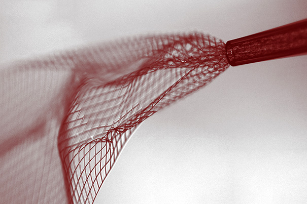
Bright-field image showing the mesh electronics being injected through sub-100 micrometer inner diameter glass needle into aqueous solution. mage courtesy of Lieber Research Group, Harvard University

Bright-field image showing the mesh electronics being injected through sub-100 micrometer inner diameter glass needle into aqueous solution. mage courtesy of Lieber Research Group, Harvard University

A June 8, 2015 Harvard University new release by Peter Reuell (also on EurekAlert), which originated the news item, describes the work in more detail,

“I do feel that this has the potential to be revolutionary,” Lieber said. “This opens up a completely new frontier where we can explore the interface between electronic structures and biology. For the past thirty years, people have made incremental improvements in micro-fabrication techniques that have allowed us to make rigid probes smaller and smaller, but no one has addressed this issue – the electronics/cellular interface – at the level at which biology works.”

The idea of merging the biological with the electronic is not a new one for Lieber.

In an earlier study, scientists in Lieber’s lab demonstrated that the scaffolds could be used to create “cyborg” tissue – when cardiac or nerve cells were grown with embedded scaffolds. [emphasis mine] Researchers were then able to use the devices to record electrical signals generated by the tissues, and to measure changes in those signals as they administered cardio- or neuro-stimulating drugs.

“We were able to demonstrate that we could make this scaffold and culture cells within it, but we didn’t really have an idea how to insert that into pre-existing tissue,” Lieber said. “But if you want to study the brain or develop the tools to explore the brain-machine interface, you need to stick something into the body. When releasing the electronics scaffold completely from the fabrication substrate, we noticed that it was almost invisible and very flexible like a polymer and could literally be sucked into a glass needle or pipette. From there, we simply asked, would it be possible to deliver the mesh electronics by syringe needle injection, a process common to delivery of many species in biology and medicine – you could go to the doctor and you inject this and you’re wired up.'”

Though not the first attempts at implanting electronics into the brain – deep brain stimulation has been used to treat a variety of disorders for decades – the nano-fabricated scaffolds operate on a completely different scale.

“Existing techniques are crude relative to the way the brain is wired,” Lieber explained. “Whether it’s a silicon probe or flexible polymers…they cause inflammation in the tissue that requires periodically changing the position or the stimulation. But with our injectable electronics, it’s as if it’s not there at all. They are one million times more flexible than any state-of-the-art flexible electronics and have subcellular feature sizes. They’re what I call “neuro-philic” – they actually like to interact with neurons..”

Despite their enormous potential, the fabrication of the injectable scaffolds is surprisingly easy.

“That’s the beauty of this – it’s compatible with conventional manufacturing techniques,” Lieber said.

The process is similar to that used to etch microchips, and begins with a dissolvable layer deposited on a substrate. To create the scaffold, researchers lay out a mesh of nanowires sandwiched in layers of organic polymer. The first layer is then dissolved, leaving the flexible mesh, which can be drawn into a syringe needle and administered like any other injection.

After injection, the input/output of the mesh can be connected to standard measurement electronics so that the integrated devices can be addressed and used to stimulate or record neural activity.

“These type of things have never been done before, from both a fundamental neuroscience and medical perspective,” Lieber said. “It’s really exciting – there are a lot of potential applications.”

Going forward, Lieber said, researchers hope to better understand how the brain and other tissues react to the injectable electronics over longer periods.

Lieber’s earlier work on “cyborg tissue” was briefly mentioned here in a Feb. 20, 2014 posting.

Getting back to the most recent work, here’s a link to and a citation for the paper,

Syringe-injectable electronics by Jia Liu, Tian-Ming Fu, Zengguang Cheng, Guosong Hong, Tao Zhou, Lihua Jin, Madhavi Duvvuri, Zhe Jiang, Peter Kruskal, Chong Xie, Zhigang Suo, Ying Fang, & Charles M. Lieber. Nature Nanotechnology (2015) doi:10.1038/nnano.2015.115 Published online 08 June 2015

This paper is behind a paywall but there is a free preview via ReadCube Access.

One final note, the researchers have tested the injectable electronics (or mesh electronics) in vivo (live animals).