Tag Archives: Daniel S. Kohane

No need for eye drops when your contact lenses can dispense your eye medication

Anyone who has difficulty getting or allowing drops into their eyes (I once slid out of an ophthalmologist’s examination chair trying to avoid the eye drops he was administering at the end of my appointment) is going appreciate this Dec. 9, 2013 news item on Nanowerk,

For nearly half a century, contact lenses have been proposed as a means of ocular drug delivery that may someday replace eye drops, but achieving controlled drug release has been a significant challenge. Researchers at Massachusetts Eye and Ear/Harvard Medical School Department of Ophthalmology, Boston Children’s Hospital, and the Massachusetts Institute of Technology are one step closer to an eye drop-free reality with the development of a drug-eluting contact lens designed for prolonged delivery of latanoprost, a common drug used for the treatment of glaucoma, the leading cause of irreversible blindness worldwide.

The Dec. 9, 2013 Massachusetts Eye and Ear Infirmary press release (also on EurekAlert), which originated the news item, notes that a lot of people have problems with eye drops and gives a general description of the research,

“In general, eye drops are an inefficient method of drug delivery that has notoriously poor patient adherence. This contact lens design can potentially be used as a treatment for glaucoma and as a platform for other ocular drug delivery applications,” said Joseph Ciolino, M.D, Mass. Eye and Ear cornea specialist and lead author of the paper.

The contacts were designed with materials that are FDA-approved for use on the eye. The latanoprost-eluting contact lenses were created by encapsulating latanoprost-polymer films in commonly used contact lens hydrogel. Their findings are described online and will be in the January 2014 printed issue of Biomaterials.

“The lens we have developed is capable of delivering large amounts of drug at substantially constant rates over weeks to months,” said Professor Daniel Kohane, director of the Laboratory for Biomaterials and Drug Delivery at Boston Children’s Hospital.

In vivo, single contact lenses were able to achieve, for one month, latanoprost concentrations in the aqueous humor that were comparable to those achieved with daily topical latanoprost solution, the current first-line treatment for glaucoma.

The lenses appeared safe in cell culture and animal studies. This is the first contact lens that has been shown to release drugs for this long in animal models.

The newly designed contact lens has a clear central aperture and contains a drug-polymer film in the periphery, which helps to control drug release. The lenses can be made with no refractive power or with the ability to correct the refractive error in near sided or far sided eyes.

Here’s a link to and a citation for the researchers’ published paper,

In vivo performance of a drug-eluting contact lens to treat glaucoma for a month by Joseph B. Ciolino, Cristina F. Stefanescu, Amy E. Ross, Borja Salvador-Culla, Priscila Cortez, Eden M. Ford, Kate A. Wymbs, Sarah L. Sprague, Daniel R. Mascoop, Shireen S. Rudina, Sunia A. Trauger, Fabiano Cade, Daniel S. Kohane. Biomaterials Volume 35, Issue 1, January 2014, Pages 432–439 DOI: S0142961213011150

This article is behind a paywall.
Medicine, nanoelectronics, social implications, and figuring it all out

Given today’s (Aug. 27, 2012) earlier posting about nanoelectronics and tissue engineering, I though it was finally time to feature Michael Berger’s Aug. 16, 2012 Nanowerk Spotlight essay, The future of nanotechnology electronics in medicine, which discusses the integration of electronics into the human body.

First, Berger offers a summary of some of the latest research (Note: I have removed  links),

In previous Nanowerk Spotlights we have already covered numerous research advances in this area: The development of a nanobioelectronic system that triggers enzyme activity and, in a similar vein, the electrically triggered drug release from smart nanomembranes; an artificial retina for color vision; nanomaterial-based breathalyzers as diagnostic tools; nanogenerators to power self-sustained biosystems and implants; future bio-nanotechnology might even use computer chips inside living cells.

A lot of nanotechnology work is going on in the area of brain research. For instance the use of a carbon nanotube rope to electrically stimlate neural stem cells; nanotechnology to repair the brain and other advances in fabricating nanomaterial-neural interfaces for signal generation.

International cooperation in this field has also picked up. Just recently, scientists have formed a global alliance for nanobioelectronics to rapidly find solutions for neurological disorders; the EuroNanoBio project is a Support Action funded under the 7th Framework Programme of the European Union; and ENIAC, the European Technology Platform on nanoelectronics, has decided to make the development of medical applications one of its main objectives.

Berger cites a recent article in the American Chemical Society’s (ACS) Nano (journal) by scientists in today’s earlier posting about tissue scaffolding and 3-D electrnonics,

In a new perspective article in the July 31, 2012, online edition of ACS Nano (“The Smartest Materials: The Future of Nanoelectronics in Medicine” [behind a paywall]), Tzahi Cohen-Karni (a researcher in Kohane’s lab), Robert Langer, and Daniel S. Kohane provide an overview of nanoelectronics’ potential in the biomedical sciences.

They write that, as with many other areas of scientific endeavor in recent decades, continued progress will require the convergence of multiple disciplines, including chemistry, biology, electrical engineering, computer science, optics, material science, drug delivery, and numerous medical disciplines. ”

Advances in this research could lead to extremely sophisticated smart materials with multifunctional capabilities that are built in – literally hard-wired. The impact of this research could cover the spectrum of biomedical possibilities from diagnostic studies to the creation of cyborgs.”

Berger finishes with this thought,

Ultimately, and here we are getting almost into science fiction territory, nanostructures could not only incorporate sensing and stimulating capabilities but also potentially introduce computational capabilities and energy-generating elements. “In this way, one could fabricate a truly independent system that senses and analyzes signals, initiates interventions, and is self-sustained. Future developments in this direction could, for example, lead to a synthetic nanoelectronic autonomic nervous system.”

This Nanowerk Spotlight essay provides a good overview of nanoelectronics  research in medicine and lots of  links to previous related essays and other related materials.

I am intrigued that there is no mention of the social implications for this research and I find social science or humanities research on social social implications of emerging technology rarely discusses the technical aspects revealing what seems to be an insurmountable gulf. I suppose that’s why we need writers, artists, musicians, dancers, pop culture, and the like to create experiences, installations, and narratives that help us examine the technologies and their social implications, up close.

The body as an electronic device—adding electronics to biological tissue

What makes this particular combination of electronic s  and living tissue special is t that it was achieved in 3-D rather than 2-D.  From the Boston Children’s Hospital Aug. 26, 2012 news release on EurekAlert,

A multi-institutional research team has developed a method for embedding networks of biocompatible nanoscale wires within engineered tissues. These networks—which mark the first time that electronics and tissue have been truly merged in 3D—allow direct tissue sensing and potentially stimulation, a potential boon for development of engineered tissues that incorporate capabilities for monitoring and stimulation, and of devices for screening new drugs.

The Aug. 27, 2012 news item on Nanowerk provides more detail about integration of the cells and electronics,

Until now, the only cellular platforms that incorporated electronic sensors consisted of flat layers of cells grown on planar metal electrodes or transistors. Those two-dimensional systems do not accurately replicate natural tissue, so the research team set out to design a 3-D scaffold that could monitor electrical activity, allowing them to see how cells inside the structure would respond to specific drugs.

The researchers built their new scaffold out of epoxy, a nontoxic material that can take on a porous, 3-D structure. Silicon nanowires embedded in the scaffold carry electrical signals to and from cells grown within the structure.

“The scaffold is not just a mechanical support for cells, it contains multiple sensors. We seed cells into the scaffold and eventually it becomes a 3-D engineered tissue,” Tian says [Bozhi Tian, a former postdoc at MIT {Massachusetts Institute of Technology} and Children’s Hospital and a lead author of the paper ].

The team chose silicon nanowires for electronic sensors because they are small, stable, can be safely implanted into living tissue and are more electrically sensitive than metal electrodes. The nanowires, which range in diameter from 30 to 80 nanometers (about 1,000 times smaller than a human hair), can detect voltages less than one-thousandth of a watt, which is the level of electricity that might be seen in a cell.

Here’s more about why the researchers want to integrate living tissue and electronics, from the Harvard University Aug. 26, 2012 news release on EurekAlert,

“The current methods we have for monitoring or interacting with living systems are limited,” said Lieber [Charles M. Lieber, the Mark Hyman, Jr. Professor of Chemistry at Harvard and one of the study’s team leaders]. “We can use electrodes to measure activity in cells or tissue, but that damages them. With this technology, for the first time, we can work at the same scale as the unit of biological system without interrupting it. Ultimately, this is about merging tissue with electronics in a way that it becomes difficult to determine where the tissue ends and the electronics begin.”

The research addresses a concern that has long been associated with work on bioengineered tissue – how to create systems capable of sensing chemical or electrical changes in the tissue after it has been grown and implanted. The system might also represent a solution to researchers’ struggles in developing methods to directly stimulate engineered tissues and measure cellular reactions.

“In the body, the autonomic nervous system keeps track of pH, chemistry, oxygen and other factors, and triggers responses as needed,” Kohane [Daniel Kohane, a Harvard Medical School professor in the Department of Anesthesia at Children’s Hospital Boston and a team leader] explained. “We need to be able to mimic the kind of intrinsic feedback loops the body has evolved in order to maintain fine control at the cellular and tissue level.”

Here’s a citation and a link to the paper (which is behind a paywall),

Macroporous nanowire nanoelectronic scaffolds for synthetic tissues by Bozhi Tian, Jia Lin, Tal Dvir, Lihua Jin, Jonathan H. Tsui, Quan  Qing, Zhigang Suo, Robert Langer, Daniel S. Kohane, and Charles M. Lieber in Nature Materials (2012) doi:10.1038/nmat3404 Published onlin26 August 2012.

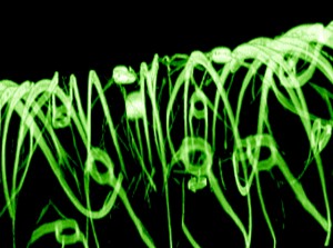
This is the image MIT included with its Aug 27, 2012 news release (which originated the news item on Nanowerk),

A 3-D reconstructed confocal fluorescence micrograph of a tissue scaffold.
Image: Charles M. Lieber and Daniel S. Kohane.

At this point they’re discussing therapeutic possibilities but I expect that ‘enhancement’ is also being considered although not mentioned for public consumption.