Tag Archives: Deutsches Elektronen-Synchrotron (DESY)

Seeing a single nanoparticle catalyst at work

https://www.desy.de/e409/e116959/e119238/media/9833/alloy_kat_np_close-up.jpg
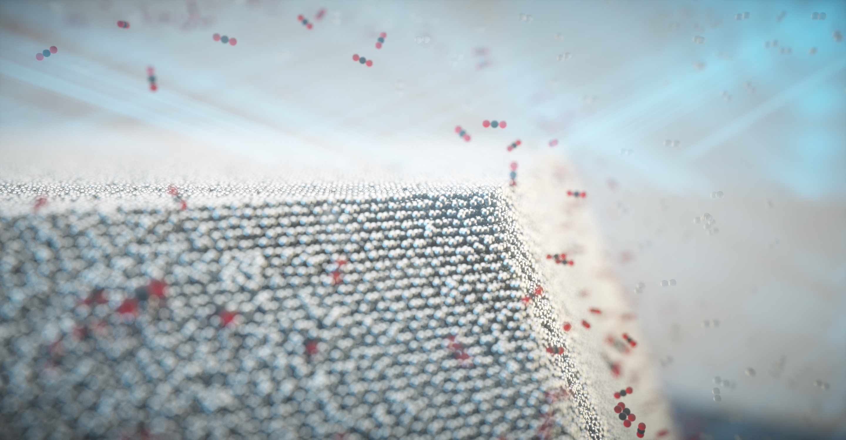
Carbon monoxide oxidises to carbon dioxide on the surface of the nanoparticle. Credit: Science Communication Lab for DESY

An October 1, 2021 news item on ScienceDaily announces research enabling scientists to observe a single nanoparticle at work,

A DESY-led research team has been using high-intensity X-rays to observe a single catalyst nanoparticle at work. The experiment has revealed for the first time how the chemical composition of the surface of an individual nanoparticle changes under reaction conditions, making it more active. The team led by DESY’s Andreas Stierle is presenting its findings in the journal Science Advances. This study marks an important step towards a better understanding of real, industrial catalytic materials.

An October 1, 2021 Deutsches Elektronen-Synchrotron (DESY) press release (also on EurekAlert), which originated the news item, explains why this research is important and provides more technical details,

Catalysts are materials that promote chemical reactions without being consumed themselves. Today, catalysts are used in numerous industrial processes, from fertiliser production to manufacturing plastics. Because of this, catalysts are of huge economic importance. A very well-known example is the catalytic converter installed in the exhaust systems of cars. These contain precious metals such as platinum, rhodium and palladium, which allow highly toxic carbon monoxide (CO) to be converted into carbon dioxide (CO2) and reduce the amount of harmful nitrogen oxides (NOx).

“In spite of their widespread use and great importance, we are still ignorant of many important details of just how the various catalysts work,” explains Stierle, head of the DESY NanoLab. “That’s why we have long wanted to study real catalysts while in operation.” This is not easy, because in order to make the active surface as large as possible, catalysts are typically used in the form of tiny nanoparticles, and the changes that affect their activity occur on their surface.

Surface strain relates to chemical composition

In the framework of the EU project Nanoscience Foundries and Fine Analysis (NFFA), the team from DESY NanoLab has developed a technique for labelling individual nanoparticles and thereby identifying them in a sample. “For the study, we grew nanoparticles of a platinum-rhodium alloy on a substrate in the lab and labelled one specific particle,” says co-author Thomas Keller from DESY NanoLab and in charge of the project at DESY. “The diameter of the labelled particle is around 100 nanometres, and it is similar to the particles used in a car’s catalytic converter.” A nanometre is a millionth of a millimetre.

Using X-rays from the European Synchrotron Radiation Facility ESRF in Grenoble, France, the team was not only able to create a detailed image of the nanoparticle; it also measured the mechanical strain within its surface. “The surface strain is related to the surface composition, in particular the ratio of platinum to rhodium atoms,” explains co-author Philipp Plessow from the Karlsruhe Institute of Technology (KIT), whose group computed strain as a function of surface composition. By comparing the observed and computed facet-dependent strain, conclusions can be drawn concerning the chemical composition at the particle surface. The different surfaces of a nanoparticle are called facets, just like the facets of a cut gemstone.

When the nanoparticle is grown, its surface consists mainly of platinum atoms, as this configuration is energetically favoured. However, the scientists studied the shape of the particle and its surface strain under different conditions, including the operating conditions of an automotive catalytic converter. To do this, they heated the particle to around 430 degrees Celsius and allowed carbon monoxide and oxygen molecules to pass over it. “Under these reaction conditions, the rhodium inside the particle becomes mobile and migrates to the surface because it interacts more strongly with oxygen than the platinum,” explains Plessow. This is also predicted by theory.

“As a result, the surface strain and the shape of the particle change,” reports co-author Ivan Vartaniants, from DESY, whose team converted the X-ray diffraction data into three-dimensional spatial images. “A facet-dependent rhodium enrichment takes place, whereby additional corners and edges are formed.” The chemical composition of the surface, and the shape and size of the particles have a significant effect on their function and efficiency. However, scientists are only just beginning to understand exactly how these are connected and how to control the structure and composition of the nanoparticles. The X-rays allow researchers to detect changes of as little as 0.1 in a thousand in the strain, which in this experiment corresponds to a precision of about 0.0003 nanometres (0.3 picometres).

Crucial step towards analysing industrial catalyst materials

“We can now, for the first time, observe the details of the structural changes in such catalyst nanoparticles while in operation,” says Stierle, Lead Scientist at DESY and professor for nanoscience at the University of Hamburg. “This is a major step forward and is helping us to understand an entire class of reactions that make use of alloy nanoparticles.” Scientists at KIT and DESY now want to explore this systematically at the new Collaborative Research Centre 1441, funded by the German Research Foundation (DFG) and entitled “Tracking the Active Sites in Heterogeneous Catalysis for Emission Control (TrackAct)”.

“Our investigation is an important step towards analysing industrial catalytic materials,” Stierle points out. Until now, scientists have had to grow model systems in the laboratory in order to conduct such investigations. “In this study, we have gone to the limit of what can be done. With DESY’s planned X-ray microscope PETRA IV, we will be able to look at ten times smaller individual particles in real catalysts, and under reaction conditions.”
 
DESY is one of the world’s leading particle accelerator centres and investigates the structure and function of matter – from the interaction of tiny elementary particles and the behaviour of novel nanomaterials and vital biomolecules to the great mysteries of the universe. The particle accelerators and detectors that DESY develops and builds at its locations in Hamburg and Zeuthen are unique research tools. They generate the most intense X-ray radiation in the world, accelerate particles to record energies and open up new windows onto the universe. DESY is a member of the Helmholtz Association, Germany’s largest scientific association, and receives its funding from the German Federal Ministry of Education and Research (BMBF) (90 per cent) and the German federal states of Hamburg and Brandenburg (10 per cent).

Here’s a link to and a citation for the paper,

Single alloy nanoparticle x-ray imaging during a catalytic reaction by Young Yong Kim, Thomas F. Keller, Tiago J. Goncalves, Manuel Abuin, Henning Runge, Luca Gelisio, Jerome Carnis, Vedran Vonk, Philipp N. Plessow, Ivan A. Vartaniants, Andreas Stierle. Science Advances • 1 Oct 2021 • Vol 7, Issue 40 • DOI: 10.1126/sciadv.abh0757

This paper is open access.
Stronger than steel and spider silk: artificial, biodegradable, cellulose nanofibres

This is an artificial and biodegradable are two adjectives you don’t usually see united by the conjunction, and. However, it is worth noting that the artificial material is initially derived from a natural material, cellulose. Here’s more from a May 16, 2018 news item on ScienceDaily,

At DESY’s [Deutsches Elektronen-Synchrotron] X-ray light source PETRA III, a team led by Swedish researchers has produced the strongest bio-material that has ever been made. The artifical, but bio-degradable cellulose fibres are stronger than steel and even than dragline spider silk, which is usually considered the strongest bio-based material. The team headed by Daniel Söderberg from the KTH Royal Institute of Technology in Stockholm reports the work in the journal ACS Nano of the American Chemical Society.

A May 16, 2018 DESY press release (also on EurekAlert), which originated the news item, provides more detail,

The ultrastrong material is made of cellulose nanofibres (CNF), the essential building blocks of wood and other plant life. Using a novel production method, the researchers have successfully transferred the unique mechanical properties of these nanofibres to a macroscopic, lightweight material that could be used as an eco-friendly alternative for plastic in airplanes, cars, furniture and other products. “Our new material even has potential for biomedicine since cellulose is not rejected by your body”, explains Söderberg.

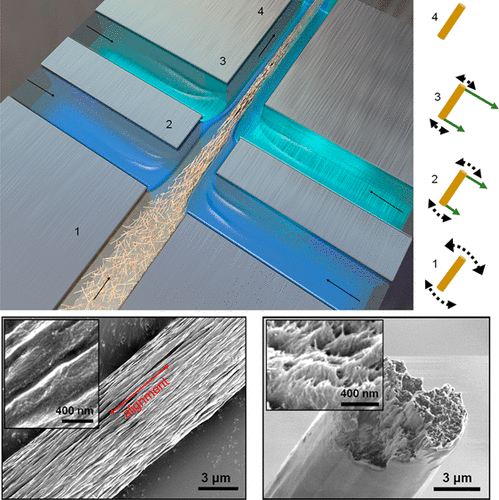
The scientists started with commercially available cellulose nanofibres that are just 2 to 5 nanometres in diameter and up to 700 nanometres long. A nanometre (nm) is a millionth of a millimetre. The nanofibres were suspended in water and fed into a small channel, just one millimetre wide and milled in steel. Through two pairs of perpendicular inflows additional deionized water and water with a low pH-value entered the channel from the sides, squeezing the stream of nanofibres together and accelerating it.

This process, called hydrodynamic focussing, helped to align the nanofibres in the right direction as well as their self-organisation into a well-packed macroscopic thread. No glue or any other component is needed, the nanofibres assemble into a tight thread held together by supramolecular forces between the nanofibres, for example electrostatic and Van der Waals forces.

With the bright X-rays from PETRA III the scientists could follow and optimise the process. “The X-rays allow us to analyse the detailed structure of the thread as it forms as well as the material structure and hierarchical order in the super strong fibres,” explains co-author Stephan Roth from DESY, head of the Micro- and Nanofocus X-ray Scattering Beamline P03 where the threads were spun. “We made threads up to 15 micrometres thick and several metres in length.”

Measurements showed a tensile stiffness of 86 gigapascals (GPa) for the material and a tensile strength of 1.57 GPa. “The bio-based nanocellulose fibres fabricated here are 8 times stiffer and have strengths higher than natural dragline spider silk fibres,” says Söderberg. “If you are looking for a bio-based material, there is nothing quite like it. And it is also stronger than steel and any other metal or alloy as well as glass fibres and most other synthetic materials.” The artificial cellulose fibres can be woven into a fabric to create materials for various applications. The researchers estimate that the production costs of the new material can compete with those of strong synthetic fabrics. “The new material can in principle be used to create bio-degradable components,” adds Roth.

The study describes a new method that mimics nature’s ability to accumulate cellulose nanofibres into almost perfect macroscale arrangements, like in wood. It opens the way for developing nanofibre material that can be used for larger structures while retaining the nanofibres’ tensile strength and ability to withstand mechanical load. “We can now transform the super performance from the nanoscale to the macroscale,” Söderberg underlines. “This discovery is made possible by understanding and controlling the key fundamental parameters essential for perfect nanostructuring, such as particle size, interactions, alignment, diffusion, network formation and assembly.” The process can also be used to control nanoscale assembly of carbon tubes and other nano-sized fibres.

(There are some terminology and spelling issues, which are described at the end of this post.)

Let’s get back to a material that rivals spider silk and steel for strength (for some reason that reminded me of an old carnival game where you’d test your strength by swinging a mallet down on a ‘teeter-totter-like’ board and sending a metal piece up a post to make a bell ring). From a May 16, 2018 DESY press release (also on EurekAlert), which originated the news item,

The ultrastrong material is made of cellulose nanofibres (CNF), the essential building blocks of wood and other plant life. Using a novel production method, the researchers have successfully transferred the unique mechanical properties of these nanofibres to a macroscopic, lightweight material that could be used as an eco-friendly alternative for plastic in airplanes, cars, furniture and other products. “Our new material even has potential for biomedicine since cellulose is not rejected by your body”, explains Söderberg.

The scientists started with commercially available cellulose nanofibres that are just 2 to 5 nanometres in diameter and up to 700 nanometres long. A nanometre (nm) is a millionth of a millimetre. The nanofibres were suspended in water and fed into a small channel, just one millimetre wide and milled in steel. Through two pairs of perpendicular inflows additional deionized water and water with a low pH-value entered the channel from the sides, squeezing the stream of nanofibres together and accelerating it.

This process, called hydrodynamic focussing, helped to align the nanofibres in the right direction as well as their self-organisation into a well-packed macroscopic thread. No glue or any other component is needed, the nanofibres assemble into a tight thread held together by supramolecular forces between the nanofibres, for example electrostatic and Van der Waals forces.

With the bright X-rays from PETRA III the scientists could follow and optimise the process. “The X-rays allow us to analyse the detailed structure of the thread as it forms as well as the material structure and hierarchical order in the super strong fibres,” explains co-author Stephan Roth from DESY, head of the Micro- and Nanofocus X-ray Scattering Beamline P03 where the threads were spun. “We made threads up to 15 micrometres thick and several metres in length.”

Measurements showed a tensile stiffness of 86 gigapascals (GPa) for the material and a tensile strength of 1.57 GPa. “The bio-based nanocellulose fibres fabricated here are 8 times stiffer and have strengths higher than natural dragline spider silk fibres,” says Söderberg. “If you are looking for a bio-based material, there is nothing quite like it. And it is also stronger than steel and any other metal or alloy as well as glass fibres and most other synthetic materials.” The artificial cellulose fibres can be woven into a fabric to create materials for various applications. The researchers estimate that the production costs of the new material can compete with those of strong synthetic fabrics. “The new material can in principle be used to create bio-degradable components,” adds Roth.

The study describes a new method that mimics nature’s ability to accumulate cellulose nanofibres into almost perfect macroscale arrangements, like in wood. It opens the way for developing nanofibre material that can be used for larger structures while retaining the nanofibres’ tensile strength and ability to withstand mechanical load. “We can now transform the super performance from the nanoscale to the macroscale,” Söderberg underlines. “This discovery is made possible by understanding and controlling the key fundamental parameters essential for perfect nanostructuring, such as particle size, interactions, alignment, diffusion, network formation and assembly.” The process can also be used to control nanoscale assembly of carbon tubes and other nano-sized fibres.

Here’s a link to and a citation for the paper,

Multiscale Control of Nanocellulose Assembly: Transferring Remarkable Nanoscale Fibril Mechanics to Macroscale Fibers by Nitesh Mittal, Farhan Ansari, Krishne Gowda V, Christophe Brouzet, Pan Chen, Per Tomas Larsson, Stephan V. Roth, Fredrik Lundell, Lars Wågberg, Nicholas A. Kotov, and L. Daniel Söderberg. ACS Nano, Article ASAP DOI: 10.1021/acsnano.8b01084 Publication Date (Web): May 9, 2018

Copyright © 2018 American Chemical Society

This paper is open access and accompanied by this image illustrating the work,

Courtesy: American Chemical Society and the researchers [Note: The bottom two images of cellulose nanofibres, which are constittuents of an artificial cellulose fibre, appear to be from a scanning tunneling microsscope. Credit: Nitesh Mittal, KTH Stockholm

This news has excited interest at General Electric (GE) (its Wikipedia entry), which has highlighted the work in a May 25, 2018 posting (The 5 Coolest Things On Earth This Week) by Tomas Kellner on the GE Reports blog.

Terminology and spelling

I’ll start with spelling since that’s the easier of the two. In some parts of the world it’s spelled ‘fibres’ and in other parts of the world it’s spelled ‘fibers’. When I write the text in my post, it tends to reflect the spelling used in the news/press releases. In other words, I swing in whichever direction the wind is blowing.

For diehards only

As i understand the terminology situation, nanocellulose and cellulose nanomaterials are interchangeable generic terms. Further, cellulose nanofibres (CNF) seems to be another generic term and it encompasses both cellulose nanocrystals (CNC) and cellulose nanofibrils (CNF). Yes, there appear to be two CNFs. Making matters more interesting is the fact that cellulose nanocrystals were originally christened nanocrystalline cellulose (NCC). For anyone who follows the science and technology scene, it becomes obvious that competing terminologies are the order of the day. Eventually the dust settles and naming conventions are resolved. More or less.

Ordinarily I would reference the Nanocellulose Wikipedia entry in my attempts to clarify the issues but it seems that the writers for the entry have not caught up to the current naming convention for cellulose nanocrystals, still referring to the material as nanocrystalline cellulose. This means, I can’t trust the rest of the entry, which has only one CNF (cellulose nanofibres).

I have paid more attention to the NCC/CNC situation and am not as familiar with the CNF situation. Using, NCC/CNC as an example of a terminology issue, I believe it was first developed in Canada and it was Canadian researchers who were pushing their NCC terminology while the international community pushed back with CNC.

In the end, NCC became a brand name, which was trademarked by CelluForce, a Canadian company in the CNC market. From the CelluForce Products page on Cellulose Nanocrystals,

CNC are not all made equal. The CNC produced by CelluForce is called CelluForce NCCTM and has specific properties and are especially easy to disperse. CelluForce NCCTM is the base material that CelluForce uses in all its products. This base material can be modified and tailored to suit the specific needs in various applications.

These, days CNC is almost universally used but NCC (not as a trademark) is a term still employed on occasion (and, oddly, the researchers are not necessarily Canadian).

Should anyone have better information about terminology issues, please feel free to comment.

MOFs (metal-organic frameworks) to clean up nuclear waste?

There’s a possibility that metal-organic frameworks could be used to clean up nuclear waste according to an Aug. 5, 2015 news item on phys.org,

One of the most versatile and widely applicable classes of materials being studied today are the metal-organic frameworks. These materials, known as MOFs, are characterized by metal ions or metal-ion clusters that are linked together with organic molecules, forming ordered crystal structures that contain tiny cage-like pores with diameters of two nanometers or less.

MOFs can be thought of as highly specialized and customizable sieves. By designing them with pores of a certain size, shape, and chemical composition, researchers can tailor them for specific purposes. A few of the many, many possible applications for MOFs are storing hydrogen in fuel cells, capturing environmental contaminants, or temporarily housing catalytic agents for chemical reactions.

At [US Department of Energy] Brookhaven National Laboratory, physicist Sanjit Ghose and his collaborators have been studying MOFs designed for use in the separation of waste from nuclear reactors, which results from the reprocessing of nuclear fuel rods. He is targeting two waste products in particular: the noble gases xenon (Xe) and krypton (Kr).

An Aug. 4, 2015 Brookhaven National Laboratory news release, which originated the news item, describes not only the research and the reasons for it but also the institutional collaborations necessary to conduct the research,

There are compelling economic and environmental reasons to separate Xe and Kr from the nuclear waste stream. For one, because they have very different half-lives – about 36 days for Xe and nearly 11 years for Kr – pulling out the Xe greatly reduces the amount of waste that needs to be stored long-term before it is safe to handle. Additionally, the extracted Xe can be used for industrial applications, such as in commercial lighting and as an anesthetic. This research may also help scientists determine how to create MOFs that can remove other materials from the nuclear waste stream and expose the remaining unreacted nuclear fuel for further re-use. This could lead to much less overall waste that must be stored long-term and a more efficient system for producing nuclear energy, which is the source of about 20 percent of the electricity in the U.S.

Because Xe and Kr are noble gases, meaning their outer electron orbitals are filled and they don’t tend to bind to other atoms, they are difficult to manipulate. The current method for extracting them from the nuclear waste stream is cryogenic distillation, a process that is energy-intensive and expensive. The MOFs studied here use a very different approach: polarizing the gas atoms dynamically, just enough to draw them in using the van der Waals force. The mechanism works at room temperature, but also at hotter temperatures, which is key if the MOFs are to be used in a nuclear environment.

Recently, Ghose co-authored two papers that describe MOFs capable of adsorbing Xe and Kr, and excel at separating the Xe from the Kr. The papers are published in the May 22 online edition of the Journal of the American Chemical Society and the April 16 online edition of the Journal of Physical Chemistry Letters.

“Only a handful of noble-gas-specific MOFs have been studied so far, and we felt there was certainly scope for improvement through the discovery of more selective materials,” said Ghose.

Both MOF studies were carried out by large multi-institution collaborations, using a combination of X-ray diffraction, theoretical modeling, and other methods. The X-ray work was performed at Brookhaven’s former National Synchrotron Light Source (permanently closed and replaced by its successor, NSLS-II) and the Advanced Photon Source at Argonne National Laboratory (ANL), both DOE Office of Science User Facilities.

The JACS paper was co-authored by researchers from Brookhaven Lab, Stony Brook University (SBU), Pacific Northwest National Laboratory (PNNL), and the University of Amsterdam. Authors on the JPCL paper include scientists from Brookhaven, SBU, PNNL, ANL, the Deutsches Elektronen-Synchrotron (DESY) in Germany, and DM Strachan, LLC.

Here’s more about the first published paper in the Journal of Physical Chemistry Letters (JCPL) (from the news release)

A nickel-based MOF

The MOF studied in the JCPL paper consists of nickel (Ni) and the organic compound dioxido-benzene-dicarboxylate (DOBC), and is thus referred to as Ni-DOBDC. Ni-DOBDC can adsorb both Xe and Kr at room temperature but is highly selective toward Xe. In fact, it boasts what may be the highest Xe adsorption capacity of a MOF discovered to date.

The group studied Ni-DOBC using two main techniques: X-ray diffraction and first-principles density functional theory (DFT). The paper is the first published report to detail the adsorption mechanism by which the MOF takes in these noble gases at room temperature and pressure.

“Our results provide a fundamental understanding of the adsorption structure and the interactions between the MOF and the gas by combining direct structural analyses from experimental X-ray diffraction data and DFT calculations,” said Ghose.

The group was also able to discover the existence of a secondary site at the pore center in addition to the six-fold primary site. The seven-atom loading scheme was initially proposed by theorist Yan Li, an co-author of the JCPL paper and formerly on staff at Brookhaven (she is now an editor at Physical Review B), which was confirmed experimentally and theoretically. Data also indicate that Xe are adsorbed more strongly than Kr, due to its higher atomic polarizability. They also discovered a temperature-dependence of the adsorption that furthers this MOF’s selectivity for Xe over Kr. As the temperature was increased above room temperature, the Kr adsorption drops more drastically than for Xe. Over the entire temperature range tested, Xe adsorption always dominates that of Kr.

“The high separation capacity of Ni-DOBDC suggests that it has great potential for removing Xe from Kr in the off-gas streams in nuclear spent fuel reprocessing, as well as filtering Xe at low concentration from other gas mixtures,” said Ghose.

Ghose and Li are now preparing a manuscript that will discuss a more in-depth investigation into the possibility of packing in even more Xe atoms.

“Because of the confinement offered by each pore, we want to see if it’s possible to fit enough Xe in each chamber to form a solid,” said Li.

Ghose and Li hope to experimentally test this idea at NSLS-II in the future, at the facility’s X-ray Powder Diffraction (XPD) beamline, which Ghose has helped develop and build. Additional future studies of these and other MOFs will also take place at XPD. For example, they want to see what happens when other gases are present, such as nitrogen oxides, to mimic what happens in an actual nuclear reactor.

Then, there was the second paper published in the Journal of the American Chemical Society (JACS),

Another MOF, Another Promising Result

In the JACS paper, Ghose and researchers from Brookhaven, SBU, PNNL, and the University of Amsterdam describe a second MOF, dubbed Stony Brook MOF-2 (SBMOF-2). It also captures both Xe and Kr at room temperature and pressure, although is about ten times as effective at taking in Xe, with Xe taking up as much as 27 percent of its weight. SBMOF-2 had been theoretically predicted to be an efficient adsorbent for Xe and Kr, but until this research there had been no experimental results to back up the prediction.

“Our study is different than MOF research done by other groups,” said chemist John Parise, a coauthor of the JACS paper who holds a joint position with Brookhaven and SBU. “We did a lot of testing and investigated the capture mechanism very closely to get clues that would help us understand why the MOF worked, and how to tailor the structure to have even better properties.”

SBMOF-2 contains calcium (Ca) ions and an organic compound with the chemical formula C34H22O8. X-ray data show that its structure is unusual among microporous MOFs. It has fewer calcium sites than expected and an excess of oxygen over calcium. The calcium and oxgyen form CaO6, which takes the form of a three-dimensional octahedron. Notably, none of the six oxygen atoms bound to the calcium ion are shared with any other nearby calcium ions. The authors believe that SBMOF-2 is the first microporous MOF with these isolated CaO6 octahedra, which are connected by organic linker molecules.

The group discovered that the preference of SBMOF-2 for Xe over Kr is due to both the geometry and chemistry of its pores. All the pores have diamond-shaped cross sections, but they come in two sizes, designated type-1 and type-2. Both sizes are a better fit for the Xe molecule. The interiors of the pores have walls made of phenyl groups – ring-shaped C6H5 molecules – along with delocalized electron clouds and H atoms pointing into the pore. The type-2 pores also have hydroxyl anions (OH-) available. All of these features provide are potential sites for adsorbed Xe and Kr atoms.

In follow-up studies, Ghose and his colleagues will use these results to guide them as they determine what changes can be made to these MOFs to improve adsorption, as well as to determine what existing MOFs may yield similar or better performance.

Here are links to and citations for both papers,

Understanding the Adsorption Mechanism of Xe and Kr in a Metal–Organic Framework from X-ray Structural Analysis and First-Principles Calculations by Sanjit K. Ghose, Yan Li, Andrey Yakovenko, Eric Dooryhee, Lars Ehm, Lynne E. Ecker, Ann-Christin Dippel, Gregory J. Halder, Denis M. Strachan, and Praveen K. Thallapally. J. Phys. Chem. Lett., 2015, 6 (10), pp 1790–1794 DOI: 10.1021/acs.jpclett.5b00440 Publication Date (Web): April 16, 2015

Copyright © 2015 American Chemical Society

Direct Observation of Xe and Kr Adsorption in a Xe-Selective Microporous Metal–Organic Framework by Xianyin Chen, Anna M. Plonka, Debasis Banerjee, Rajamani Krishna, Herbert T. Schaef, Sanjit Ghose, Praveen K. Thallapally, and John B. Parise. J. Am. Chem. Soc., 2015, 137 (22), pp 7007–7010 DOI: 10.1021/jacs.5b02556 Publication Date (Web): May 22, 2015
Copyright © 2015 American Chemical Society

Both papers are behind a paywall.