Tag Archives: diagnostic tests

Detect lung cancer early by inhaling a nanosensor

The technology described in a January 5, 2024 news item on Nanowerk has not been tried in human clinical trials but early pre-clinical trial testing offers promise,

Using a new technology developed at MIT, diagnosing lung cancer could become as easy as inhaling nanoparticle sensors and then taking a urine test that reveals whether a tumor is present.

Key Takeaways

*This non-invasive approach may serve as an alternative or supplement to traditional CT scans, particularly beneficial in areas with limited access to advanced medical equipment.

*The technology focuses on detecting cancer-linked proteins in the lungs, with results obtainable through a simple paper test strip.

*Designed for early-stage lung cancer detection, the method has shown promise in animal models and may soon advance to human clinical trials.

*This innovation holds potential for significantly improving lung cancer screening and early detection, especially in low-resource settings.

A January 5, 2024 Massachusetts Institute of Technology (MIT) news release (also on EurkeAlert), which originated the news item, goes on to provide some technical details,

The new diagnostic is based on nanosensors that can be delivered by an inhaler or a nebulizer. If the sensors encounter cancer-linked proteins in the lungs, they produce a signal that accumulates in the urine, where it can be detected with a simple paper test strip.

This approach could potentially replace or supplement the current gold standard for diagnosing lung cancer, low-dose computed tomography (CT). It could have an especially significant impact in low- and middle-income countries that don’t have widespread availability of CT scanners, the researchers say.

“Around the world, cancer is going to become more and more prevalent in low- and middle-income countries. The epidemiology of lung cancer globally is that it’s driven by pollution and smoking, so we know that those are settings where accessibility to this kind of technology could have a big impact,” says Sangeeta Bhatia, the John and Dorothy Wilson Professor of Health Sciences and Technology and of Electrical Engineering and Computer Science at MIT, and a member of MIT’s Koch Institute for Integrative Cancer Research and the Institute for Medical Engineering and Science.

Bhatia is the senior author of the paper, which appears today [January 5, 2024] in Science Advances. Qian Zhong, an MIT research scientist, and Edward Tan, a former MIT postdoc, are the lead authors of the study.

Inhalable particles

To help diagnose lung cancer as early as possible, the U.S. Preventive Services Task Force recommends that heavy smokers over the age of 50 undergo annual CT scans. However, not everyone in this target group receives these scans, and the high false-positive rate of the scans can lead to unnecessary, invasive tests.

Bhatia has spent the last decade developing nanosensors for use in diagnosing cancer and other diseases, and in this study, she and her colleagues explored the possibility of using them as a more accessible alternative to CT screening for lung cancer.

These sensors consist of polymer nanoparticles coated with a reporter, such as a DNA barcode, that is cleaved from the particle when the sensor encounters enzymes called proteases, which are often overactive in tumors. Those reporters eventually accumulate in the urine and are excreted from the body.

Previous versions of the sensors, which targeted other cancer sites such as the liver and ovaries, were designed to be given intravenously. For lung cancer diagnosis, the researchers wanted to create a version that could be inhaled, which could make it easier to deploy in lower resource settings.

“When we developed this technology, our goal was to provide a method that can detect cancer with high specificity and sensitivity, and also lower the threshold for accessibility, so that hopefully we can improve the resource disparity and inequity in early detection of lung cancer,” Zhong says.

To achieve that, the researchers created two formulations of their particles: a solution that can be aerosolized and delivered with a nebulizer, and a dry powder that can be delivered using an inhaler.

Once the particles reach the lungs, they are absorbed into the tissue, where they encounter any proteases that may be present. Human cells can express hundreds of different proteases, and some of them are overactive in tumors, where they help cancer cells to escape their original locations by cutting through proteins of the extracellular matrix. These cancerous proteases cleave DNA barcodes from the sensors, allowing the barcodes to circulate in the bloodstream until they are excreted in the urine.

In the earlier versions of this technology, the researchers used mass spectrometry to analyze the urine sample and detect DNA barcodes. However, mass spectrometry requires equipment that might not be available in low-resource areas, so for this version, the researchers created a lateral flow assay, which allows the barcodes to be detected using a paper test strip.

The researchers designed the strip to detect up to four different DNA barcodes, each of which indicates the presence of a different protease. No pre-treatment or processing of the urine sample is required, and the results can be read about 20 minutes after the sample is obtained.

“We were really pushing this assay to be point-of-care available in a low-resource setting, so the idea was to not do any sample processing, not do any amplification, just to be able to put the sample right on the paper and read it out in 20 minutes,” Bhatia says.

Accurate diagnosis

The researchers tested their diagnostic system in mice that are genetically engineered to develop lung tumors similar to those seen in humans. The sensors were administered 7.5 weeks after the tumors started to form, a time point that would likely correlate with stage 1 or 2 cancer in humans.

In their first set of experiments in the mice, the researchers measured the levels of 20 different sensors designed to detect different proteases. Using a machine learning algorithm to analyze those results, the researchers identified a combination of just four sensors that was predicted to give accurate diagnostic results. They then tested that combination in the mouse model and found that it could accurately detect early-stage lung tumors.

For use in humans, it’s possible that more sensors might be needed to make an accurate diagnosis, but that could be achieved by using multiple paper strips, each of which detects four different DNA barcodes, the researchers say.

The researchers now plan to analyze human biopsy samples to see if the sensor panels they are using would also work to detect human cancers. In the longer term, they hope to perform clinical trials in human patients. A company called Sunbird Bio has already run phase 1 trials on a similar sensor developed by Bhatia’s lab, for use in diagnosing liver cancer and a form of hepatitis known as nonalcoholic steatohepatitis (NASH).

In parts of the world where there is limited access to CT scanning, this technology could offer a dramatic improvement in lung cancer screening, especially since the results can be obtained during a single visit.

“The idea would be you come in and then you get an answer about whether you need a follow-up test or not, and we could get patients who have early lesions into the system so that they could get curative surgery or lifesaving medicines,” Bhatia says.

Here’s a link to and a citation for the paper,

Inhalable point-of-care urinary diagnostic platform by Qian Zhong, Edward K. W. Tan, Carmen Martin-Alonso, Tiziana Parisi, Liangliang Hao, Jesse D. Kirkpatrick, Tarek Fadel, Heather E. Fleming, Tyler Jacks, and Sangeeta N. Bhatia. Science Advances 5 Jan 2024 Vol 10, Issue 1 DOI: 10.1126/sciadv.adj9591

This paper is open access.

Sunbird Bio (the company mentioned in the news release) can be found here.
Detecting COVID-19 in under five minutes with paper-based sensor made of graphene

A Dec. 7, 2020 news item on Nanowerk announced a new technology for rapid COVID-19 testing (Note: A link has been removed),

As the COVID-19 pandemic continues to spread across the world, testing remains a key strategy for tracking and containing the virus. Bioengineering graduate student, Maha Alafeef, has co-developed a rapid, ultrasensitive test using a paper-based electrochemical sensor that can detect the presence of the virus in less than five minutes.

The team led by professor Dipanjan Pan reported their findings in ACS Nano (“Rapid, Ultrasensitive, and Quantitative Detection of SARS-CoV-2 Using Antisense Oligonucleotides Directed Electrochemical Biosensor Chip”).

“Currently, we are experiencing a once-in-a-century life-changing event,” said Alafeef. “We are responding to this global need from a holistic approach by developing multidisciplinary tools for early detection and diagnosis and treatment for SARS-CoV-2.”

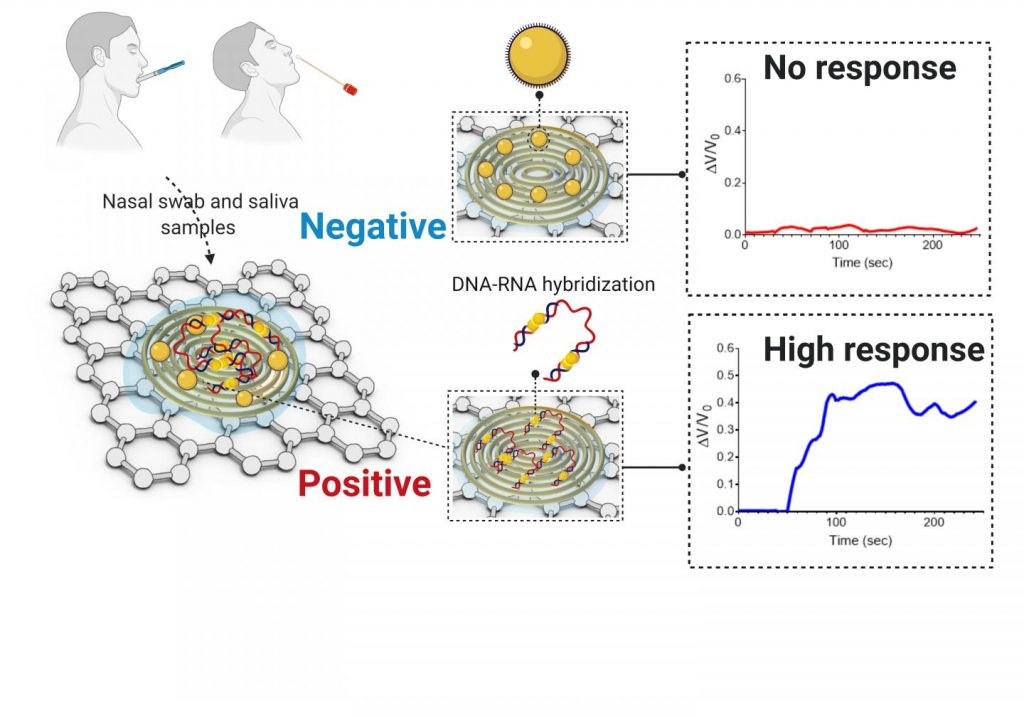
I wonder why they didn’t think to provide a caption for the graphene substrate (the square surface) underlying the gold electrode (the round thing) or provide a caption for the electrode. Maybe they assumed anyone knowledgeable about graphene would be able to identify it?

Caption: COVID-19 electrochemical sensing platform. Credit: University of Illinois

A Dec. 7, 2020 University of Illinois Grainger College of Engineering news release (also on EurekAlert) by Huan Song, which originated the news item, provides more technical detail including a description of the graphene substrate and the gold electrode, which make up the cheaper, faster COVID-19 sensing platform,

There are two broad categories of COVID-19 tests on the market. The first category uses reverse transcriptase real-time polymerase chain reaction (RT-PCR) and nucleic acid hybridization strategies to identify viral RNA. Current FDA [US Food and Drug Administration]-approved diagnostic tests use this technique. Some drawbacks include the amount of time it takes to complete the test, the need for specialized personnel and the availability of equipment and reagents.

The second category of tests focuses on the detection of antibodies. However, there could be a delay of a few days to a few weeks after a person has been exposed to the virus for them to produce detectable antibodies.

In recent years, researchers have had some success with creating point-of-care biosensors using 2D nanomaterials such as graphene to detect diseases. The main advantages of graphene-based biosensors are their sensitivity, low cost of production and rapid detection turnaround. “The discovery of graphene opened up a new era of sensor development due to its properties. Graphene exhibits unique mechanical and electrochemical properties that make it ideal for the development of sensitive electrochemical sensors,” said Alafeef. The team created a graphene-based electrochemical biosensor with an electrical read-out setup to selectively detect the presence of SARS-CoV-2 genetic material.

There are two components [emphasis mine] to this biosensor: a platform to measure an electrical read-out and probes to detect the presence of viral RNA. To create the platform, researchers first coated filter paper with a layer of graphene nanoplatelets to create a conductive film [emphasis mine]. Then, they placed a gold electrode with a predefined design on top of the graphene [emphasis mine] as a contact pad for electrical readout. Both gold and graphene have high sensitivity and conductivity which makes this platform ultrasensitive to detect changes in electrical signals.

Current RNA-based COVID-19 tests screen for the presence of the N-gene (nucleocapsid phosphoprotein) on the SARS-CoV-2 virus. In this research, the team designed antisense oligonucleotide (ASOs) probes to target two regions of the N-gene. Targeting two regions ensures the reliability of the senor in case one region undergoes gene mutation. Furthermore, gold nanoparticles (AuNP) are capped with these single-stranded nucleic acids (ssDNA), which represents an ultra-sensitive sensing probe for the SARS-CoV-2 RNA.

The researchers previously showed the sensitivity of the developed sensing probes in their earlier work published in ACS Nano. The hybridization of the viral RNA with these probes causes a change in the sensor electrical response. The AuNP caps accelerate the electron transfer and when broadcasted over the sensing platform, results in an increase in the output signal and indicates the presence of the virus.

The team tested the performance of this sensor by using COVID-19 positive and negative samples. The sensor showed a significant increase in the voltage of positive samples compared to the negative ones and confirmed the presence of viral genetic material in less than five minutes. Furthermore, the sensor was able to differentiate viral RNA loads in these samples. Viral load is an important quantitative indicator of the progress of infection and a challenge to measure using existing diagnostic methods.

This platform has far-reaching applications due to its portability and low cost. The sensor, when integrated with microcontrollers and LED screens or with a smartphone via Bluetooth or wifi, could be used at the point-of-care in a doctor’s office or even at home. Beyond COVID-19, the research team also foresees the system to be adaptable for the detection of many different diseases.

“The unlimited potential of bioengineering has always sparked my utmost interest with its innovative translational applications,” Alafeef said. “I am happy to see my research project has an impact on solving a real-world problem. Finally, I would like to thank my Ph.D. advisor professor Dipanjan Pan for his endless support, research scientist Dr. Parikshit Moitra, and research assistant Ketan Dighe for their help and contribution toward the success of this study.”

Here’s a link to and a citation for the paper,

Rapid, Ultrasensitive, and Quantitative Detection of SARS-CoV-2 Using Antisense Oligonucleotides Directed Electrochemical Biosensor Chip by Maha Alafeef, Ketan Dighe, Parikshit Moitra, and Dipanjan Pan. ACS Nano 2020, 14, 12, 17028–17045 DOI: https://doi.org/10.1021/acsnano.0c06392 Publication Date:October 20, 2020 Copyright © 2020 American Chemical Society

I’m not sure where I found this notice but it is most definitely from the American Chemical Society: “This paper is freely accessible, at this time, for unrestricted RESEARCH re-use and analyses in any form or by any means with acknowledgement of the original source. These permissions are granted for the duration of the World Health Organization (WHO) declaration of COVID-19 as a global pandemic.”

Gold nanoparticles could help detect the presence of COVID-19 in ten minutes

If this works out, it would make testing for COVID-19 an infinitely easier task. From a May 29, 2020 news item on phys.org,

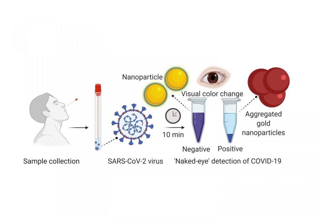
Scientists from the University of Maryland School of Medicine (UMSOM) developed an experimental diagnostic test for COVID-19 that can visually detect the presence of the virus in 10 minutes. It uses a simple assay containing plasmonic gold nanoparticles to detect a color change when the virus is present. The test does not require the use of any advanced laboratory techniques, such as those commonly used to amplify DNA, for analysis. The authors published their work last week [May 21, 2020] in the American Chemical Society’s nanotechnology journal ACS Nano.

“Based on our preliminary results, we believe this promising new test may detect RNA [ribonucleic acid] material from the virus as early as the first day of infection. Additional studies are needed, however, to confirm whether this is indeed the case,” said study leader Dipanjan Pan, PhD, Professor of Diagnostic Radiology and Nuclear Medicine and Pediatrics at the UMSOM.

Caption: A nasal swab containing a test sample is mixed with a simple lab test. It contains a liquid mixed with gold nanoparticles attached to a molecule that binds to the novel coronavirus. If the virus is present, the gold nanoparticles turns the solution a deep blue color (bottom of the tube) and a precipitation is noticed. If it is not present, the solution retains its original purple color. Credit: University of Maryland School of Medicine

A May 28, 2020 University of Maryland news release (also on EurekAlert), which originated the news item, provides more detail,

Once a nasal swab or saliva sample is obtained from a patient, the RNA is extracted from the sample via a simple process that takes about 10 minutes. The test uses a highly specific molecule attached to the gold nanoparticles to detect a particular protein. This protein is part of the genetic sequence that is unique to the novel coronavirus. When the biosensor binds to the virus’s gene sequence, the gold nanoparticles respond by turning the liquid reagent from purple to blue.

“The accuracy of any COVID-19 test is based on being able to reliably detect any virus. This means it does not give a false negative result if the virus actually is present, nor a false positive result if the virus is not present,” said Dr. Pan. “Many of the diagnostic tests currently on the market cannot detect the virus until several days after infection. For this reason, they have a significant rate of false negative results.”

Dr. Pan created a company called VitruVian Bio to develop the test for commercial application. He plans to have a pre-submission meeting with the U.S. Food and Drug Administration (FDA) within the next month to discuss requirements for getting an emergency use authorization for the test. New FDA policy allows for the marketing of COVID-19 tests without requiring them to go through the usual approval or clearance process. These tests do, however, need to meet certain validation testing requirements to ensure that they provide reliable results.

“This RNA-based test appears to be very promising in terms of detecting the virus. The innovative approach provides results without the need for a sophisticated laboratory facility,” said study co-author Matthew Frieman, PhD, Associate Professor of Microbiology and Immunology at UMSOM.

Although more clinical studies are warranted, this test could be far less expensive to produce and process than a standard COVID-19 lab test; it does not require laboratory equipment or trained personnel to run the test and analyze the results. If this new test meets FDA expectations, it could potentially be used in daycare centers, nursing homes, college campuses, and work places as a surveillance technique to monitor any resurgence of infections.

In Dr. Pan’s laboratory, research scientist Parikshit Moitra, PhD, and UMSOM research fellow Maha Alafeef conducted the studies along with research fellow Ketan Dighe from UMBC.

Dr. Pan holds a joint appointment with the College of Engineering at the University of Maryland Baltimore County and is also a faculty member of the Center for Blood Oxygen Transport and Hemostasis (CBOTH).

“This is another example of how our faculty is driving innovation to fulfill a vital need to expand the capacity of COVID-19 testing,” said Dean E. Albert Reece, MD, PhD, MBA, who is also Executive Vice President for Medical Affairs, UM Baltimore, and the John Z. and Akiko K. Bowers Distinguished Professor, University of Maryland School of Medicine. “Our nation will be relying on inexpensive, rapid tests that can be dispersed widely and used often until we have effective vaccines against this pandemic.”

Here’s a link to and a citation for the paper,

Selective Naked-Eye Detection of SARS-CoV-2 Mediated by N Gene Targeted Antisense Oligonucleotide Capped Plasmonic Nanoparticles by Parikshit Moitra, Maha Alafeef, Ketan Dighe, Matthew B. Frieman, and Dipanjan Pan. ACS Nano 2020, XXXX, XXX, XXX-XXX DOI: https://doi.org/10.1021/acsnano.0c03822 Publication Date:May 21, 2020 Copyright © 2020 American Chemical Society

This paper appears to be open access.

I tried to find Dr. Pan’s company, VitruVian Bio and found a business with an almost identical name, Vitruvian Biomedical, which does not include Dr. Pan on its management team list and this company’s focus is on Alzheimer’s Disease. Finally, there is no mention of the COVID-19 test anywhere on the Vitruvian Biomedical website.

Eliminate cold storage for diagnostic tests?

There’s a nanoparticle coating that could eliminate the need for cold storage and/or refrigeration for diagnostic testing according to a Jan. 4, 2017 news item on Nanowerk,

Many diagnostic tests use antibodies to help confirm a myriad of medical conditions, from Zika infections to heart ailments and even some forms of cancer. Antibodies capture and help detect proteins, enzymes, bacteria and viruses present in injuries and illnesses, and must be kept at a constant low temperature to ensure their viability — often requiring refrigeration powered by electricity. This can make diagnostic testing in underdeveloped countries, disaster or remote areas and even war zones extremely expensive and difficult.

A team of engineers from Washington University in St. Louis and Air Force Research Laboratory have discovered an inexpensive work-around: a protective coating that could completely eliminate the need for cold storage and change the scope of medical diagnostic testing in places where it’s often needed the most.

“In many developing countries, electricity is not guaranteed,” said Srikanth Singamaneni, associate professor of mechanical engineering and materials science in Engineering & Applied Science at Washington University in St. Louis.

“So how do we best get them medical diagnostics? We did not know how to solve this problem previously.”

A Jan. 4, 2016 Washington University in St. Louis news release by Erika Ebsworth-Goold, which originated the news item, describes how previous research helped lead to a solution,

Singamaneni’s team previously used tiny gold nanorods in bio-diagnostic research, measuring changes in their optical properties to quantify protein concentrations in bio-fluids: the higher a concentration, the higher the likelihood of injury or disease.

In this new research, published in Advanced Materials, Singamaneni worked with faculty from Washington University’s School of Medicine and researchers from the Air Force Research Lab to grow metal-organic frameworks (MOFs) around antibodies attached to gold nanorods. The crystalline MOFs formed a protective layer around the antibodies and prevented them from losing activity at elevated temperatures. The protective effect lasted for a week even when the samples were stored at 60°C.

“This technology would allow point-of-care screening for biomarkers of diseases in urban and rural clinic settings where immediate patient follow-up is critical to treatment and wellbeing,” said Dr. Jeremiah J. Morrissey, professor of anesthesiology, Division of Clinical and Translational Research, Washington University School of Medicine and a co-author on the paper.

“On the spot testing eliminates the time lag in sending blood/urine samples to a central lab for testing and in tracking down patients to discuss test results. In addition, it may reduce costs associated with refrigerated shipping and storage.”

The protective MOF layer can be quickly and easily removed from the antibodies with a simple rinse of slightly acidic water, making a diagnostic strip or paper immediately ready to use. Singamaneni says this proof of concept research is now ready to be tested for clinical samples.

“As long as you are using antibodies, you can use this technology,” said Congzhou Wang, a postdoctoral researcher in Singamaneni’s lab and the paper’s lead author. “In bio-diagnostics from here on out, we will no longer need refrigeration.”

“The MOF-based protection of antibodies on sensor surfaces is ideal for preserving biorecognition abilities of sensors that are designed for deployment in the battlefield,” said Dr. Rajesh R. Naik, 711th Human Performance Wing of the Air Force Research Laboratory, Wright-Patterson Air Force Base, and a co-corresponding author of the paper.  “It provides remarkable stability and extremely easy to remove right before use.”

Here’s a link to and a citation for the paper,

Metal-Organic Framework as a Protective Coating for Biodiagnostic Chips by Congzhou Wang, Sirimuvva Tadepalli, Jingyi Luan, Keng-Ku Liu, Jeremiah J. Morrissey, Evan D. Kharasch, Rajesh R. Naik, and Srikanth Singamaneni. Advanced Materials DOI: 10.1002/adma.201604433 Version of Record online: 7 DEC 2016

© 2016 WILEY-VCH Verlag GmbH & Co. KGaA, Weinheim

This paper is behind a paywall.

A final observation, there’s at least one other project aimed at eliminating the need for refrigeration in the field of medical applications and that’s the nanopatch, a replacement for syringes used for liquid medications and vaccines (see my Dec. 16, 2016 posting for a description).

What colour is your diagnosis?

Mark Lorch has written an April 16, 2015 piece for The Conversation (h/t the Guardian’s April 17, 2015 posting) about a very appealing approach to diagnostics (Note: A link has been removed),

If you’ve ever sat opposite a doctor and wondered what she was scribbling on her notepad, the answer may soon not only be medical notes on your condition, but real-time chemical preparations for an instant diagnostic test.

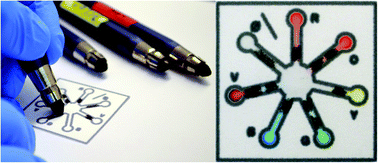
Thanks to the work of a team of researchers from California Polytechnic State University, recently published in the journal Lab on a Chip, chemicals formed into pencils can be made to react with one another by simply drawing with them on paper. The team may have taken inspiration from colouring books for their take on a chemical toolkit, but their approach could make carrying out simple but common diagnostic tests based on chemical reactions – for example diabetes, HIV, or tests for environmental pollutants – much easier.

Here’s a picture of the pens,

ReagentPencilsDiagnostics

Courtesy: Lab on a Chip

Lorch provides a good description of the technology giving descriptions of reagents and paper-based microfluidics, as well as, describing how the researchers turned the concept of colouring pencils into a diagnostic tool.

Lorch also provides a description of a specific test (Note: Links have been removed),

The team demonstrated a potential use of the reagent pencil technique by using it in place of a common test used by diabetics to check their blood glucose levels, which involves reacting a pinprick blood sample with a chemical solution and examining the result.

One pencil was constructed with a mixture of enzymes, one called horseradish peroxidase (HRP) and the other glucose oxidase (GOx). A second pencil contained a reagent called ABTS. When combined in the presence of glucose these react together to give a blue-coloured product. Comparing the results from their pencils on the pad with the more traditional dropper method used by diabetics the team found the results were identical.

This new ‘pencil kit’ diagnostic technology is easy to use and features a big improvement over the current diagnostic tests,

This is of course extremely easy to set up. Traditional diagnostic tests require training, while this pad and pencil system requires no more than skill than required to colour within the lines. The reagents are extremely stable once made into pencils – usually they would degrade in a matter of days as liquids, limiting how and where the tests can be made. However the reagent pencils showed no sign of degrading after two months.

Being able to use the pencils for two months as opposed to liquids that remain viable for a few days? That’s a huge jump and it makes me wonder about using these kits in harsh conditions such as desert climates and/or emergency situations. Materials that don’t need to be refrigerated and could be used for up to two months and don’t require intensive training could be very helpful. Lorch suggests some other possibilities as well,

… There’s scope to monitor environmental pollutants, carry out diagnostic tests in remote locations – not to mention teach chemistry in primary schools.

Here’s a link to and a citation for the study on the ‘colouring pencil kit’,

Reagent pencils: a new technique for solvent-free deposition of reagents onto paper-based microfluidic devices by Haydn T. Mitchell, Isabelle C. Noxon, Cory A. Chaplan, Samantha J. Carlton, Cheyenne H. Liu, Kirsten A. Ganaja, Nathaniel W. Martinez, Chad E. Immoos, Philip J. Costanzo, and Andres W. Martinez. Lab Chip, 2015, Advance Article DOI: 10.1039/C5LC00297D First published online 08 Apr 2015

This paper is open access but you do have to register on the site unless you have another means of access.