Tag Archives: DNA

Cloaking devices made from DNA and gold nanoparticles using top-down lithography

This new technique seems promising but there’ve been a lot of ‘cloaking’ devices announced in the years I’ve been blogging and, in all likelihood, I was late to the party so I’m exercising a little caution before getting too excited. For the latest development in cloaking devices, there’s a January 18, 2018 news item on Nanowerk,

Northwestern University researchers have developed a first-of-its-kind technique for creating entirely new classes of optical materials and devices that could lead to light bending and cloaking devices — news to make the ears of Star Trek’s Spock perk up.

Using DNA [deoxyribonucleic acid] as a key tool, the interdisciplinary team took gold nanoparticles of different sizes and shapes and arranged them in two and three dimensions to form optically active superlattices. Structures with specific configurations could be programmed through choice of particle type and both DNA-pattern and sequence to exhibit almost any color across the visible spectrum, the scientists report.

A January 18, 2018 Northwestern University news release (also on EurekAlert) by Megan Fellman, which originated the news item, delves into more detail (Note: Links have been removed),

“Architecture is everything when designing new materials, and we now have a new way to precisely control particle architectures over large areas,” said Chad A. Mirkin, the George B. Rathmann Professor of Chemistry in the Weinberg College of Arts and Sciences at Northwestern. “Chemists and physicists will be able to build an almost infinite number of new structures with all sorts of interesting properties. These structures cannot be made by any known technique.”

The technique combines an old fabrication method — top-down lithography, the same method used to make computer chips — with a new one — programmable self-assembly driven by DNA. The Northwestern team is the first to combine the two to achieve individual particle control in three dimensions.

The study was published online by the journal Science today (Jan. 18). Mirkin and Vinayak P. Dravid and Koray Aydin, both professors in Northwestern’s McCormick School of Engineering, are co-corresponding authors.

Scientists will be able to use the powerful and flexible technique to build metamaterials — materials not found in nature — for a range of applications including sensors for medical and environmental uses.

The researchers used a combination of numerical simulations and optical spectroscopy techniques to identify particular nanoparticle superlattices that absorb specific wavelengths of visible light. The DNA-modified nanoparticles — gold in this case — are positioned on a pre-patterned template made of complementary DNA. Stacks of structures can be made by introducing a second and then a third DNA-modified particle with DNA that is complementary to the subsequent layers.

In addition to being unusual architectures, these materials are stimuli-responsive: the DNA strands that hold them together change in length when exposed to new environments, such as solutions of ethanol that vary in concentration. The change in DNA length, the researchers found, resulted in a change of color from black to red to green, providing extreme tunability of optical properties.

“Tuning the optical properties of metamaterials is a significant challenge, and our study achieves one of the highest tunability ranges achieved to date in optical metamaterials,” said Aydin, assistant professor of electrical engineering and computer science at McCormick.

“Our novel metamaterial platform — enabled by precise and extreme control of gold nanoparticle shape, size and spacing — holds significant promise for next-generation optical metamaterials and metasurfaces,” Aydin said.

The study describes a new way to organize nanoparticles in two and three dimensions. The researchers used lithography methods to drill tiny holes — only one nanoparticle wide — in a polymer resist, creating “landing pads” for nanoparticle components modified with strands of DNA. The landing pads are essential, Mirkin said, since they keep the structures that are grown vertical.

The nanoscopic landing pads are modified with one sequence of DNA, and the gold nanoparticles are modified with complementary DNA. By alternating nanoparticles with complementary DNA, the researchers built nanoparticle stacks with tremendous positional control and over a large area. The particles can be different sizes and shapes (spheres, cubes and disks, for example).

“This approach can be used to build periodic lattices from optically active particles, such as gold, silver and any other material that can be modified with DNA, with extraordinary nanoscale precision,” said Mirkin, director of Northwestern’s International Institute for Nanotechnology.

Mirkin also is a professor of medicine at Northwestern University Feinberg School of Medicine and professor of chemical and biological engineering, biomedical engineering and materials science and engineering in the McCormick School.

The success of the reported DNA programmable assembly required expertise with hybrid (soft-hard) materials and exquisite nanopatterning and lithographic capabilities to achieve the requisite spatial resolution, definition and fidelity across large substrate areas. The project team turned to Dravid, a longtime collaborator of Mirkin’s who specializes in nanopatterning, advanced microscopy and characterization of soft, hard and hybrid nanostructures.

Dravid contributed his expertise and assisted in designing the nanopatterning and lithography strategy and the associated characterization of the new exotic structures. He is the Abraham Harris Professor of Materials Science and Engineering in McCormick and the founding director of the NUANCE center, which houses the advanced patterning, lithography and characterization used in the DNA-programmed structures.

Here’s a link to and a citation for the paper,

Building superlattices from individual nanoparticles via template-confined DNA-mediated assembly by Qing-Yuan Lin, Jarad A. Mason, Zhongyang, Wenjie Zhou, Matthew N. O’Brien, Keith A. Brown, Matthew R. Jones, Serkan Butun, Byeongdu Lee, Vinayak P. Dravid, Koray Aydin, Chad A. Mirkin. Science 18 Jan 2018: eaaq0591 DOI: 10.1126/science.aaq0591

This paper is behind a paywall.

As noted earlier, it could be a while before cloaking devices are made available. In the meantime, you may find this image inspiring,

Caption: Northwestern University researchers have developed a new method to precisely arrange nanoparticles of different sizes and shapes in two and three dimensions, resulting in optically active superlattices. Credit: Northwestern University

New nanomapping technology: CRISPR-CAS9 as a programmable nanoparticle

A November 21, 2017 news item on Nanowerk describes a rather extraordinary (to me, anyway) approach to using CRRISP ( Clustered Regularly Interspaced Short Palindromic Repeats)-CAS9 (Note: A link has been removed),

A team of scientists led by Virginia Commonwealth University physicist Jason Reed, Ph.D., have developed new nanomapping technology that could transform the way disease-causing genetic mutations are diagnosed and discovered. Described in a study published today [November 21, 2017] in the journal Nature Communications (“DNA nanomapping using CRISPR-Cas9 as a programmable nanoparticle”), this novel approach uses high-speed atomic force microscopy (AFM) combined with a CRISPR-based chemical barcoding technique to map DNA nearly as accurately as DNA sequencing while processing large sections of the genome at a much faster rate. What’s more–the technology can be powered by parts found in your run-of-the-mill DVD player.

A November 21, 2017 Virginia Commonwealth University news release by John Wallace, which originated the news item, provides more detail,

The human genome is made up of billions of DNA base pairs. Unraveled, it stretches to a length of nearly six feet long. When cells divide, they must make a copy of their DNA for the new cell. However, sometimes various sections of the DNA are copied incorrectly or pasted together at the wrong location, leading to genetic mutations that cause diseases such as cancer. DNA sequencing is so precise that it can analyze individual base pairs of DNA. But in order to analyze large sections of the genome to find genetic mutations, technicians must determine millions of tiny sequences and then piece them together with computer software. In contrast, biomedical imaging techniques such as fluorescence in situ hybridization, known as FISH, can only analyze DNA at a resolution of several hundred thousand base pairs.

Reed’s new high-speed AFM method can map DNA to a resolution of tens of base pairs while creating images up to a million base pairs in size. And it does it using a fraction of the amount of specimen required for DNA sequencing.

“DNA sequencing is a powerful tool, but it is still quite expensive and has several technological and functional limitations that make it difficult to map large areas of the genome efficiently and accurately,” said Reed, principal investigator on the study. Reed is a member of the Cancer Molecular Genetics research program at VCU Massey Cancer Center and an associate professor in the Department of Physics in the College of Humanities and Sciences.

“Our approach bridges the gap between DNA sequencing and other physical mapping techniques that lack resolution,” he said. “It can be used as a stand-alone method or it can complement DNA sequencing by reducing complexity and error when piecing together the small bits of genome analyzed during the sequencing process.”

IBM scientists made headlines in 1989 when they developed AFM technology and used a related technique to rearrange molecules at the atomic level to spell out “IBM.” AFM achieves this level of detail by using a microscopic stylus — similar to a needle on a record player — that barely makes contact with the surface of the material being studied. The interaction between the stylus and the molecules creates the image. However, traditional AFM is too slow for medical applications and so it is primarily used by engineers in materials science.

“Our device works in the same fashion as AFM but we move the sample past the stylus at a much greater velocity and use optical instruments to detect the interaction between the stylus and the molecules. We can achieve the same level of detail as traditional AFM but can process material more than a thousand times faster,” said Reed, whose team proved the technology can be mainstreamed by using optical equipment found in DVD players. “High-speed AFM is ideally suited for some medical applications as it can process materials quickly and provide hundreds of times more resolution than comparable imaging methods.”

Increasing the speed of AFM was just one hurdle Reed and his colleagues had to overcome. In order to actually identify genetic mutations in DNA, they had to develop a way to place markers or labels on the surface of the DNA molecules so they could recognize patterns and irregularities. An ingenious chemical barcoding solution was developed using a form of CRISPR technology.

CRISPR has made a lot of headlines recently in regard to gene editing. CRISPR is an enzyme that scientists have been able to “program” using targeting RNA in order to cut DNA at precise locations that the cell then repairs on its own. Reed’s team altered the chemical reaction conditions of the CRISPR enzyme so that it only sticks to the DNA and does not actually cut it.

“Because the CRISPR enzyme is a protein that’s physically bigger than the DNA molecule, it’s perfect for this barcoding application,” Reed said. “We were amazed to discover this method is nearly 90 percent efficient at bonding to the DNA molecules. And because it’s easy to see the CRISPR proteins, you can spot genetic mutations among the patterns in DNA.”

To demonstrate the technique’s effectiveness, the researchers mapped genetic translocations present in lymph node biopsies of lymphoma patients. Translocations occur when one section of the DNA gets copied and pasted to the wrong place in the genome. They are especially prevalent in blood cancers such as lymphoma but occur in other cancers as well.

While there are many potential uses for this technology, Reed and his team are focusing on medical applications. They are currently developing software based on existing algorithms that can analyze patterns in sections of DNA up to and over a million base pairs in size. Once completed, it would not be hard to imagine this shoebox-sized instrument in pathology labs assisting in the diagnosis and treatment of diseases linked to genetic mutations.

Here’s a link to and a citation for the paper,

DNA nanomapping using CRISPR-Cas9 as a programmable nanoparticle by Andrey Mikheikin, Anita Olsen, Kevin Leslie, Freddie Russell-Pavier, Andrew Yacoot, Loren Picco, Oliver Payton, Amir Toor, Alden Chesney, James K. Gimzewski, Bud Mishra, & Jason Reed. Nature Communications 8, Article number: 1665 (2017) doi:10.1038/s41467-017-01891-9 Published online: 21 November 2017

This paper is open access.

World heritage music stored in DNA

It seems a Swiss team from the École Polytechnique de Lausanne (EPFL) have collaborated with American companies Twist Bioscience and Microsoft, as well as, the University of Washington (state) to preserve two iconic jazz pieces on DNA (deoxyribonucleic acid) according to a Sept. 29, 2017 news item on phys.org,,

Thanks to an innovative technology for encoding data in DNA strands, two items of world heritage – songs recorded at the Montreux Jazz Festival [held in Switzerland] and digitized by EPFL – have been safeguarded for eternity. This marks the first time that cultural artifacts granted UNESCO heritage status have been saved in such a manner, ensuring they are preserved for thousands of years. The method was developed by US company Twist Bioscience and is being unveiled today in a demonstrator created at the EPFL+ECAL Lab.

“Tutu” by Miles Davis and “Smoke on the Water” by Deep Purple have already made their mark on music history. Now they have entered the annals of science, for eternity. Recordings of these two legendary songs were digitized by the Ecole Polytechnique Fédérale de Lausanne (EPFL) as part of the Montreux Jazz Digital Project, and they are the first to be stored in the form of a DNA sequence that can be subsequently decoded and listened to without any reduction in quality.

A Sept. 29, 2017 EPFL press release by Emmanuel Barraud, which originated the news item, provides more details,

This feat was achieved by US company Twist Bioscience working in association with Microsoft Research and the University of Washington. The pioneering technology is actually based on a mechanism that has been at work on Earth for billions of years: storing information in the form of DNA strands. This fundamental process is what has allowed all living species, plants and animals alike, to live on from generation to generation.

The entire world wide web in a shoe box

All electronic data storage involves encoding data in binary format – a series of zeros and ones – and then recording it on a physical medium. DNA works in a similar way, but is composed of long strands of series of four nucleotides (A, T, C and G) that make up a “code.” While the basic principle may be the same, the two methods differ greatly in terms of efficiency: if all the information currently on the internet was stored in the form of DNA, it would fit in a shoe box!

Recent advances in biotechnology now make it possible for humans to do what Mother Nature has always done. Today’s scientists can create artificial DNA strands, “record” any kind of genetic code on them and then analyze them using a sequencer to reconstruct the original data. What’s more, DNA is extraordinarily stable, as evidenced by prehistoric fragments that have been preserved in amber. Artificial strands created by scientists and carefully encapsulated should likewise last for millennia.

To help demonstrate the feasibility of this new method, EPFL’s Metamedia Center provided recordings of two famous songs played at the Montreux Jazz Festival: “Tutu” by Miles Davis, and “Smoke on the Water” by Deep Purple. Twist Bioscience and its research partners encoded the recordings, transformed them into DNA strands and then sequenced and decoded them and played them again – without any reduction in quality.

The amount of artificial DNA strands needed to record the two songs is invisible to the naked eye, and the amount needed to record all 50 years of the Festival’s archives, which have been included in UNESCO’s [United Nations Educational, Scientific and Cultural Organization] Memory of the World Register, would be equal in size to a grain of sand. “Our partnership with EPFL in digitizing our archives aims not only at their positive exploration, but also at their preservation for the next generations,” says Thierry Amsallem, president of the Claude Nobs Foundation. “By taking part in this pioneering experiment which writes the songs into DNA strands, we can be certain that they will be saved on a medium that will never become obsolete!”

A new concept of time

At EPFL’s first-ever ArtTech forum, attendees got to hear the two songs played after being stored in DNA, using a demonstrator developed at the EPFL+ECAL Lab. The system shows that being able to store data for thousands of years is a revolutionary breakthrough that can completely change our relationship with data, memory and time. “For us, it means looking into radically new ways of interacting with cultural heritage that can potentially cut across civilizations,” says Nicolas Henchoz, head of the EPFL+ECAL Lab.

Quincy Jones, a longstanding Festival supporter, is particularly enthusiastic about this technological breakthrough: “With advancements in nanotechnology, I believe we can expect to see people living prolonged lives, and with that, we can also expect to see more developments in the enhancement of how we live. For me, life is all about learning where you came from in order to get where you want to go, but in order to do so, you need access to history! And with the unreliability of how archives are often stored, I sometimes worry that our future generations will be left without such access… So, it absolutely makes my soul smile to know that EPFL, Twist Bioscience and their partners are coming together to preserve the beauty and history of the Montreux Jazz Festival for our future generations, on DNA! I’ve been a part of this festival for decades and it truly is a magnificent representation of what happens when different cultures unite for the sake of music. Absolute magic. And I’m proud to know that the memory of this special place will never be lost.

A Sept. 29, 2017 Twist Bioscience news release is repetitive in some ways but interesting nonetheless,

Twist Bioscience, a company accelerating science and innovation through rapid, high-quality DNA synthesis, today announced that, working with Microsoft and University of Washington researchers, they have successfully stored archival-quality audio recordings of two important music performances from the archives of the world-renowned Montreux Jazz Festival.
These selections are encoded and stored in nature’s preferred storage medium, DNA, for the first time. These tiny specks of DNA will preserve a part of UNESCO’s Memory of the World Archive, where valuable cultural heritage collections are recorded. This is the first time DNA has been used as a long-term archival-quality storage medium.
Quincy Jones, world-renowned Entertainment Executive, Music Composer and Arranger, Musician and Music Producer said, “With advancements in nanotechnology, I believe we can expect to see people living prolonged lives, and with that, we can also expect to see more developments in the enhancement of how we live. For me, life is all about learning where you came from in order to get where you want to go, but in order to do so, you need access to history! And with the unreliability of how archives are often stored, I sometimes worry that our future generations will be left without such access…So, it absolutely makes my soul smile to know that EPFL, Twist Bioscience and others are coming together to preserve the beauty and history of the Montreux Jazz Festival for our future generations, on DNA!…I’ve been a part of this festival for decades and it truly is a magnificent representation of what happens when different cultures unite for the sake of music. Absolute magic. And I’m proud to know that the memory of this special place will never be lost.”
“Our partnership with EPFL in digitizing our archives aims not only at their positive exploration, but also at their preservation for the next generations,” says Thierry Amsallem, president of the Claude Nobs Foundation. “By taking part in this pioneering experiment which writes the songs into DNA strands, we can be certain that they will be saved on a medium that will never become obsolete!”
The Montreux Jazz Digital Project is a collaboration between the Claude Nobs Foundation, curator of the Montreux Jazz Festival audio-visual collection and the École Polytechnique Fédérale de Lausanne (EPFL) to digitize, enrich, store, show, and preserve this notable legacy created by Claude Nobs, the Festival’s founder.
In this proof-of-principle project, two quintessential music performances from the Montreux Jazz Festival – Smoke on the Water, performed by Deep Purple and Tutu, performed by Miles Davis – have been encoded onto DNA and read back with 100 percent accuracy. After being decoded, the songs were played on September 29th [2017] at the ArtTech Forum (see below) in Lausanne, Switzerland. Smoke on the Water was selected as a tribute to Claude Nobs, the Montreux Jazz Festival’s founder. The song memorializes a fire and Funky Claude’s rescue efforts at the Casino Barrière de Montreux during a Frank Zappa concert promoted by Claude Nobs. Miles Davis’ Tutu was selected for the role he played in music history and the Montreux Jazz Festival’s success. Miles Davis died in 1991.
“We archived two magical musical pieces on DNA of this historic collection, equating to 140MB of stored data in DNA,” said Karin Strauss, Ph.D., a Senior Researcher at Microsoft, and one of the project’s leaders.  “The amount of DNA used to store these songs is much smaller than one grain of sand. Amazingly, storing the entire six petabyte Montreux Jazz Festival’s collection would result in DNA smaller than one grain of rice.”
Luis Ceze, Ph.D., a professor in the Paul G. Allen School of Computer Science & Engineering at the University of Washington, said, “DNA, nature’s preferred information storage medium, is an ideal fit for digital archives because of its durability, density and eternal relevance. Storing items from the Montreux Jazz Festival is a perfect way to show how fast DNA digital data storage is becoming real.”
Nature’s Preferred Storage Medium
Nature selected DNA as its hard drive billions of years ago to encode all the genetic instructions necessary for life. These instructions include all the information necessary for survival. DNA molecules encode information with sequences of discrete units. In computers, these discrete units are the 0s and 1s of “binary code,” whereas in DNA molecules, the units are the four distinct nucleotide bases: adenine (A), cytosine (C), guanine (G) and thymine (T).
“DNA is a remarkably efficient molecule that can remain stable for millennia,” said Bill Peck, Ph.D., chief technology officer of Twist Bioscience.  “This is a very exciting project: we are now in an age where we can use the remarkable efficiencies of nature to archive master copies of our cultural heritage in DNA.   As we develop the economies of this process new performances can be added any time.  Unlike current storage technologies, nature’s media will not change and will remain readable through time. There will be no new technology to replace DNA, nature has already optimized the format.”
DNA: Far More Efficient Than a Computer 
Each cell within the human body contains approximately three billion base pairs of DNA. With 75 trillion cells in the human body, this equates to the storage of 150 zettabytes (1021) of information within each body. By comparison, the largest data centers can be hundreds of thousands to even millions of square feet to hold a comparable amount of stored data.
The Elegance of DNA as a Storage Medium
Like music, which can be widely varied with a finite number of notes, DNA encodes individuality with only four different letters in varied combinations. When using DNA as a storage medium, there are several advantages in addition to the universality of the format and incredible storage density. DNA can be stable for thousands of years when stored in a cool dry place and is easy to copy using polymerase chain reaction to create back-up copies of archived material. In addition, because of PCR, small data sets can be targeted and recovered quickly from a large dataset without needing to read the entire file.
How to Store Digital Data in DNA
To encode the music performances into archival storage copies in DNA, Twist Bioscience worked with Microsoft and University of Washington researchers to complete four steps: Coding, synthesis/storage, retrieval and decoding. First, the digital files were converted from the binary code using 0s and 1s into sequences of A, C, T and G. For purposes of the example, 00 represents A, 10 represents C, 01 represents G and 11 represents T. Twist Bioscience then synthesizes the DNA in short segments in the sequence order provided. The short DNA segments each contain about 12 bytes of data as well as a sequence number to indicate their place within the overall sequence. This is the process of storage. And finally, to ensure that the file is stored accurately, the sequence is read back to ensure 100 percent accuracy, and then decoded from A, C, T or G into a two-digit binary representation.
Importantly, to encapsulate and preserve encoded DNA, the collaborators are working with Professor Dr. Robert Grass of ETH Zurich. Grass has developed an innovative technology inspired by preservation of DNA within prehistoric fossils.  With this technology, digital data encoded in DNA remains preserved for millennia.
About UNESCO’s Memory of the World Register
UNESCO established the Memory of the World Register in 1992 in response to a growing awareness of the perilous state of preservation of, and access to, documentary heritage in various parts of the world.  Through its National Commissions, UNESCO prepared a list of endangered library and archive holdings and a world list of national cinematic heritage.
A range of pilot projects employing contemporary technology to reproduce original documentary heritage on other media began. These included, for example, a CD-ROM of the 13th Century Radzivill Chronicle, tracing the origins of the peoples of Europe, and Memoria de Iberoamerica, a joint newspaper microfilming project involving seven Latin American countries. These projects enhanced access to this documentary heritage and contributed to its preservation.
“We are incredibly proud to be a part of this momentous event, with the first archived songs placed into the UNESCO Memory of the World Register,” said Emily Leproust, Ph.D., CEO of Twist Bioscience.
About ArtTech
The ArtTech Foundation, created by renowned scientists and dignitaries from Crans-Montana, Switzerland, wishes to stimulate reflection and support pioneering and innovative projects beyond the known boundaries of culture and science.
Benefitting from the establishment of a favorable environment for the creation of technology companies, the Foundation aims to position itself as key promoter of ideas and innovative endeavors within a landscape of “Culture and Science” that is still being shaped.
Several initiatives, including our annual global platform launched in the spring of 2017, are helping to create a community that brings together researchers, celebrities in the world of culture and the arts, as well as investors and entrepreneurs from Switzerland and across the globe.
 
About EPFL
EPFL, one of the two Swiss Federal Institutes of Technology, based in Lausanne, is Europe’s most cosmopolitan technical university with students, professors and staff from over 120 nations. A dynamic environment, open to Switzerland and the world, EPFL is centered on its three missions: teaching, research and technology transfer. EPFL works together with an extensive network of partners including other universities and institutes of technology, developing and emerging countries, secondary schools and colleges, industry and economy, political circles and the general public, to bring about real impact for society.
About Twist Bioscience
At Twist Bioscience, our expertise is accelerating science and innovation by leveraging the power of scale. We have developed a proprietary semiconductor-based synthetic DNA manufacturing process featuring a high throughput silicon platform capable of producing synthetic biology tools, including genes, oligonucleotide pools and variant libraries. By synthesizing DNA on silicon instead of on traditional 96-well plastic plates, our platform overcomes the current inefficiencies of synthetic DNA production, and enables cost-effective, rapid, high-quality and high throughput synthetic gene production, which in turn, expedites the design, build and test cycle to enable personalized medicines, pharmaceuticals, sustainable chemical production, improved agriculture production, diagnostics and biodetection. We are also developing new technologies to address large scale data storage. For more information, please visit www.twistbioscience.com. Twist Bioscience is on Twitter. Sign up to follow our Twitter feed @TwistBioscience at https://twitter.com/TwistBioscience.

If you hadn’t read the EPFL press release first, it might have taken a minute to figure out why EPFL is being mentioned in the Twist Bioscience news release. Presumably someone was rushing to make a deadline. Ah well, I’ve seen and written worse.

I haven’t been able to find any video or audio recordings of the DNA-preserved performances but there is an informational video (originally published July 7, 2016) from Microsoft and the University of Washington describing the DNA-based technology,

I also found this description of listening to the DNA-preserved music in an Oct. 6, 2017 blog posting for the Canadian Broadcasting Corporation’s (CBC) Day 6 radio programme,

To listen to them, one must first suspend the DNA holding the songs in a solution. Next, one can use a DNA sequencer to read the letters of the bases forming the molecules. Then, algorithms can determine the digital code those letters form. From that code, comes the music.

It’s complicated but Ceze says his team performed this process without error.

You can find out more about UNESCO’s Memory of the World and its register here , more about the EPFL+ECAL Lab here, and more about Twist Bioscience here.

Nondirect relationships between the number of hydrogen bonds in DNA pairs and their relative strengths (three can be less than two)

Michael Berger’s Oct. 10, 2017 Nanowerk Spotlight article features research from the Institut Català de Nanociència i Nanotecnologia, (Catalan Institute of Nanoscience and Nanotechnology and acronym ICN2) which has nothing to do with recent vote on independence (for more, see this Oct. 23, 2017 article about how countries such as ‘Catalonia’ and others do and don’t gain independence in The Atlantic),

This is the first report on the electrical characterization of DNA with intrabond resolution.

Specifically, it quantifies electrical forces due to a single hydrogen bond in DNA and provides proof of a non-direct relationship between the number of hydrogen bonds in DNA pairs and the relative strengths of such pairs.

Such understanding of the relative strengths of the forces involved in the specific bonding of DNA as well as its electrical origin provides physical foundations to control mechanisms associated with DNA stability. It could help to develop new methods for the understanding, characterization; and control of relevant events originated at intrabond scales e.g. controlled DNA repair and damage; controlled modification of the expression of the genome; and communications below the single bond limit.

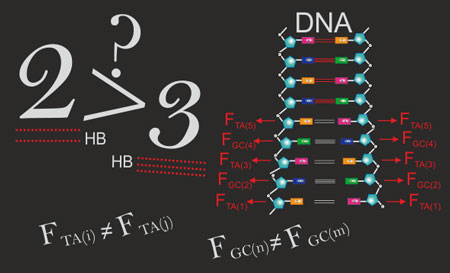
A blackboard representation of the manuscript’s key message: the quantification of the relative strengths between base pairs in DNA due to zipping hydrogen bonds might place on doubt such mechanisms regarding the interpretation of thermodynamic properties of DNA based on the assumption that A/T pairs are weaker than G/C pairs due to the sole difference in the number of hydrogen bonds, 2 and 3 respectively. (Image: Dr. Yamila García-Martínez)

Generally, being able to control DNA stability at the single bond level by means of electromagnetic interactions opens new avenues to induce modifications of the replication and transcription processes of DNA by means of noncontact methods.

….

Going forward, the researchers will study the effects of external electromagnetic fields on DNA at the level of single bond events. This will have not only an enormous interest in the medical field but also in nanotechnology where it would open the door to non-contact atomic manipulation of DNA – the analogue to the CRISPR gene editing method [emphasis mine] but using electromagnetic fields to drive changes in DNA.

Interesting stuff, eh?

Here’s a link to and a citation for the paper,

Unveiled electric profiles within hydrogen bonds suggest DNA base pairs with similar bond strengths by Y. B. Ruiz-Blanco, Y. Almeida, C. M. Sotomayor-Torres, Y. García. PLOS [Public Library of Science] https://doi.org/10.1371/journal.pone.0185638 Published: October 5, 2017

This paper is open access.

CRISPR and editing the germline in the US (part 3 of 3): public discussions and pop culture

After giving a basic explanation of the technology and some of the controversies in part 1 and offering more detail about the technology and about the possibility of designer babies in part 2; this part covers public discussion, a call for one and the suggestion that one is taking place in popular culture.

But a discussion does need to happen

In a move that is either an exquisite coincidence or has been carefully orchestrated (I vote for the latter), researchers from the University of Wisconsin-Madison have released a study about attitudes in the US to human genome editing. From an Aug. 11, 2017 University of Wisconsin-Madison news release (also on EurekAllert),

In early August 2017, an international team of scientists announced they had successfully edited the DNA of human embryos. As people process the political, moral and regulatory issues of the technology — which nudges us closer to nonfiction than science fiction — researchers at the University of Wisconsin-Madison and Temple University show the time is now to involve the American public in discussions about human genome editing.

In a study published Aug. 11 in the journal Science, the researchers assessed what people in the United States think about the uses of human genome editing and how their attitudes may drive public discussion. They found a public divided on its uses but united in the importance of moving conversations forward.

“There are several pathways we can go down with gene editing,” says UW-Madison’s Dietram Scheufele, lead author of the study and member of a National Academy of Sciences committee that compiled a report focused on human gene editing earlier this year. “Our study takes an exhaustive look at all of those possible pathways forward and asks where the public stands on each one of them.”

Compared to previous studies on public attitudes about the technology, the new study takes a more nuanced approach, examining public opinion about the use of gene editing for disease therapy versus for human enhancement, and about editing that becomes hereditary versus editing that does not.

The research team, which included Scheufele and Dominique Brossard — both professors of life sciences communication — along with Michael Xenos, professor of communication arts, first surveyed study participants about the use of editing to treat disease (therapy) versus for enhancement (creating so-called “designer babies”). While about two-thirds of respondents expressed at least some support for therapeutic editing, only one-third expressed support for using the technology for enhancement.

Diving even deeper, researchers looked into public attitudes about gene editing on specific cell types — somatic or germline — either for therapy or enhancement. Somatic cells are non-reproductive, so edits made in those cells do not affect future generations. Germline cells, however, are heritable, and changes made in these cells would be passed on to children.

Public support of therapeutic editing was high both in cells that would be inherited and those that would not, with 65 percent of respondents supporting therapy in germline cells and 64 percent supporting therapy in somatic cells. When considering enhancement editing, however, support depended more upon whether the changes would affect future generations. Only 26 percent of people surveyed supported enhancement editing in heritable germline cells and 39 percent supported enhancement of somatic cells that would not be passed on to children.

“A majority of people are saying that germline enhancement is where the technology crosses that invisible line and becomes unacceptable,” says Scheufele. “When it comes to therapy, the public is more open, and that may partly be reflective of how severe some of those genetically inherited diseases are. The potential treatments for those diseases are something the public at least is willing to consider.”

Beyond questions of support, researchers also wanted to understand what was driving public opinions. They found that two factors were related to respondents’ attitudes toward gene editing as well as their attitudes toward the public’s role in its emergence: the level of religious guidance in their lives, and factual knowledge about the technology.

Those with a high level of religious guidance in their daily lives had lower support for human genome editing than those with low religious guidance. Additionally, those with high knowledge of the technology were more supportive of it than those with less knowledge.

While respondents with high religious guidance and those with high knowledge differed on their support for the technology, both groups highly supported public engagement in its development and use. These results suggest broad agreement that the public should be involved in questions of political, regulatory and moral aspects of human genome editing.

“The public may be split along lines of religiosity or knowledge with regard to what they think about the technology and scientific community, but they are united in the idea that this is an issue that requires public involvement,” says Scheufele. “Our findings show very nicely that the public is ready for these discussions and that the time to have the discussions is now, before the science is fully ready and while we have time to carefully think through different options regarding how we want to move forward.”

Here’s a  link to and a citation for the paper,

U.S. attitudes on human genome editing by Dietram A. Scheufele, Michael A. Xenos, Emily L. Howell, Kathleen M. Rose, Dominique Brossard1, and Bruce W. Hardy. Science 11 Aug 2017: Vol. 357, Issue 6351, pp. 553-554 DOI: 10.1126/science.aan3708

This paper is behind a paywall.

A couple of final comments

Briefly, I notice that there’s no mention of the ethics of patenting this technology in the news release about the study.

Moving on, it seems surprising that the first team to engage in germline editing in the US is in Oregon; I would have expected the work to come from Massachusetts, California, or Illinois where a lot of bleeding edge medical research is performed. However, given the dearth of financial support from federal funding institutions, it seems likely that only an outsider would dare to engage i the research. Given the timing, Mitalipov’s work was already well underway before the recent about-face from the US National Academy of Sciences (Note: Kaiser’s Feb. 14, 2017 article does note that for some the recent recommendations do not represent any change).

As for discussion on issues such as editing of the germline, I’ve often noted here that popular culture (including advertising with the science fiction and other dramas laid in various media) often provides an informal forum for discussion. Joelle Renstrom in an Aug. 13, 2017 article for slate.com writes that Orphan Black (a BBC America series featuring clones) opened up a series of questions about science and ethics in the guise of a thriller about clones. She offers a précis of the first four seasons (Note: A link has been removed),

If you stopped watching a few seasons back, here’s a brief synopsis of how the mysteries wrap up. Neolution, an organization that seeks to control human evolution through genetic modification, began Project Leda, the cloning program, for two primary reasons: to see whether they could and to experiment with mutations that might allow people (i.e., themselves) to live longer. Neolution partnered with biotech companies such as Dyad, using its big pharma reach and deep pockets to harvest people’s genetic information and to conduct individual and germline (that is, genetic alterations passed down through generations) experiments, including infertility treatments that result in horrifying birth defects and body modification, such as tail-growing.

She then provides the article’s thesis (Note: Links have been removed),

Orphan Black demonstrates Carl Sagan’s warning of a time when “awesome technological powers are in the hands of a very few.” Neolutionists do whatever they want, pausing only to consider whether they’re missing an opportunity to exploit. Their hubris is straight out of Victor Frankenstein’s playbook. Frankenstein wonders whether he ought to first reanimate something “of simpler organisation” than a human, but starting small means waiting for glory. Orphan Black’s evil scientists embody this belief: if they’re going to play God, then they’ll control not just their own destinies, but the clones’ and, ultimately, all of humanity’s. Any sacrifices along the way are for the greater good—reasoning that culminates in Westmoreland’s eugenics fantasy to genetically sterilize 99 percent of the population he doesn’t enhance.

Orphan Black uses sci-fi tropes to explore real-world plausibility. Neolution shares similarities with transhumanism, the belief that humans should use science and technology to take control of their own evolution. While some transhumanists dabble in body modifications, such as microchip implants or night-vision eye drops, others seek to end suffering by curing human illness and aging. But even these goals can be seen as selfish, as access to disease-eradicating or life-extending technologies would be limited to the wealthy. Westmoreland’s goal to “sell Neolution to the 1 percent” seems frighteningly plausible—transhumanists, who statistically tend to be white, well-educated, and male, and their associated organizations raise and spend massive sums of money to help fulfill their goals. …

On Orphan Black, denial of choice is tantamount to imprisonment. That the clones have to earn autonomy underscores the need for ethics in science, especially when it comes to genetics. The show’s message here is timely given the rise of gene-editing techniques such as CRISPR. Recently, the National Academy of Sciences gave germline gene editing the green light, just one year after academy scientists from around the world argued it would be “irresponsible to proceed” without further exploring the implications. Scientists in the United Kingdom and China have already begun human genetic engineering and American scientists recently genetically engineered a human embryo for the first time. The possibility of Project Leda isn’t farfetched. Orphan Black warns us that money, power, and fear of death can corrupt both people and science. Once that happens, loss of humanity—of both the scientists and the subjects—is inevitable.

In Carl Sagan’s dark vision of the future, “people have lost the ability to set their own agendas or knowledgeably question those in authority.” This describes the plight of the clones at the outset of Orphan Black, but as the series continues, they challenge this paradigm by approaching science and scientists with skepticism, ingenuity, and grit. …

I hope there are discussions such as those Scheufele and Brossard are advocating but it might be worth considering that there is already some discussion underway, as informal as it is.

-30-

Part 1: CRISPR and editing the germline in the US (part 1 of 3): In the beginning

Part 2: CRISPR and editing the germline in the US (part 2 of 3): ‘designer babies’?

CRISPR and editing the germline in the US (part 2 of 3): ‘designer babies’?

Having included an explanation of CRISPR-CAS9 technology along with the news about the first US team to edit the germline and bits and pieces about ethics and a patent fight (part 1), this part hones in on the details of the work and worries about ‘designer babies’.

The interest flurry

I found three articles addressing the research and all three concur that despite some of the early reporting, this is not the beginning of a ‘designer baby’ generation.

First up was Nick Thieme in a July 28, 2017 article for Slate,

MIT Technology Review reported Thursday that a team of researchers from Portland, Oregon were the first team of U.S.-based scientists to successfully create a genetically modified human embryo. The researchers, led by Shoukhrat Mitalipov of Oregon Health and Science University, changed the DNA of—in MIT Technology Review’s words—“many tens” of genetically-diseased embryos by injecting the host egg with CRISPR, a DNA-based gene editing tool first discovered in bacteria, at the time of fertilization. CRISPR-Cas9, as the full editing system is called, allows scientists to change genes accurately and efficiently. As has happened with research elsewhere, the CRISPR-edited embryos weren’t implanted—they were kept sustained for only a couple of days.

In addition to being the first American team to complete this feat, the researchers also improved upon the work of the three Chinese research teams that beat them to editing embryos with CRISPR: Mitalipov’s team increased the proportion of embryonic cells that received the intended genetic changes, addressing an issue called “mosaicism,” which is when an embryo is comprised of cells with different genetic makeups. Increasing that proportion is essential to CRISPR work in eliminating inherited diseases, to ensure that the CRISPR therapy has the intended result. The Oregon team also reduced the number of genetic errors introduced by CRISPR, reducing the likelihood that a patient would develop cancer elsewhere in the body.

Separate from the scientific advancements, it’s a big deal that this work happened in a country with such intense politicization of embryo research. …

But there are a great number of obstacles between the current research and the future of genetically editing all children to be 12-foot-tall Einsteins.

Ed Yong in an Aug. 2, 2017 article for The Atlantic offered a comprehensive overview of the research and its implications (unusually for Yong, there seems to be mildly condescending note but it’s worth ignoring for the wealth of information in the article; Note: Links have been removed),

… the full details of the experiment, which are released today, show that the study is scientifically important but much less of a social inflection point than has been suggested. “This has been widely reported as the dawn of the era of the designer baby, making it probably the fifth or sixth time people have reported that dawn,” says Alta Charo, an expert on law and bioethics at the University of Wisconsin-Madison. “And it’s not.”

Given the persistent confusion around CRISPR and its implications, I’ve laid out exactly what the team did, and what it means.

Who did the experiments?

Shoukhrat Mitalipov is a Kazakhstani-born cell biologist with a history of breakthroughs—and controversy—in the stem cell field. He was the scientist to clone monkeys. He was the first to create human embryos by cloning adult cells—a move that could provide patients with an easy supply of personalized stem cells. He also pioneered a technique for creating embryos with genetic material from three biological parents, as a way of preventing a group of debilitating inherited diseases.

Although MIT Tech Review name-checked Mitalipov alone, the paper splits credit for the research between five collaborating teams—four based in the United States, and one in South Korea.

What did they actually do?

The project effectively began with an elevator conversation between Mitalipov and his colleague Sanjiv Kaul. Mitalipov explained that he wanted to use CRISPR to correct a disease-causing gene in human embryos, and was trying to figure out which disease to focus on. Kaul, a cardiologist, told him about hypertrophic cardiomyopathy (HCM)—an inherited heart disease that’s commonly caused by mutations in a gene called MYBPC3. HCM is surprisingly common, affecting 1 in 500 adults. Many of them lead normal lives, but in some, the walls of their hearts can thicken and suddenly fail. For that reason, HCM is the commonest cause of sudden death in athletes. “There really is no treatment,” says Kaul. “A number of drugs are being evaluated but they are all experimental,” and they merely treat the symptoms. The team wanted to prevent HCM entirely by removing the underlying mutation.

They collected sperm from a man with HCM and used CRISPR to change his mutant gene into its normal healthy version, while simultaneously using the sperm to fertilize eggs that had been donated by female volunteers. In this way, they created embryos that were completely free of the mutation. The procedure was effective, and avoided some of the critical problems that have plagued past attempts to use CRISPR in human embryos.

Wait, other human embryos have been edited before?

There have been three attempts in China. The first two—in 2015 and 2016—used non-viable embryos that could never have resulted in a live birth. The third—announced this March—was the first to use viable embryos that could theoretically have been implanted in a womb. All of these studies showed that CRISPR gene-editing, for all its hype, is still in its infancy.

The editing was imprecise. CRISPR is heralded for its precision, allowing scientists to edit particular genes of choice. But in practice, some of the Chinese researchers found worrying levels of off-target mutations, where CRISPR mistakenly cut other parts of the genome.

The editing was inefficient. The first Chinese team only managed to successfully edit a disease gene in 4 out of 86 embryos, and the second team fared even worse.

The editing was incomplete. Even in the successful cases, each embryo had a mix of modified and unmodified cells. This pattern, known as mosaicism, poses serious safety problems if gene-editing were ever to be used in practice. Doctors could end up implanting women with embryos that they thought were free of a disease-causing mutation, but were only partially free. The resulting person would still have many tissues and organs that carry those mutations, and might go on to develop symptoms.

What did the American team do differently?

The Chinese teams all used CRISPR to edit embryos at early stages of their development. By contrast, the Oregon researchers delivered the CRISPR components at the earliest possible point—minutes before fertilization. That neatly avoids the problem of mosaicism by ensuring that an embryo is edited from the very moment it is created. The team did this with 54 embryos and successfully edited the mutant MYBPC3 gene in 72 percent of them. In the other 28 percent, the editing didn’t work—a high failure rate, but far lower than in previous attempts. Better still, the team found no evidence of off-target mutations.

This is a big deal. Many scientists assumed that they’d have to do something more convoluted to avoid mosaicism. They’d have to collect a patient’s cells, which they’d revert into stem cells, which they’d use to make sperm or eggs, which they’d edit using CRISPR. “That’s a lot of extra steps, with more risks,” says Alta Charo. “If it’s possible to edit the embryo itself, that’s a real advance.” Perhaps for that reason, this is the first study to edit human embryos that was published in a top-tier scientific journal—Nature, which rejected some of the earlier Chinese papers.

Is this kind of research even legal?

Yes. In Western Europe, 15 countries out of 22 ban any attempts to change the human germ line—a term referring to sperm, eggs, and other cells that can transmit genetic information to future generations. No such stance exists in the United States but Congress has banned the Food and Drug Administration from considering research applications that make such modifications. Separately, federal agencies like the National Institutes of Health are banned from funding research that ultimately destroys human embryos. But the Oregon team used non-federal money from their institutions, and donations from several small non-profits. No taxpayer money went into their work. [emphasis mine]

Why would you want to edit embryos at all?

Partly to learn more about ourselves. By using CRISPR to manipulate the genes of embryos, scientists can learn more about the earliest stages of human development, and about problems like infertility and miscarriages. That’s why biologist Kathy Niakan from the Crick Institute in London recently secured a license from a British regulator to use CRISPR on human embryos.

Isn’t this a slippery slope toward making designer babies?

In terms of avoiding genetic diseases, it’s not conceptually different from PGD, which is already widely used. The bigger worry is that gene-editing could be used to make people stronger, smarter, or taller, paving the way for a new eugenics, and widening the already substantial gaps between the wealthy and poor. But many geneticists believe that such a future is fundamentally unlikely because complex traits like height and intelligence are the work of hundreds or thousands of genes, each of which have a tiny effect. The prospect of editing them all is implausible. And since genes are so thoroughly interconnected, it may be impossible to edit one particular trait without also affecting many others.

“There’s the worry that this could be used for enhancement, so society has to draw a line,” says Mitalipov. “But this is pretty complex technology and it wouldn’t be hard to regulate it.”

Does this discovery have any social importance at all?

“It’s not so much about designer babies as it is about geographical location,” says Charo. “It’s happening in the United States, and everything here around embryo research has high sensitivity.” She and others worry that the early report about the study, before the actual details were available for scrutiny, could lead to unnecessary panic. “Panic reactions often lead to panic-driven policy … which is usually bad policy,” wrote Greely [bioethicist Hank Greely].

As I understand it, despite the change in stance, there is no federal funding available for the research performed by Mitalipov and his team.

Finally, University College London (UCL) scientists Joyce Harper and Helen O’Neill wrote about CRISPR, the Oregon team’s work, and the possibilities in an Aug. 3, 2017 essay for The Conversation (Note: Links have been removed),

The genome editing tool used, CRISPR-Cas9, has transformed the field of biology in the short time since its discovery in that it not only promises, but delivers. CRISPR has surpassed all previous efforts to engineer cells and alter genomes at a fraction of the time and cost.

The technology, which works like molecular scissors to cut and paste DNA, is a natural defence system that bacteria use to fend off harmful infections. This system has the ability to recognise invading virus DNA, cut it and integrate this cut sequence into its own genome – allowing the bacterium to render itself immune to future infections of viruses with similar DNA. It is this ability to recognise and cut DNA that has allowed scientists to use it to target and edit specific DNA regions.

When this technology is applied to “germ cells” – the sperm and eggs – or embryos, it changes the germline. That means that any alterations made would be permanent and passed down to future generations. This makes it more ethically complex, but there are strict regulations around human germline genome editing, which is predominantly illegal. The UK received a licence in 2016 to carry out CRISPR on human embryos for research into early development. But edited embryos are not allowed to be inserted into the uterus and develop into a fetus in any country.

Germline genome editing came into the global spotlight when Chinese scientists announced in 2015 that they had used CRISPR to edit non-viable human embryos – cells that could never result in a live birth. They did this to modify the gene responsible for the blood disorder β-thalassaemia. While it was met with some success, it received a lot of criticism because of the premature use of this technology in human embryos. The results showed a high number of potentially dangerous, off-target mutations created in the procedure.

Impressive results

The new study, published in Nature, is different because it deals with viable human embryos and shows that the genome editing can be carried out safely – without creating harmful mutations. The team used CRISPR to correct a mutation in the gene MYBPC3, which accounts for approximately 40% of the myocardial disease hypertrophic cardiomyopathy. This is a dominant disease, so an affected individual only needs one abnormal copy of the gene to be affected.

The researchers used sperm from a patient carrying one copy of the MYBPC3 mutation to create 54 embryos. They edited them using CRISPR-Cas9 to correct the mutation. Without genome editing, approximately 50% of the embryos would carry the patients’ normal gene and 50% would carry his abnormal gene.

After genome editing, the aim would be for 100% of embryos to be normal. In the first round of the experiments, they found that 66.7% of embryos – 36 out of 54 – were normal after being injected with CRIPSR. Of the remaining 18 embryos, five had remained unchanged, suggesting editing had not worked. In 13 embryos, only a portion of cells had been edited.

The level of efficiency is affected by the type of CRISPR machinery used and, critically, the timing in which it is put into the embryo. The researchers therefore also tried injecting the sperm and the CRISPR-Cas9 complex into the egg at the same time, which resulted in more promising results. This was done for 75 mature donated human eggs using a common IVF technique called intracytoplasmic sperm injection. This time, impressively, 72.4% of embryos were normal as a result. The approach also lowered the number of embryos containing a mixture of edited and unedited cells (these embryos are called mosaics).

Finally, the team injected a further 22 embryos which were grown into blastocyst – a later stage of embryo development. These were sequenced and the researchers found that the editing had indeed worked. Importantly, they could show that the level of off-target mutations was low.

A brave new world?

So does this mean we finally have a cure for debilitating, heritable diseases? It’s important to remember that the study did not achieve a 100% success rate. Even the researchers themselves stress that further research is needed in order to fully understand the potential and limitations of the technique.

In our view, it is unlikely that genome editing would be used to treat the majority of inherited conditions anytime soon. We still can’t be sure how a child with a genetically altered genome will develop over a lifetime, so it seems unlikely that couples carrying a genetic disease would embark on gene editing rather than undergoing already available tests – such as preimplantation genetic diagnosis or prenatal diagnosis – where the embryos or fetus are tested for genetic faults.

-30-

As might be expected there is now a call for public discussion about the ethics about this kind of work. See Part 3.

For anyone who started in the middle of this series, here’s Part 1 featuring an introduction to the technology and some of the issues.

CRISPR and editing the germline in the US (part 1 of 3): In the beginning

There’s been a minor flurry of interest in CRISPR (Clustered Regularly Interspaced Short Palindromic Repeats; also known as CRISPR-CAS9), a gene-editing technique, since a team in Oregon announced a paper describing their work editing the germline. Since I’ve been following the CRISPR-CAS9 story for a while this seems like a good juncture for a more in-depth look at the topic. In this first part I’m including an introduction to CRISPR, some information about the latest US work, and some previous writing about ethics issues raised when Chinese scientists first announced their work editing germlines in 2015 and during the patent dispute between the University of California at Berkeley and Harvard University’s Broad Institute.

Introduction to CRISPR

I’ve been searching for a good description of CRISPR and this helped to clear up some questions for me (Thank you to MIT Review),

For anyone who’s been reading about science for a while, this upbeat approach to explaining how a particular technology will solve all sorts of problems will seem quite familiar. It’s not the most hyperbolic piece I’ve seen but it barely mentions any problems associated with research (for some of the problems see: ‘The interest flurry’ later in part 2).

Oregon team

Steve Connor’s July 26, 2017 article for the MIT (Massachusetts Institute of Technology) Technology Review breaks the news (Note: Links have been removed),

The first known attempt at creating genetically modified human embryos in the United States has been carried out by a team of researchers in Portland, Oregon, MIT Technology Review has learned.

The effort, led by Shoukhrat Mitalipov of Oregon Health and Science University, involved changing the DNA of a large number of one-cell embryos with the gene-editing technique CRISPR, according to people familiar with the scientific results.

Until now, American scientists have watched with a combination of awe, envy, and some alarm as scientists elsewhere were first to explore the controversial practice. To date, three previous reports of editing human embryos were all published by scientists in China.

Now Mitalipov is believed to have broken new ground both in the number of embryos experimented upon and by demonstrating that it is possible to safely and efficiently correct defective genes that cause inherited diseases.

Although none of the embryos were allowed to develop for more than a few days—and there was never any intention of implanting them into a womb—the experiments are a milestone on what may prove to be an inevitable journey toward the birth of the first genetically modified humans.

In altering the DNA code of human embryos, the objective of scientists is to show that they can eradicate or correct genes that cause inherited disease, like the blood condition beta-thalassemia. The process is termed “germline engineering” because any genetically modified child would then pass the changes on to subsequent generations via their own germ cells—the egg and sperm.

Some critics say germline experiments could open the floodgates to a brave new world of “designer babies” engineered with genetic enhancements—a prospect bitterly opposed by a range of religious organizations, civil society groups, and biotech companies.

The U.S. intelligence community last year called CRISPR a potential “weapon of mass destruction.”

Here’s a link to a citation for the groundbreaking paper,

Correction of a pathogenic gene mutation in human embryos by Hong Ma, Nuria Marti-Gutierrez, Sang-Wook Park, Jun Wu, Yeonmi Lee, Keiichiro Suzuki, Amy Koski, Dongmei Ji, Tomonari Hayama, Riffat Ahmed, Hayley Darby, Crystal Van Dyken, Ying Li, Eunju Kang, A.-Reum Park, Daesik Kim, Sang-Tae Kim, Jianhui Gong, Ying Gu, Xun Xu, David Battaglia, Sacha A. Krieg, David M. Lee, Diana H. Wu, Don P. Wolf, Stephen B. Heitner, Juan Carlos Izpisua Belmonte, Paula Amato, Jin-Soo Kim, Sanjiv Kaul, & Shoukhrat Mitalipov. Nature (2017) doi:10.1038/nature23305 Published online 02 August 2017

This paper appears to be open access.

CRISPR Issues: ethics and patents

In my May 14, 2015 posting I mentioned a ‘moratorium’ on germline research, the Chinese research paper, and the stance taken by the US National Institutes of Health (NIH),

The CRISPR technology has reignited a discussion about ethical and moral issues of human genetic engineering some of which is reviewed in an April 7, 2015 posting about a moratorium by Sheila Jasanoff, J. Benjamin Hurlbut and Krishanu Saha for the Guardian science blogs (Note: A link has been removed),

On April 3, 2015, a group of prominent biologists and ethicists writing in Science called for a moratorium on germline gene engineering; modifications to the human genome that will be passed on to future generations. The moratorium would apply to a technology called CRISPR/Cas9, which enables the removal of undesirable genes, insertion of desirable ones, and the broad recoding of nearly any DNA sequence.

Such modifications could affect every cell in an adult human being, including germ cells, and therefore be passed down through the generations. Many organisms across the range of biological complexity have already been edited in this way to generate designer bacteria, plants and primates. There is little reason to believe the same could not be done with human eggs, sperm and embryos. Now that the technology to engineer human germlines is here, the advocates for a moratorium declared, it is time to chart a prudent path forward. They recommend four actions: a hold on clinical applications; creation of expert forums; transparent research; and a globally representative group to recommend policy approaches.

The authors go on to review precedents and reasons for the moratorium while suggesting we need better ways for citizens to engage with and debate these issues,

An effective moratorium must be grounded in the principle that the power to modify the human genome demands serious engagement not only from scientists and ethicists but from all citizens. We need a more complex architecture for public deliberation, built on the recognition that we, as citizens, have a duty to participate in shaping our biotechnological futures, just as governments have a duty to empower us to participate in that process. Decisions such as whether or not to edit human genes should not be left to elite and invisible experts, whether in universities, ad hoc commissions, or parliamentary advisory committees. Nor should public deliberation be temporally limited by the span of a moratorium or narrowed to topics that experts deem reasonable to debate.

I recommend reading the post in its entirety as there are nuances that are best appreciated in the entirety of the piece.

Shortly after this essay was published, Chinese scientists announced they had genetically modified (nonviable) human embryos. From an April 22, 2015 article by David Cyranoski and Sara Reardon in Nature where the research and some of the ethical issues discussed,

In a world first, Chinese scientists have reported editing the genomes of human embryos. The results are published1 in the online journal Protein & Cell and confirm widespread rumours that such experiments had been conducted — rumours that sparked a high-profile debate last month2, 3 about the ethical implications of such work.

In the paper, researchers led by Junjiu Huang, a gene-function researcher at Sun Yat-sen University in Guangzhou, tried to head off such concerns by using ‘non-viable’ embryos, which cannot result in a live birth, that were obtained from local fertility clinics. The team attempted to modify the gene responsible for β-thalassaemia, a potentially fatal blood disorder, using a gene-editing technique known as CRISPR/Cas9. The researchers say that their results reveal serious obstacles to using the method in medical applications.

“I believe this is the first report of CRISPR/Cas9 applied to human pre-implantation embryos and as such the study is a landmark, as well as a cautionary tale,” says George Daley, a stem-cell biologist at Harvard Medical School in Boston, Massachusetts. “Their study should be a stern warning to any practitioner who thinks the technology is ready for testing to eradicate disease genes.”

….

Huang says that the paper was rejected by Nature and Science, in part because of ethical objections; both journals declined to comment on the claim. (Nature’s news team is editorially independent of its research editorial team.)

He adds that critics of the paper have noted that the low efficiencies and high number of off-target mutations could be specific to the abnormal embryos used in the study. Huang acknowledges the critique, but because there are no examples of gene editing in normal embryos he says that there is no way to know if the technique operates differently in them.

Still, he maintains that the embryos allow for a more meaningful model — and one closer to a normal human embryo — than an animal model or one using adult human cells. “We wanted to show our data to the world so people know what really happened with this model, rather than just talking about what would happen without data,” he says.

This, too, is a good and thoughtful read.

There was an official response in the US to the publication of this research, from an April 29, 2015 post by David Bruggeman on his Pasco Phronesis blog (Note: Links have been removed),

In light of Chinese researchers reporting their efforts to edit the genes of ‘non-viable’ human embryos, the National Institutes of Health (NIH) Director Francis Collins issued a statement (H/T Carl Zimmer).

“NIH will not fund any use of gene-editing technologies in human embryos. The concept of altering the human germline in embryos for clinical purposes has been debated over many years from many different perspectives, and has been viewed almost universally as a line that should not be crossed. Advances in technology have given us an elegant new way of carrying out genome editing, but the strong arguments against engaging in this activity remain. These include the serious and unquantifiable safety issues, ethical issues presented by altering the germline in a way that affects the next generation without their consent, and a current lack of compelling medical applications justifying the use of CRISPR/Cas9 in embryos.” …

The US has modified its stance according to a February 14, 2017 article by Jocelyn Kaiser for Science Magazine (Note: Links have been removed),

Editing the DNA of a human embryo to prevent a disease in a baby could be ethically allowable one day—but only in rare circumstances and with safeguards in place, says a widely anticipated report released today.

The report from an international committee convened by the U.S. National Academy of Sciences (NAS) and the National Academy of Medicine in Washington, D.C., concludes that such a clinical trial “might be permitted, but only following much more research” on risks and benefits, and “only for compelling reasons and under strict oversight.” Those situations could be limited to couples who both have a serious genetic disease and for whom embryo editing is “really the last reasonable option” if they want to have a healthy biological child, says committee co-chair Alta Charo, a bioethicist at the University of Wisconsin in Madison.

Some researchers are pleased with the report, saying it is consistent with previous conclusions that safely altering the DNA of human eggs, sperm, or early embryos—known as germline editing—to create a baby could be possible eventually. “They have closed the door to the vast majority of germline applications and left it open for a very small, well-defined subset. That’s not unreasonable in my opinion,” says genome researcher Eric Lander of the Broad Institute in Cambridge, Massachusetts. Lander was among the organizers of an international summit at NAS in December 2015 who called for more discussion before proceeding with embryo editing.

But others see the report as lowering the bar for such experiments because it does not explicitly say they should be prohibited for now. “It changes the tone to an affirmative position in the absence of the broad public debate this report calls for,” says Edward Lanphier, chairman of the DNA editing company Sangamo Therapeutics in Richmond, California. Two years ago, he co-authored a Nature commentary calling for a moratorium on clinical embryo editing.

One advocacy group opposed to embryo editing goes further. “We’re very disappointed with the report. It’s really a pretty dramatic shift from the existing and widespread agreement globally that human germline editing should be prohibited,” says Marcy Darnovsky, executive director of the Center for Genetics and Society in Berkeley, California.

Interestingly, this change of stance occurred just prior to a CRISPR patent decision (from my March 15, 2017 posting),

I have written about the CRISPR patent tussle (Harvard & MIT’s [Massachusetts Institute of Technology] Broad Institute vs the University of California at Berkeley) previously in a Jan. 6, 2015 posting and in a more detailed May 14, 2015 posting. I also mentioned (in a Jan. 17, 2017 posting) CRISPR and its patent issues in the context of a posting about a Slate.com series on Frankenstein and the novel’s applicability to our own time. This patent fight is being bitterly fought as fortunes are at stake.

It seems a decision has been made regarding the CRISPR patent claims. From a Feb. 17, 2017 article by Charmaine Distor for The Science Times,

After an intense court battle, the US Patent and Trademark Office (USPTO) released its ruling on February 15 [2017]. The rights for the CRISPR-Cas9 gene editing technology was handed over to the Broad Institute of Harvard University and the Massachusetts Institute of Technology (MIT).

According to an article in Nature, the said court battle was between the Broad Institute and the University of California. The two institutions are fighting over the intellectual property right for the CRISPR patent. The case between the two started when the patent was first awarded to the Broad Institute despite having the University of California apply first for the CRISPR patent.

Heidi Ledford’s Feb. 17, 2017 article for Nature provides more insight into the situation (Note: Links have been removed),

It [USPTO] ruled that the Broad Institute of Harvard and MIT in Cambridge could keep its patents on using CRISPR–Cas9 in eukaryotic cells. That was a blow to the University of California in Berkeley, which had filed its own patents and had hoped to have the Broad’s thrown out.

The fight goes back to 2012, when Jennifer Doudna at Berkeley, Emmanuelle Charpentier, then at the University of Vienna, and their colleagues outlined how CRISPR–Cas9 could be used to precisely cut isolated DNA1. In 2013, Feng Zhang at the Broad and his colleagues — and other teams — showed2 how it could be adapted to edit DNA in eukaryotic cells such as plants, livestock and humans.

Berkeley filed for a patent earlier, but the USPTO granted the Broad’s patents first — and this week upheld them. There are high stakes involved in the ruling. The holder of key patents could make millions of dollars from CRISPR–Cas9’s applications in industry: already, the technique has sped up genetic research, and scientists are using it to develop disease-resistant livestock and treatments for human diseases.

….

I also noted this eyebrow-lifting statistic,  “As for Ledford’s 3rd point, there are an estimated 763 patent families (groups of related patents) claiming CAS9 leading to the distinct possibility that the Broad Institute will be fighting many patent claims in the future.)

-30-

Part 2 covers three critical responses to the reporting and between them describe the technology in more detail and the possibility of ‘designer babies’.  CRISPR and editing the germline in the US (part 2 of 3): ‘designer babies’?

Part 3 is all about public discussion or, rather, the lack of and need for according to a couple of social scientists. Informally, there is some discussion via pop culture and Joelle Renstrom notes although she is focused on the larger issues touched on by the television series, Orphan Black and as I touch on in my final comments. CRISPR and editing the germline in the US (part 3 of 3): public discussions and pop culture

DNA sunscreen: the longer you wear it, the better it gets due to its sacrificial skin

Using this new sunscreen does mean slathering on salmon sperm, more or lees, (read the Methods section of the academic paper cited later in this post). Considering that you’ve likely eaten (insect parts in chocolate) and slathered on more discomfiting stuff already and this development gives you access to an all natural, highly effective sunscreen, if it ever makes its way out of the laboratory, it might not be so bad. From a July 26, 2017 article by Sarah Knapton for The Telegraph,

Sunscreen made from DNA [deoxyribonucleic acid] which acts like a second skin to prevent sun damage is on the horizon.

Scientists in the US have developed a film from the DNA of salmon which gets better at protecting the skin from ultraviolet light the more it is exposed to the Sun.

It also helps lock in moisture beneath the surface which is usually lost during tanning.

Exciting, yes? A July 27, 2017 Binghamton University news release (also on EurekAlert but dated July 26, 2017) provides more detail,

“Ultraviolet (UV) light can actually damage DNA, and that’s not good for the skin,” said Guy German, assistant professor of biomedical engineering at Binghamton University. “We thought, let’s flip it. What happens instead if we actually used DNA as a sacrificial layer? So instead of damaging DNA within the skin, we damage a layer on top of the skin.”

German and a team of researchers developed thin and optically transparent crystalline DNA films and irradiated them with UV light. They found that the more they exposed the film to UV light, the better the film got at absorbing it.

“If you translate that, it means to me that if you use this as a topical cream or sunscreen, the longer that you stay out on the beach, the better it gets at being a sunscreen,” said German.

As an added bonus, the DNA coatings are also hygroscopic, meaning that skin coated with the DNA films can store and hold water much more than uncoated skin. When applied to human skin, they are capable of slowing water evaporation and keeping the tissue hydrated for extended periods of time.

German intends to see next if these materials might be good as a wound covering for hostile environments where 1) you want to be able to see the wound healing without removing the dressing, 2) you want to protect the wound from the sun and 3) you want to keep the wound in a moist environment, known to promote faster wound healing rates.

“Not only do we think this might have applications for sunscreen and moisturizers directly, but if it’s optically transparent and prevents tissue damage from the sun and it’s good at keeping the skin hydrated, we think this might be potentially exploitable as a wound covering for extreme environments,” he said.

Here’s a link to and a citation for the paper,

Non-ionising UV light increases the optical density of hygroscopic self assembled DNA crystal films by Alexandria E. Gasperini, Susy Sanchez, Amber L. Doiron, Mark Lyles & Guy K. German. Scientific Reports 7, Article number: 6631 (2017) doi:10.1038/s41598-017-06884-8 Published online: 26 July 2017

This paper is open access.

Using CRISPR to reverse retinosa pigmentosa (eye disease)

Years ago I worked as a publicist for the BC (British Columbia) Motorcycle Federation’s Ride for Sight; they were raising funds for research into retinitis pigmentosa (RP). I hadn’t thought about that in years but it all came back when I saw this April 21, 2017 news item on ScienceDaily,

Using the gene-editing tool CRISPR/Cas9, researchers at University of California San Diego [UCSD] School of Medicine and Shiley Eye Institute at UC San Diego Health, with colleagues in China, have reprogrammed mutated rod photoreceptors to become functioning cone photoreceptors, reversing cellular degeneration and restoring visual function in two mouse models of retinitis pigmentosa.

Caption: This is a confocal micrograph of mouse retina depicting optic fiber layer. Credit: Image courtesy of National Center for Microscopy and Imaging Research, UC San Diego.

An April 21, 2017 UCSD news release by Scott LaFee (also on EurekAlert), which originated the news item, delves further into retinitis pigmentosa and this CRISPR research,

Retinitis pigmentosa (RP) is a group of inherited vision disorders caused by numerous mutations in more than 60 genes. The mutations affect the eyes’ photoreceptors, specialized cells in the retina that sense and convert light images into electrical signals sent to the brain. There are two types: rod cells that function for night vision and peripheral vision, and cone cells that provide central vision (visual acuity) and discern color. The human retina typically contains 120 million rod cells and 6 million cone cells.

In RP, which affects approximately 100,000 Americans and 1 in 4,000 persons worldwide, rod-specific genetic mutations cause rod photoreceptor cells to dysfunction and degenerate over time. Initial symptoms are loss of peripheral and night vision, followed by diminished visual acuity and color perception as cone cells also begin to fail and die. There is no treatment for RP. The eventual result may be legal blindness.

In their published research, a team led by senior author Kang Zhang, MD, PhD, chief of ophthalmic genetics, founding director of the Institute for Genomic Medicine and co-director of biomaterials and tissue engineering at the Institute of Engineering in Medicine, both at UC San Diego School of Medicine, used CRISPR/Cas9 to deactivate a master switch gene called Nrl and a downstream transcription factor called Nr2e3.

CRISPR, which stands for Clustered Regularly Interspaced Short Palindromic Repeats, allows researchers to target specific stretches of genetic code and edit DNA at precise locations, modifying select gene functions. Deactivating either Nrl or Nr2e3 reprogrammed rod cells to become cone cells.

“Cone cells are less vulnerable to the genetic mutations that cause RP,” said Zhang. “Our strategy was to use gene therapy to make the underlying mutations irrelevant, resulting in the preservation of tissue and vision.”

The scientists tested their approach in two different mouse models of RP. In both cases, they found an abundance of reprogrammed cone cells and preserved cellular architecture in the retinas. Electroretinography testing of rod and cone receptors in live mice show improved function.

Zhang said a recent independent study led by Zhijian Wu, PhD, at National Eye Institute, part of the National Institutes of Health, also reached similar conclusions.

The researchers used adeno-associated virus (AAV) to perform the gene therapy, which they said should help advance their work to human clinical trials quicker. “AAV is a common cold virus and has been used in many successful gene therapy treatments with a relatively good safely profile,” said Zhang. “Human clinical trials could be planned soon after completion of preclinical study. There is no treatment for RP so the need is great and pressing. In addition, our approach of reprogramming mutation-sensitive cells to mutation-resistant cells may have broader application to other human diseases, including cancer.”

Here’s a link to and a citation for the paper,

Gene and mutation independent therapy via CRISPR-Cas9 mediated cellular reprogramming in rod photoreceptors by Jie Zhu, Chang Ming, Xin Fu, Yaou Duan, Duc Anh Hoang, Jeffrey Rutgard, Runze Zhang, Wenqiu Wang, Rui Hou, Daniel Zhang, Edward Zhang, Charlotte Zhang, Xiaoke Hao, Wenjun Xiong, and Kang Zhang. Cell Research advance online publication 21 April 2017; doi: 10.1038/cr.2017.57

This paper (it’s in the form of a letter to the editor) is open access.

Shooting drugs to an infection site with a slingshot

It seems as if I’ve been writing up nanomedicine research a lot lately, so I would have avoided this piece. However, since I do try to cover Canadian nanotechnology regardless of the topic and this work features researchers from l’Université de Montréal (Québec, Canada), here’s one of the latest innovations in the field of nanomedicine. (I have some additional comments about the nano scene in Canada and one major issue concerning nanomedicine at the end of this posting.) From a May 8, 2017 news item on ScienceDaily,

An international team of researchers from the University of Rome Tor Vergata and the University of Montreal has reported, in a paper published this week in Nature Communications, the design and synthesis of a nanoscale molecular slingshot made of DNA that is 20,000 times smaller than a human hair. This molecular slingshot could “shoot” and deliver drugs at precise locations in the human body once triggered by specific disease markers.

A May 8, 2017 University of Montreal news release (also on EurekAlert), which originated the news item, delves further into the research (Note: A link has been removed),

The molecular slingshot is only a few nanometres long and is composed of a synthetic DNA strand that can load a drug and then effectively act as the rubber band of the slingshot. The two ends of this DNA “rubber band” contain two anchoring moieties that can specifically stick to a target antibody, a Y-shaped protein expressed by the body in response to different pathogens such as bacteria and viruses. When the anchoring moieties of the slingshot recognize and bind to the arms of the target antibody the DNA “rubber band” is stretched and the loaded drug is released.

“One impressive feature about this molecular slingshot,” says Francesco Ricci, Associate Professor of Chemistry at the University of Rome Tor Vergata, “is that it can only be triggered by the specific antibody recognizing the anchoring tags of the DNA ‘rubber band’. By simply changing these tags, one can thus program the slingshot to release a drug in response to a variety of specific antibodies. Since different antibodies are markers of different diseases, this could become a very specific weapon in the clinician’s hands.”

“Another great property of our slingshot,” adds Alexis Vallée-Bélisle, Assistant Professor in the Department of Chemistry at the University of Montreal, “is its high versatility. For example, until now we have demonstrated the working principle of the slingshot using three different trigger antibodies, including an HIV antibody, and employing nucleic acids as model drugs. But thanks to the high programmability of DNA chemistry, one can now design the DNA slingshot to ‘shoot’ a wide range of threrapeutic molecules.”

“Designing this molecular slingshot was a great challenge,” says Simona Ranallo, a postdoctoral researcher in Ricci’s team and principal author of the new study. “It required a long series of experiments to find the optimal design, which keeps the drug loaded in ‘rubber band’ in the absence of the antibody, without affecting too much its shooting efficiency once the antibody triggers the slingshot.”

The group of researchers is now eager to adapt the slingshot for the delivery of clinically relevant drugs, and to demonstrate its clinical efficiency. [emphasis mine] “We envision that similar molecular slingshots may be used in the near future to deliver drugs to specific locations in the body. This would drastically improve the efficiency of drugs as well as decrease their toxic secondary effects,” concludes Ricci.

Here’s a link to and a citation for the paper,

Antibody-powered nucleic acid release using a DNA-based nanomachine by Simona Ranallo, Carl Prévost-Tremblay, Andrea Idili, Alexis Vallée-Bélisle, & Francesco Ricci. Nature Communications 8, Article number: 15150 (2017) doi:10.1038/ncomms15150 Published online: 08 May 2017

This is an open access paper.

A couple of comments

The Canadian nanotechnology scene is pretty much centered in Alberta and Québec. The two provinces have invested a fair amount of money in their efforts. Despite the fact that the province of Alberta also hosts the federal government’s National Institute of Nanotechnology, it seems that the province of Québec is the one making the most progress in its various ‘nano’ fields of endeavour. Another province that should be mentioned with regard to its ‘nano’ efforts is Ontario. As far as I can tell, nanotechnology there doesn’t enjoy the same level of provincial funding support as the other two but there is some important work coming out of Ontario.

My other comment has to do with nanomedicine. While it is an exciting field, there is a tendency toward a certain hyperbole. For anyone who got excited about the ‘slingshot’, don’t forget this hasn’t been tested on any conditions close to the conditions found in a human body nor have they even used, “... clinically relevant drugs,  … .”  It’s also useful to know that less than 1% of the drugs used in nanoparticle-delivery systems make their way to the affected site (from an April 27, 2016 posting about research investigating the effectiveness of nanoparticle-based drug delivery systems). By the way, it was a researcher at the University of Toronto (Ontario, Canada) who first noted this phenomenon after a meta-analysis of the research,

More generally, the authors argue that, in order to increase nanoparticle delivery efficiency, a systematic and coordinated long-term strategy is necessary. To build a strong foundation for the field of cancer nanomedicine, researchers will need to understand a lot more about the interactions between nanoparticles and the body’s various organs than they do today. …

It’s not clear from the news release, the paper, or the May 8, 2017 article by Sherry Noik for the Canadian Broadcasting Corporation’s News Online website, how this proposed solution would be administered but presumably the same factors which affect other nano-based drug deliveries could affect this new one,

Scientists have for many years been working on improving therapies like chemo and radiation on that score, but most efforts have focused on modifying the chemistry rather than altering the delivery of the drug.

“It’s all about tuning the concentration of the drug optimally in the body: high concentration where you want it to be active, and low concentration where you don’t want to affect other healthy parts,” says Prof. Alexis Vallée-Bélisle of the University of Montreal, co-author of the report published this week in Nature Communications.

“If you can increase the concentration of that drug at the specific location, that drug will be more efficient,” he told CBC News in an interview.

‘Like a weapon’

Restricting the movement of the drug also reduces potentially harmful secondary effects on other parts of the body — for instance, the hair loss that can result from toxic cancer treatments, or the loss of so-called good bacteria due to antibiotic use.

The idea of the slingshot is to home in on the target cells at a molecular level.

The two ends of the strand anchor themselves to the antibody, stretching the strand taut and catapulting the drug to its target.

“Imagine our slingshot like a weapon, and this weapon is being used by our own antibody,” said Vallée-Bélisle, who heads the Laboratory of Biosensors & Nanomachines at U of M. “We design a specific weapon targeting, for example, HIV. We provide the weapon in the body with the bullet — the drug. If the right solider is there, the soldier can use the weapon and shoot the problem.”

Equally important: if the wrong soldier is present, the weapon won’t be deployed.

So rather than delay treatment for an unidentified infection that could be either viral or bacterial, a patient could receive the medication for both and their body would only use the one it needed.

Getting back to my commentary, how does the drug get to its target? Through the bloodstream?  Does it get passed through various organs? How do we increase the amount of medication (in nano-based drug delivery systems) reaching affected areas from less than 1%?

The researchers deserve to be congratulated for this work and given much encouragement and thanks as they grapple with the questions I’ve posed and with all of the questions I don’t know how to ask.