Tag Archives: European Synchrotron Radiation Facility

New electrochromic material for ‘smart’ windows

Given that it’s summer, I seem to be increasingly obsessed with windows that help control the heat from the sun. So, this Aug. 22, 2016 news item on ScienceDaily hit my sweet spot,

Researchers in the Cockrell School of Engineering at The University of Texas at Austin have invented a new flexible smart window material that, when incorporated into windows, sunroofs, or even curved glass surfaces, will have the ability to control both heat and light from the sun. …

Delia Milliron, an associate professor in the McKetta Department of Chemical Engineering, and her team’s advancement is a new low-temperature process for coating the new smart material on plastic, which makes it easier and cheaper to apply than conventional coatings made directly on the glass itself. The team demonstrated a flexible electrochromic device, which means a small electric charge (about 4 volts) can lighten or darken the material and control the transmission of heat-producing, near-infrared radiation. Such smart windows are aimed at saving on cooling and heating bills for homes and businesses.

An Aug. 22, 2016 University of Texas at Austin news release (also on EurekAlert), which originated the news item, describes the international team behind this research and offers more details about the research itself,

The research team is an international collaboration, including scientists at the European Synchrotron Radiation Facility and CNRS in France, and Ikerbasque in Spain. Researchers at UT Austin’s College of Natural Sciences provided key theoretical work.

Milliron and her team’s low-temperature process generates a material with a unique nanostructure, which doubles the efficiency of the coloration process compared with a coating produced by a conventional high-temperature process. It can switch between clear and tinted more quickly, using less power.

The new electrochromic material, like its high-temperature processed counterpart, has an amorphous structure, meaning the atoms lack any long-range organization as would be found in a crystal. However, the new process yields a unique local arrangement of the atoms in a linear, chain-like structure. Whereas conventional amorphous materials produced at high temperature have a denser three-dimensionally bonded structure, the researchers’ new linearly structured material, made of chemically condensed niobium oxide, allows ions to flow in and out more freely. As a result, it is twice as energy efficient as the conventionally processed smart window material.

At the heart of the team’s study is their rare insight into the atomic-scale structure of the amorphous materials, whose disordered structures are difficult to characterize. Because there are few techniques for characterizing the atomic-scale structure sufficiently enough to understand properties, it has been difficult to engineer amorphous materials to enhance their performance.

“There’s relatively little insight into amorphous materials and how their properties are impacted by local structure,” Milliron said. “But, we were able to characterize with enough specificity what the local arrangement of the atoms is, so that it sheds light on the differences in properties in a rational way.”

Graeme Henkelman, a co-author on the paper and chemistry professor in UT Austin’s College of Natural Sciences, explains that determining the atomic structure for amorphous materials is far more difficult than for crystalline materials, which have an ordered structure. In this case, the researchers were able to use a combination of techniques and measurements to determine an atomic structure that is consistent in both experiment and theory.

“Such collaborative efforts that combine complementary techniques are, in my view, the key to the rational design of new materials,” Henkelman said.

Milliron believes the knowledge gained here could inspire deliberate engineering of amorphous materials for other applications such as supercapacitors that store and release electrical energy rapidly and efficiently.

The Milliron lab’s next challenge is to develop a flexible material using their low-temperature process that meets or exceeds the best performance of electrochromic materials made by conventional high-temperature processing.

“We want to see if we can marry the best performance with this new low-temperature processing strategy,” she said.

Here’s a link to and a citation for the paper,

Linear topology in amorphous metal oxide electrochromic networks obtained via low-temperature solution processing by Anna Llordés, Yang Wang, Alejandro Fernandez-Martinez, Penghao Xiao, Tom Lee, Agnieszka Poulain, Omid Zandi, Camila A. Saez Cabezas, Graeme Henkelman, & Delia J. Milliron. Nature Materials (2016)  doi:10.1038/nmat4734 Published online 22 August 2016

This paper is behind a paywall.
Discovering why your teeth aren’t perfectly crack-resistant

This helps make your teeth crack-resistant?

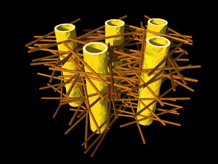
Caption: Illustration shows complex biostructure of dentin: the dental tubuli (yellow hollow cylinders, diameters appr. 1 micrometer) are surrounded by layers of mineralized collagen fibers (brown rods). The tiny mineral nanoparticles are embedded in the mesh of collagen fibers and not visible here. Credit: JB Forien @Charité

Caption: Illustration shows complex biostructure of dentin: the dental tubuli (yellow hollow cylinders, diameters appr. 1 micrometer) are surrounded by layers of mineralized collagen fibers (brown rods). The tiny mineral nanoparticles are embedded in the mesh of collagen fibers and not visible here. Credit: JB Forien @Charité

A June 10, 2015 Helmholtz Zentrum Berlin (HZB) press release (also on EurekAlert) explains how the illustration above relates to the research,

Human teeth have to serve for a lifetime, despite being subjected to huge forces. But the high failure resistance of dentin in teeth is not fully understood. An interdisciplinary team led by scientists of Charite Universitaetsmedizin Berlin has now analyzed the complex structure of dentin. At the synchrotron sources BESSY II at HZB, Berlin, Germany, and the European Synchrotron Radiation Facility ESRF, Grenoble, France, they could reveal that the mineral particles are precompressed.

The internal stress works against crack propagation and increases resistance of the biostructure.

Engineers use internal stresses to strengthen materials for specific technical purposes. Now it seems that evolution has long ‘known’ about this trick, and has put it to use in our natural teeth. Unlike bones, which are made partly of living cells, human teeth are not able to repair damage. Their bulk is made of dentin, a bonelike material consisting of mineral nanoparticles. These mineral nanoparticles are embedded in collagen protein fibres, with which they are tightly connected. In every tooth, such fibers can be found, and they lie in layers, making teeth tough and damage resistant. Still, it was not well understood, how crack propagation in teeth can be stopped.

The press release goes on to describe the new research and the teams which investigated the role of the mineral nanoparticles with regard to compression and cracking,

Now researchers from Charite Julius-Wolff-Institute, Berlin have been working with partners from Materials Engineering Department of Technische Universitaets Berlin, MPI of Colloids and Interfaces, Potsdam and Technion – Israel Institute of Technology, Haifa, to examine these biostructures more closely. They performed Micro-beam in-situ stress experiments in the mySpot BESSY facility of HZB, Berlin, Germany and analyzed the local orientation of the mineral nanoparticles using the nano-imaging facility of the European Synchrotron Radiation Facility (ESRF) in Grenoble, France.

When the tiny collagen fibers shrink, the attached mineral particles become increasingly compressed, the science team found out. “Our group was able to use changes in humidity to demonstrate how stress appears in the mineral in the collagen fibers, Dr. Paul Zaslansky from Julius Wolff-Institute of Charite Berlin explains. “The compressed state helps to prevents cracks from developing and we found that compression takes place in such a way that cracks cannot easily reach the tooth inner parts, which could damage the sensitive pulp. In this manner, compression stress helps to prevent cracks from rushing through the tooth.

The scientists also examined what happens if the tight mineral-protein link is destroyed by heating: In that case, dentin in teeth becomes much weaker. We therefore believe that the balance of stresses between the particles and the protein is important for the extended survival of teeth in the mouth, Charite scientist Jean-Baptiste Forien says. Their results may explain why artificial tooth replacements usually do not work as well as healthy teeth do: they are simply too passive, lacking the mechanisms found in the natural tooth structures, and consequently fillings cannot sustain the stresses in the mouth as well as teeth do. “Our results might inspire the development of tougher ceramic structures for tooth repair or replacement, Zaslansky hopes.

Experiments took place as part of the DFG project “Biomimetic Materials Research: Functionality by Hierarchical Structuring of Materials (SPP1420).

Here’s a link to and a citation for the paper,

Compressive Residual Strains in Mineral Nanoparticles as a Possible Origin of Enhanced Crack Resistance in Human Tooth Dentin by Jean-Baptiste Forien, Claudia Fleck, Peter Cloetens, Georg Duda, Peter Fratzl, Emil Zolotoyabko, and Paul Zaslansky. Nano Lett., 2015, 15 (6), pp 3729–3734 DOI: 10.1021/acs.nanolett.5b00143 Publication Date (Web): May 26, 2015

Copyright © 2015 American Chemical Society

This paper is behind a paywall.

Cerium dixoide nanoparticle sponges and their electron clouds

This research team is very excited about what they’ve accomplished (from a Nov. 12, 3013 news item on ScienceDaily),

A new chapter has been opened in our understanding of the chemical activity of nanoparticles says a team of international scientists. Using the X-ray beams of The European Synchrotron (ESRF) they showed that the electrons absorbed and released by cerium dioxide nanoparticles during chemical reactions behave in a completely different way than previously thought: the electrons are not bound to individual atoms but, like a cloud, distribute themselves over the whole nanoparticle. Inspired by the similarity of its shape, the scientists call this spatial distribution of particles an “electron sponge.”

The Nov. 12, 2013 European Synchrotron Radiation Facility news release (also on EurekAlert), which originated the news item, explains the scientists’ interest in cerium dioxide in more detail,

Today, cerium dioxide nanoparticles are widely used in industrial processes and also in consumer products. They are present, for example, in the walls of self-cleaning ovens and act as a hydrocarbon catalyst during the high temperature cleaning process. They are also a hot candidate for the next generation of lithium-ion batteries which will exhibit higher voltages and a greater storage capacity compared to today’s energy cells.

The element Cerium is abundant in the Earth’s crust and can easily be mined and purified. However, without a thorough understanding of the chemical processes happening on the surface of cerium dioxide nanoparticles, it is impossible to optimise their current and future use. And to address a more complex issue, it is also impossible to assess the limits of their safe use.

Most chemical reactions involve the transfer of an electron from one atom to another. In the past, it was believed that the electrons involved in a chemical reaction on the surface of a nanoparticle were localised in one of the atoms at the surface. To determine the behaviour of the electrons during the reaction, the scientists used the intense X-ray beams at the ESRF [European Synchrotron Radiation Facility] to probe solutions of nanoparticles in water and ethanol. The nanoparticles had a diameter of 3 nm and consisted of several thousands of molecules of cerium dioxide.

It is known that nanoparticles can change their behaviour under vacuum when studied with an electron microscope, for example. The scientists therefore carried out their experiment under realistic conditions, studying the nanoparticles in solution and in real time as the chemical reaction was taking place. “It was only possible to conduct these experiments in a liquid rather than under vacuum because we used X-rays as probes for the electron distribution.” says Jean Daniel Cafun [first author is Jean-Daniel Cafun from the ESRF].

In their experiment, the scientists were successful in observing the creation of the nanoparticles in solution and then how these nanoparticles eliminated highly reactive molecules (reactive oxygen species, or ROS) from the solution. This elimination process mimics the role of an important enzyme in living organisms – catalase – that protects cells from these aggressive molecules. Cancer patients undergoing radiation therapy have high levels of ROS in their bodies and ceria nanoparticles have been proposed as a way of reducing the levels of ROS and thus alleviating the negative impacts of the therapy on the patients. Throughout the chemical reaction, the electronic structure of the cerium atoms and thus the redistribution of the electron cloud was monitored. “It is crucial to be able to study the chemical processes of the particles in an environment that is close to conditions found in biological systems.” emphasizes Victor Puntes [Victor Puntes from the Universitá Autònoma of Barcelona, Catalan Institute of Nanotechnologies {Spain}].

“Scientists have been discussing the question: What happens when electrons are added to ceria nanoparticles? The work by Cafun et al. is a key study because it questions the present, widely accepted model and will lead the research in a new direction.” says Frank de Groot, an expert on nanomaterials at Utrecht University who did not take part in the experiment.

The next step, which has already been initiated, will be to assess whether non-localised electrons are a property of cerium dioxide only or also of other widely used nanoparticles like titanium dioxide. “In parallel, chemists have to revisit their theoretical models to explain the chemical behaviour of nanoparticles and to better understand how electrons are transferred in chemical reactions taking place on their surface.” concludes Pieter Glatzel [team leader Pieter Glatzel from The European Synchrotron {ESRF} in Grenoble {France}].

For anyone who’d like to explore this topic further,

Absence of Ce3+ Sites in Chemically Active Colloidal Ceria Nanoparticles by Jean-Daniel Cafun, Kristina O. Kvashnina, Eudald Casals, Victor F. Puntes, and Pieter Glatzel. ACS Nano, Article ASAP DOI: 10.1021/nn403542p Publication Date (Web): November 12, 2013
Copyright © 2013 American Chemical Society

This article is behind a paywall.

Soybeans and nanoparticles redux

If you read the Feb. 6, 2013 news release on EurekAlert too quickly you might not realize that only one of the two types of the tested nanoparticles adversely affects soybean plants,

Two of the most widely used nanoparticles (NPs) accumulate in soybeans — second only to corn as a key food crop in the United States — in ways previously shown to have the potential to adversely affect the crop yields and nutritional quality, a new study has found. It appears in the journal ACS Nano. [emphasis mine]

Jorge L. Gardea-Torresdey and colleagues cite rapid increases in commercial and industrial uses of NPs, the building blocks of a nanotechnology industry projected to put $1 trillion worth of products on the market by 2015. Zinc oxide and cerium dioxide are among today’s most widely used NPs. Both are used in cosmetics, lotions, sunscreens and other products. They eventually go down the drain, through municipal sewage treatment plants, and wind up in the sewage sludge that some farmers apply to crops as fertilizer. Gardea-Torresdey’s team previously showed that soybean plants grown in hydroponic solutions accumulated zinc and cerium dioxide in ways that alter plant growth and could have health implications.

The question remained, however, as to whether such accumulation would occur in the real-world conditions in which farmers grow soybeans in soil, rather than nutrient solution. Other important questions included the relationship of soybean plants and NPs, the determination of their entrance into the food chain, their biotransformation and toxicity and the possible persistence of these products into the next plant generation. Their new study, performed at two world-class synchrotron facilities — the SLAC National Accelerator Laboratory in California and the European Synchrotron Radiation Facility in Grenoble, France, addressed those questions. “To our knowledge, this is the first report on the presence of cerium dioxide and zinc compounds in the reproductive/edible portions of the soybean plant grown in farm soil with cerium dioxide and zinc oxide nanoparticles. In addition, our results have shown that cerium dioxide NPs in soil can be taken up by food crops and are not biotransformed in soybeans. [emphasis mine] This suggests that cerium dioxide NPs can reach the food chain and the next soybean plant generation, with potential health implications,” the study notes.

The University of Texas El Paso Feb. 6, 2013 news release provides more detail and more clarity about the results of the research ,

Experiments led by Jorge Gardea-Torresdey, Ph.D., of The University of Texas at El Paso (UTEP) have shown that certain man-made nanoparticles that land in soil can be transferred from the roots of plants to the grains, thus entering the food supply via crops grown for human consumption.

Cerium dioxide, which is commonly used in sunscreens and oil refining, remained intact when it was absorbed by the plant, and was transferred all the way into the edible soybean grains. [emphasis mine]

On the other hand, zinc oxide – commonly used in sunscreens and cosmetics – was transferred to the grain, but had broken down to a nontoxic form. [emphasis mine]

To track the nanoparticles’ route within the plants, the researchers used the intense beams of X-rays from the SLAC National Accelerator Laboratory’s Stanford Synchrotron Radiation Lightsource (SSRL) and the European Synchrotron Radiation Facility (ESRF) in Grenoble, France. The X-rays also helped reveal whether or not the nanoparticles were chemically transformed in the process.

While studies are under way, Gardea-Torresdey says there is currently little information on the potential health implications of nanoparticles.

UTEP has produced a video titled, UTEP Study Shows Engineered Nanoparticles Can Enter Food Supply. This piece, which features Gardea-Torresdey and a student,  seems to be less about the study and more about the benefits of studying at UTEP and the impact of the Latino community in the US,


Here’s a citation and a link to the article (Note: This work bears a remarkable resemblance to the work mentioned in my Aug. 20, 2012 posting about soybeans and nanoparticles, not least because the studies share three or more authors),

In Situ Synchrotron X-ray Fluorescence Mapping and Speciation of CeO2 and ZnO Nanoparticles in Soil Cultivated Soybean (Glycine max) by Jose A. Hernandez-Viezcas, Hiram Castillo-Michel, Joy Cooke Andrews , Marine Cotte , Cyren Rico, Jose R. Peralta-Videa, Yuan Ge, John H. Priester, Patricia Ann Holden, and Jorge L. Gardea-Torresdey. ACS Nano, DOI: 10.1021/nn305196q Publication Date (Web): January 15, 2013

Copyright © 2013 American Chemical Society

The article is behind a paywall.