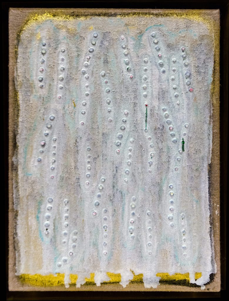
One of Joseph Cohen’s painting incorporating carbon nanotubes photographed in normal light. Photo courtesy of Joseph Cohen. [downloaded from https://news.artnet.com/art-world/carbon-nanotube-cancer-paint-1638340?utm_content=from_&utm_source=Sailthru&utm_medium=email&utm_campaign=Global%20September%202%20PM&utm_term=artnet%20News%20Daily%20Newsletter%20USE%20%2830%20Day%20Engaged%20Only%29]
The artist credited with the work seen in the above, Joseph Cohen, has done something remarkable with carbon nanotubes (CNTs). Something even more remarkable than the painting as Sarah Cascone recounts in her August 30, 2019 article for artnet.com (Note: A link has been removed),
Not every artist can say that his or her work is helping in the fight against cancer. But over the past several years, Joseph Cohen has done just that, working to develop a new, high-tech paint that can be used not only on canvas, but also to detect cancers and medical conditions such as hypertension and diabetes.
Sloan Kettering Institute scientist Daniel Heller first suggested that Cohen come work at his lab after seeing the artist’s work, which is often made with pigments that incorporate diamond dust and gold, at the DeBuck Gallery in New York.
“We initially thought that in working with an artist, we would make art to shed a little light on our science for the public,” Heller told the Memorial Sloan Kettering blog. “But the collaboration actually taught us something that could help us shine a light on cancer.”
For Cohen, the project was initially intended to develop a new way of art-making. In Heller’s lab, he worked with carbon nanotubes, which Heller was already employing in cancer research, for their optical properties. “They fluoresce in the infrared spectrum,” Cohen says. “That gives artists the opportunity to create paintings in a new spectrum, with a whole new palette of colors.”
Because human eyesight is limited, we can’t actually see infrared fluorescence. But using a special short-wave infrared camera, Cohen is able to document otherwise invisible effects, revealing the carbon nanotube paint’s hidden colors.
“What you’re perceiving as a static painting is actually in motion,” Cohen says. “I’m creating paintings that exist outside of the visible experience.”
Art Supplies—and a Diagnostic Tool
That same imaging technique can be used by doctors looking for microalbuminuria, a condition that causes the kidneys to leak trace amounts of albumin into urine, which is an early sign of of several cancers, diabetes, and high blood pressure.
Cohen helped co-author a paper published this month in Nature Communications about using the nanosensor paint in litmus paper tests with patient urine samples. The study found that the paint, when viewed through infrared light, was able to reveal the presence of albumin based on changes in the paint’s fluorescence after being exposed to the urine sample.
“It’s easy to detect albumen with a dipstick if there’s a lot of levels in the urine, but that would be like looking at stage four cancer,” Cohen says. “This is early detection.”
What’s more, a nanosensor paint can be easily used around the world, even in poor areas that don’t have access to the best diagnostic technologies. Doctors may even be able to view the urine samples using an infrared imaging attachments on their smartphones.
…
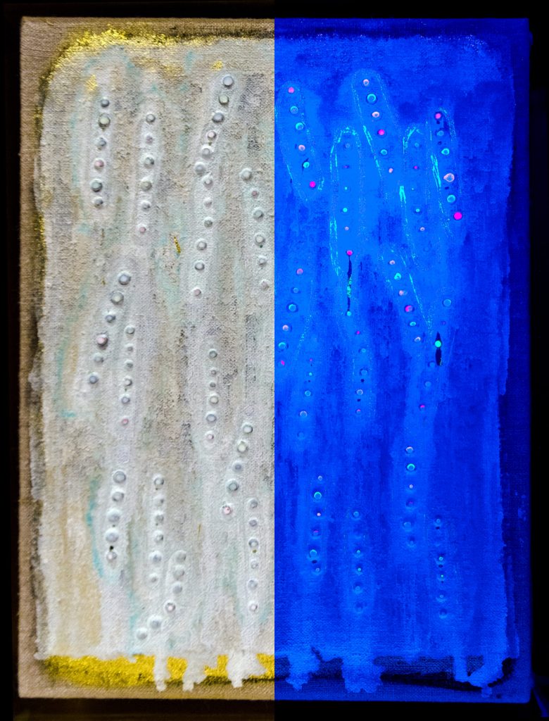
One of Joseph Cohen’s painting incorporating carbon nanotubes shown in both the visible light (left) and in UV fluorescence (right). Photo courtesy of Joseph Cohen. [downloaded from https://news.artnet.com/art-world/carbon-nanotube-cancer-paint-1638340?utm_content=from_&utm_source=Sailthru&utm_medium=email&utm_campaign=Global%20September%202%20PM&utm_term=artnet%20News%20Daily%20Newsletter%20USE%20%2830%20Day%20Engaged%20Only%29]
Amazing, eh? If you have the time, do read Cascone’s article in its entirety and should your curiosity be insatiable, there’s also an August 22, 2019 posting by Jim Stallard on the Memorial Sloan Kettering Cancer Center blog,
Here’s a link to and a citation for the paper,
Synthetic molecular recognition nanosensor paint for microalbuminuria by Januka Budhathoki-Uprety, Janki Shah, Joshua A. Korsen, Alysandria E. Wayne, Thomas V. Galassi, Joseph R. Cohen, Jackson D. Harvey, Prakrit V. Jena, Lakshmi V. Ramanathan, Edgar A. Jaimes & Daniel A. Heller. Nature Communicationsvolume 10, Article number: 3605 (2019) DOI: https://doi.org/10.1038/s41467-019-11583-1 Published: 09 August 2019
This paper is open access.
Joseph Cohen has graced this blog before in a May 3, 2019 posting titled, Where do I stand? a graphene artwork. It seems Cohen is very invested in using nanoscale carbon particles for his art.
Cotton that’s grown with molecules that endow appealing properties – like fluorescence or magnetism – may one day eliminate the need for applying chemical treatments to fabrics to achieve such qualities, a new study suggests. Applying synthetic polymers to fabrics can result in a range of appealing properties, but anything added to a fabric can get washed or worn away. Furthermore, while many fibers used in fabrics are synthetic (e.g., polyester), some consumers prefer natural fibers to avoid issues related to sensation, skin irritation, smoothness, and weight. Here, Filipe Natalio and colleagues created cotton fibers that incorporate composites with fluorescent and magnetic properties. They synthesized glucose derivatives that deliver the desirable molecules into the growing ovules of the cotton plant, Gossypium hirsutum. Thus, the molecules are embedded into the cotton fibers themselves, rather than added in the form of a chemical treatment. The resulting fibers exhibited fluorescent or magnetic properties, respectively, although they were weaker than raw fibers lacking the embedded composites, the authors report. They propose that similar techniques could be expanded to other biological systems such as bacteria, bamboo, silk, and flax – essentially opening a new era of “material farming.”
Robert Service’s Sept. 14, 2017 article for Science explores the potential of growing cotton with new properties (Note: A link has been removed),
You may have heard about smartphones and smart homes. But scientists are also designing smart clothes, textiles that can harvest energy, light up, detect pollution, and even communicate with the internet. The problem? Even when they work, these often chemically treated fabrics wear out rapidly over time. Now, researchers have figured out a way to “grow” some of these functions directly into cotton fibers. If the work holds, it could lead to stronger, lighter, and brighter textiles that don’t wear out.
Yet, as the new paper went to press today in Science, editors at the journal were made aware of mistakes in a figure in the supplemental material that prompted them to issue an Editorial Expression of Concern, at least until they receive clarification from the authors. Filipe Natalio, lead author and chemist at the Weizmann Institute of Science in Rehovot, Israel, says the mistakes were errors in the names of pigments used in control experiments, which he is working with the editors to fix.
That hasn’t dampened enthusiasm for the work. “I like this paper a lot,” says Michael Strano, a chemical engineer at the Massachusetts Institute of Technology in Cambridge. The study, he says, lays out a new way to add new functions into plants without changing their genes through genetic engineering. Those approaches face steep regulatory hurdles for widespread use. “Assuming the methods claimed are correct, that’s a big advantage,” Strano says.
…
Sam Lemonick’s Sept. 14, 2017 article for forbes.com describes how the researchers introduced new properties (in this case, glowing colours) into the cotton plants,
…
His [Filipe Natalio] team of researchers in Israel, Germany, and Austria used sugar molecules to sneak new properties into cotton. Like a Trojan horse, Natalio says. They tested the method by tagging glucose with a fluorescent dye molecule that glows green when hit with the right kind of light.
They bathed cotton ovules—the part of the plant that makes the fibers—in the glucose. And just like flowers suck up dyed water in grade school experiments, the ovules absorbed the sugar solution and piped the tagged glucose molecules to their cells. As the fibers grew, they took on a yellowish tinge—and glowed bright green under ultraviolet light.
Glowing cotton wasn’t enough for Natalio. It took his group about six months to be sure they were actually delivering the fluorescent protein into the cotton cells and not just coating the fibers in it. Once they were certain, they decided to push the envelope with something very unnatural: magnets.
This time, Natalio’s team modified glucose with the rare earth metal dysprosium, making a molecule that acts like a magnet. And just like they did with the dye, the researchers fed it to cotton ovules and ended up with fibers with magnetic properties.
…
Both Service and Lemonwick note that the editor of the journal Science (where the research paper was published) Jeremy Berg has written an expression of editorial concern as of Sept. 14, 2017,
In the 15 September [2017] issue, Science published the Report “Biological fabrication of cellulose fibers with tailored properties” by F. Natalio et al. (1). After the issue went to press, we became aware of errors in the labeling and/or identification of the pigments used for the control experiments detailed in figs. S1 and S2 of the supplementary materials. Science is publishing this Editorial Expression of Concern to alert our readers to this information as we await full explanation and clarification from the authors.
The problem seems to be one of terminology (from the Lemonwick article),
… Filipe Natalio, lead author and chemist at the Weizmann Institute of Science in Rehovot, Israel, says the mistakes were errors in the names of pigments used in control experiments, which he is working with the editors to fix.
…
These things happen. Terminology and spelling aren’t always the same from one country to the next and it can result in confusion. I’m glad to see the discussion is being held openly.
Here’s a link to and a citation for the paper,
Biological fabrication of cellulose fibers with tailored properties by Filipe Natalio, Regina Fuchs, Sidney R. Cohen, Gregory Leitus, Gerhard Fritz-Popovski, Oskar Paris, Michael Kappl, Hans-Jürgen Butt. Science 15 Sep 2017: Vol. 357, Issue 6356, pp. 1118-1122 DOI: 10.1126/science.aan5830
First, animals that flow in the dark and then, updates on other ‘glow in the dark’ projects.
The American Chemical Society (ACS) has produced a video about animals that glow in the dark, here’s more from their August 14, 2017 news release on EurekAlert,
Fireflies, frogs, jellyfish, mushrooms and even parrots have the ability to emit light from their bodies. These creatures use either bioluminescence or fluorescence to put on their light shows.
To learn more about Marc Zimmer’s research at Connecticut College and to find more great info and images involving GFP, visit his website: http://www.conncoll.edu/ccacad/zimmer…
Updates on a previous glowing plants and animals posting
In a May 5, 2013 posting I featured a Kickstarter campaign for a synthetic biology project focused on plants that emit light in the dark. I also mentioned Eduardo Kac (pronounced Katz) and his art project/transgenic bunny called Alba. At the time, I did not realize that Alba had been declared dead in 2002 adding more controversy to an already controversial topice according to Kristen Philipkoski in an Aug. 12, 2002 article (how did I miss this article in 2013?) for Wired magazine (Note: Links have been removed),
Alba, the glowing rabbit that made headlines two years ago for being, well, a glowing rabbit, has met an untimely death, according to the French researcher who genetically engineered her.
Alba the glowing rabbit was 4 years old. Or 2-1/2, depending on who’s talking.
The bunny died about a month ago for reasons that are not clear, said Louis-Marie Houdebine, a genetic researcher at France’s National Institute of Agronomic Research.
“I was informed one day that bunny was dead without any reason,” Houdebine said. “So, rabbits die often. It was about 4 years old, which is a normal lifespan in our facilities.”
Alba was an albino rabbit engineered by splicing the green fluorescent protein (GFP) of a jellyfish into her genome. Houdebine said he did not believe the GFP gene played a role in the animal’s demise.
Eduardo Kac, the artist who created a flurry by making her a work of art, doesn’t buy it, however.
First, Alba’s not 4, she’s 2-1/2, Kac says (a rabbit’s lifespan is up to 12 years), because she was bred by Houdebine specifically for him in January 2000.
Houdebine says he simply picked a rabbit with a gentle disposition that was already in his lab.
Second, he believes Houdebine might be declaring the bunny gone in order to put an end to a two-year, unwelcome barrage of media attention.
If she really is dead, Kac will never realize the final phase of his project, which was to take Alba home and keep her as a pet.
Kac says he and Houdebine originally collaborated on the GFP bunny project, until Houdebine’s director put the kibosh on it.
“My director did not understand,” Houdebine said. “He said I should not give the rabbit (to someone) outside the lab.”
Houdebine said that yes, they spoke about preliminary plans for Kac to use the bunny for his project and take it to an art show in Avignon. But he denies he bred an animal specifically for Kac.
…
Houdebine says he would not have agreed to engineer one animal specifically for any artist.
This disputed point has led fellow artists and critics to question whether Kac can rightly take credit for the Alba project.
But Kac insists that Houdebine did, in fact, agree to make the bunny specifically for him.
Kac found out sometime in mid-2000 that Houdebine’s director had a problem with the project and would not allow the rabbit to be taken from the lab.
Houdebine was initially apologetic, Kac said. But after an article ran on the front page of the Boston Globe on Sept. 17, 2000, their relationship cooled.
…
Houdebine and his director were opposed to the now-famous, brilliantly glowing photograph of Alba. They and other researchers say the rabbit doesn’t actually glow so brightly and uniformly.
“Kac fabricated data for his personal use,” Houdebine said. “This is why we totally stopped any contact with him.”
“The scientific fact is that the rabbit is not green,” he said. “He should have never published that. This was very disagreeable for me.”
Kac believes the scientists were simply afraid of public criticism. Meanwhile, he wanted to do the opposite – to encourage discourse on the transgenic rabbit.
“This director refuses to participate openly in a debate about what is done with public money,” he said. “It’s very easy to fear and reject what you don’t know. As long as they continue to isolate themselves, this mistrust will continue.”
The eyes and ears of the rabbit are green under ultraviolet light, Houdebine said, but the fur does not glow, because it’s dead tissue that doesn’t express the gene. Only if the rabbit were shaved would the body glow, he said.
…
Philipkosk’s article provides some insight into the interface between art and science and is worth reading in its entirety if you have the time.
I’ve also found an update for the glowing plants Kickstarter campaign in an April 20, 2017 article by Sarah Zhang for The Atlantic (Note: Links have been removed),
The latest update came quietly on Tuesday night [April 18, 2017?]. “We’re sorry to say that we have reached a significant transition point,” wrote the Glowing Plant project’s creator, Antony Evans. This “transition point” was more of an endpoint: The project had run out of money. The quest to genetically engineer a glow-in-the-dark plant was no more.
Four years ago, the Glowing Plant project raised nearly half a million dollars on Kickstarter, easily blowing past its initial ask of $65,000. Of course it did. The vision it presented was such potent fantasy. “What if,” Evans asked over swelling music in the pitch video, “we use trees to light our streets instead of street lamps?” What if you could get lighting without electricity? What if the natural world glowed like in Avatar?
This romantic vision so perfectly encapsulated the promises of synthetic biology, a field that treats the natural world as another system to be designed and engineered. In this case, synthetic biology became a possible solution to one of the world’s most pressing energy problems: electricity generation. Plus, it sounded really damn cool.
The Kickstarter campaign only promised a small, potted glowing plant to it backers, and I doubt many backers actually harbored illusions about trees lighting up the night sky soon. But backing the project was a small way to buy into a much grander vision.
…
At a time when “genetically modified organism,” or GMO, is such a poisoned phrase, the project’s crowdfunding success seemed to suggest that a pervasive if vague distrust of genetic modification might be countered by the sense of wonder for a glowing plant. (As the Kickstarter campaign grew, though, environmental groups raised questions and the crowdfunding site later banned giving away genetically modified organisms.)
The team also encountered the hard realities of engineering even a small plant that glows. “We did not anticipate some of the unknown technical challenges that we would get into,” Evans told me. (Plenty of scientists at the time were skeptical of the project’s timeline, though.) Evans is an MBA with a background in mobile apps, though his two original cofounders, who have both since left the project, had backgrounds in synthetic biology.
To get the plant to glow well, the research team had to insert six genes. But they never could get all six in at once. At best, some plants glowed very dimly. (The photo above of the glowing plant is a long exposure, making it appear much brighter than it actually is.) Evans says that he realizes now trying to insert six genes into a complex organism like a plant—rather than single-celled bacteria or yeast—was premature.
…
“I’m really afraid of disappointing that 16-year-old who saw this and imagined a bright wonderful future, of jading and disappointing people,” he says. Despite a few angry backers asking for a refund, most of the comments under the Kickstarter update so far have been supportive. The project had been providing regular, detailed updates on the difficulty of engineering the plants. The latest update was its 67th.
…
Zhang’s article goes on to detail other synthetic biology projects, which are showing some promise.
When you take this work into consideration with CRISPR-CAS9 and the beginnings of genetic germline editing, the question has to be asked: Will public discussion (if there’s any) be considered upstream (early in the process) or downstream (after the work has been done)? Public engagement professionals tend to favour upstream discussions, i.e., before people start demanding fear-based policy.
The extraordinary effort to colonize our brains continues apace with a new sensor from Vanderbilt University. From an Oct. 27, 2016 news item on ScienceDaily,
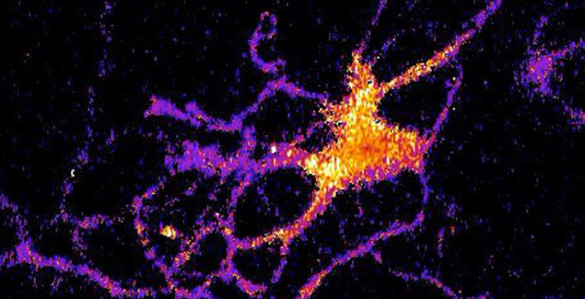
A new kind of bioluminescent sensor causes individual brain cells to imitate fireflies and glow in the dark.
The probe, which was developed by a team of Vanderbilt scientists, is a genetically modified form of luciferase, the enzyme that a number of other species including fireflies use to produce light. …
The scientists created the technique as a new and improved method for tracking the interactions within large neural networks in the brain.
“For a long time neuroscientists relied on electrical techniques for recording the activity of neurons. These are very good at monitoring individual neurons but are limited to small numbers of neurons. The new wave is to use optical techniques to record the activity of hundreds of neurons at the same time,” said Carl Johnson, Stevenson Professor of Biological Sciences, who headed the effort.
Individual neuron glowing with bioluminescent light produced by a new genetically engineered sensor. (Johnson Lab / Vanderbilt University)
“Most of the efforts in optical recording use fluorescence, but this requires a strong external light source which can cause the tissue to heat up and can interfere with some biological processes, particularly those that are light sensitive,” he [Carl Johnson] said.
Based on their research on bioluminescence in “a scummy little organism, the green alga Chlamydomonas, that nobody cares much about” Johnson and his colleagues realized that if they could combine luminescence with optogenetics – a new biological technique that uses light to control cells, particularly neurons, in living tissue – they could create a powerful new tool for studying brain activity.
“There is an inherent conflict between fluorescent techniques and optogenetics. The light required to produce the fluorescence interferes with the light required to control the cells,” said Johnson. “Luminescence, on the other hand, works in the dark!”
Johnson and his collaborators – Associate Professor Donna Webb, Research Assistant Professor Shuqun Shi, post-doctoral student Jie Yang and doctoral student Derrick Cumberbatch in biological sciences and Professor Danny Winder and postdoctoral student Samuel Centanni in molecular physiology and biophysics – genetically modified a type of luciferase obtained from a luminescent species of shrimp so that it would light up when exposed to calcium ions. Then they hijacked a virus that infects neurons and attached it to their sensor molecule so that the sensors are inserted into the cell interior.
The researchers picked calcium ions because they are involved in neuron activation. Although calcium levels are high in the surrounding area, normally they are very low inside the neurons. However, the internal calcium level spikes briefly when a neuron receives an impulse from one of its neighbors.
They tested their new calcium sensor with one of the optogenetic probes (channelrhodopsin) that causes the calcium ion channels in the neuron’s outer membrane to open, flooding the cell with calcium. Using neurons grown in culture they found that the luminescent enzyme reacted visibly to the influx of calcium produced when the probe was stimulated by brief light flashes of visible light.
To determine how well their sensor works with larger numbers of neurons, they inserted it into brain slices from the mouse hippocampus that contain thousands of neurons. In this case they flooded the slices with an increased concentration of potassium ions, which causes the cell’s ion channels to open. Again, they found that the sensor responded to the variations in calcium concentrations by brightening and dimming.
“We’ve shown that the approach works,” Johnson said. “Now we have to determine how sensitive it is. We have some indications that it is sensitive enough to detect the firing of individual neurons, but we have to run more tests to determine if it actually has this capability.”
Here’s a link to and a citation for the paper,
Coupling optogenetic stimulation with NanoLuc-based luminescence (BRET) Ca++ sensing by Jie Yang, Derrick Cumberbatch, Samuel Centanni, Shu-qun Shi, Danny Winder, Donna Webb, & Carl Hirschie Johnson. Nature Communications 7, Article number: 13268 (2016) doi:10.1038/ncomms13268 Published online: 27 October 2016
This work comes from the US Naval Research Laboratory according to a Nov. 17, 2015 news item on Nanowerk (Note: A link has been removed),
Research biologists, chemists and theoreticians at the U.S. Naval Research Laboratory (NRL), are on pace to develop the next generation of functional materials that could enable the mapping of the complex neural connections in the brain (“Electric Field Modulation of Semiconductor Quantum Dot Photoluminescence: Insights Into the Design of Robust Voltage-Sensitive Cellular Imaging Probes”). The ultimate goal is to better understand how the billions of neurons in the brain communicate with one another during normal brain function, or dysfunction, as result of injury or disease.
“There is tremendous interest in mapping all the neuron connections in the human brain,” said Dr. James Delehanty, research biologist, Center for Biomolecular Science and Engineering. “To do that we need new tools or materials that allow us to see how large groups of neurons communicate with one another while, at the same time, being able to focus in on a single neuron’s activity. Our most recent work potentially opens the integration of voltage-sensitive nanomaterials into live cells and tissues in a variety of configurations to achieve real-time imaging capabilities not currently possible.”
The basis of neuron communication is the time-dependent modulation of the strength of the electric field that is maintained across the cell’s plasma membrane. This is called an action potential. Among the nanomaterials under consideration for application in neuronal action potential imaging are quantum dots (QDs) — crystalline semiconductor nanomaterials possessing a number of advantageous photophysical attributes.
“QDs are very bright and photostable so you can look at them for long times and they allow for tissue imaging configurations that are not compatible with current materials, for example, organic dyes,” Delehanty added. “Equally important, we’ve shown here that QD brightness tracks, with very high fidelity, the time-resolved electric field strength changes that occur when a neuron undergoes an action potential. Their nanoscale size make them ideal nanoscale voltage sensing materials for interfacing with neurons and other electrically active cells for voltage sensing.”
QDs are small, bright, photo-stable materials that possess nanosecond fluorescence lifetimes. They can be localized within or on cellular plasma membranes and have low cytotoxicity when interfaced with experimental brain systems. Additionally, QDs possess two-photon action cross-section orders of magnitude larger than organic dyes or fluorescent proteins. Two-photon imaging is the preferred imaging modality for imaging deep (millimeters) into the brain and other tissues of the body.
In their most recent work, the NRL researchers showed that an electric field typical of those found in neuronal membranes results in suppression of the QD photoluminescence (PL) and, for the first time, that QD PL is able to track the action potential profile of a firing neuron with millisecond time resolution. This effect is shown to be connected with electric-field-driven QD ionization and consequent QD PL quenching, in contradiction with conventional wisdom that suppression of the QD PL is attributable to the quantum confined Stark effect — the shifting and splitting of spectral lines of atoms and molecules due to presence of an external electric field.
“The inherent superior photostability properties of QDs coupled with their voltage sensitivity could prove advantageous to long-term imaging capabilities that are not currently attainable using traditional organic voltage sensitive dyes,” Delehanty said. “We anticipate that continued research will facilitate the rational design and synthesis of voltage-sensitive QD probes that can be integrated in a variety of imaging configurations for the robust functional imaging and sensing of electrically active cells.”