Tag Archives: fruit flies

Of puke, CRISPR, fruit flies, and monarch butterflies

I’ve never seen an educational institution use a somewhat vulgar slang term such as ‘puke’ before. Especially not in a news release. You’ll find that elsewhere online ‘puke’ has been replaced, in the headline, with the more socially acceptable ‘vomit’.

Since I wanted to catch this historic moment amid concerns that the original version of the news release will disappear, I’m including the entire news release as i saw it on EurekAlert.com (from an October 2, 2019 University of California at Berkeley news release),

News Release 2-Oct-2019

CRISPRed fruit flies mimic monarch butterfly — and could make you puke
Scientists recreate in flies the mutations that let monarch butterfly eat toxic milkweed with impunity

University of California – Berkeley

The fruit flies in Noah Whiteman’s lab may be hazardous to your health.

Whiteman and his University of California, Berkeley, colleagues have turned perfectly palatable fruit flies — palatable, at least, to frogs and birds — into potentially poisonous prey that may cause anything that eats them to puke. In large enough quantities, the flies likely would make a human puke, too, much like the emetic effect of ipecac syrup.

That’s because the team genetically engineered the flies, using CRISPR-Cas9 gene editing, to be able to eat milkweed without dying and to sequester its toxins, just as America’s most beloved butterfly, the monarch, does to deter predators.

This is the first time anyone has recreated in a multicellular organism a set of evolutionary mutations leading to a totally new adaptation to the environment — in this case, a new diet and new way of deterring predators.

Like monarch caterpillars, the CRISPRed fruit fly maggots thrive on milkweed, which contains toxins that kill most other animals, humans included. The maggots store the toxins in their bodies and retain them through metamorphosis, after they turn into adult flies, which means the adult “monarch flies” could also make animals upchuck.

The team achieved this feat by making three CRISPR edits in a single gene: modifications identical to the genetic mutations that allow monarch butterflies to dine on milkweed and sequester its poison. These mutations in the monarch have allowed it to eat common poisonous plants other insects could not and are key to the butterfly’s thriving presence throughout North and Central America.

Flies with the triple genetic mutation proved to be 1,000 times less sensitive to milkweed toxin than the wild fruit fly, Drosophila melanogaster.

Whiteman and his colleagues will describe their experiment in the Oct. 2 [2019] issue of the journal Nature.

Monarch flies

The UC Berkeley researchers created these monarch flies to establish, beyond a shadow of a doubt, which genetic changes in the genome of monarch butterflies were necessary to allow them to eat milkweed with impunity. They found, surprisingly, that only three single-nucleotide substitutions in one gene are sufficient to give fruit flies the same toxin resistance as monarchs.

“All we did was change three sites, and we made these superflies,” said Whiteman, an associate professor of integrative biology. “But to me, the most amazing thing is that we were able to test evolutionary hypotheses in a way that has never been possible outside of cell lines. It would have been difficult to discover this without having the ability to create mutations with CRISPR.”

Whiteman’s team also showed that 20 other insect groups able to eat milkweed and related toxic plants – including moths, beetles, wasps, flies, aphids, a weevil and a true bug, most of which sport the color orange to warn away predators – independently evolved mutations in one, two or three of the same amino acid positions to overcome, to varying degrees, the toxic effects of these plant poisons.

In fact, his team reconstructed the one, two or three mutations that led to each of the four butterfly and moth lineages, each mutation conferring some resistance to the toxin. All three mutations were necessary to make the monarch butterfly the king of milkweed.
Resistance to milkweed toxin comes at a cost, however. Monarch flies are not as quick to recover from upsets, such as being shaken — a test known as “bang” sensitivity.

“This shows there is a cost to mutations, in terms of recovery of the nervous system and probably other things we don’t know about,” Whiteman said. “But the benefit of being able to escape a predator is so high … if it’s death or toxins, toxins will win, even if there is a cost.”

Plant vs. insect

Whiteman is interested in the evolutionary battle between plants and parasites and was intrigued by the evolutionary adaptations that allowed the monarch to beat the milkweed’s toxic defense. He also wanted to know whether other insects that are resistant — though all less resistant than the monarch — use similar tricks to disable the toxin.

“Since plants and animals first invaded land 400 million years ago, this coevolutionary arms race is thought to have given rise to a lot of the plant and animal diversity that we see, because most animals are insects, and most insects are herbivorous: they eat plants,” he said.

Milkweeds and a variety of other plants, including foxglove, the source of digitoxin and digoxin, contain related toxins — called cardiac glycosides — that can kill an elephant and any creature with a beating heart. Foxglove’s effect on the heart is the reason that an extract of the plant, in the genus Digitalis, has been used for centuries to treat heart conditions, and why digoxin and digitoxin are used today to treat congestive heart failure.

These plants’ bitterness alone is enough to deter most animals, but a small minority of insects, including the monarch (Danaus plexippus) and its relative, the queen butterfly (Danaus gilippus), have learned to love milkweed and use it to repel predators.

Whiteman noted that the monarch is a tropical lineage that invaded North America after the last ice age, in part enabled by the three mutations that allowed it to eat a poisonous plant other animals could not, giving it a survival edge and a natural defense against predators.

“The monarch resists the toxin the best of all the insects, and it has the biggest population size of any of them; it’s all over the world,” he said.

The new paper reveals that the mutations had to occur in the right sequence, or else the flies would never have survived the three separate mutational events.

Thwarting the sodium pump

The poisons in these plants, most of them a type of cardenolide, interfere with the sodium/potassium pump (Na+/K+-ATPase) that most of the body’s cells use to move sodium ions out and potassium ions in. The pump creates an ion imbalance that the cell uses to its favor. Nerve cells, for example, transmit signals along their elongated cell bodies, or axons, by opening sodium and potassium gates in a wave that moves down the axon, allowing ions to flow in and out to equilibrate the imbalance. After the wave passes, the sodium pump re-establishes the ionic imbalance.

Digitoxin, from foxglove, and ouabain, the main toxin in milkweed, block the pump and prevent the cell from establishing the sodium/potassium gradient. This throws the ion concentration in the cell out of whack, causing all sorts of problems. In animals with hearts, like birds and humans, heart cells begin to beat so strongly that the heart fails; the result is death by cardiac arrest.

Scientists have known for decades how these toxins interact with the sodium pump: they bind the part of the pump protein that sticks out through the cell membrane, clogging the channel. They’ve even identified two specific amino acid changes or mutations in the protein pump that monarchs and the other insects evolved to prevent the toxin from binding.

But Whiteman and his colleagues weren’t satisfied with this just so explanation: that insects coincidentally developed the same two identical mutations in the sodium pump 14 separate times, end of story. With the advent of CRISPR-Cas9 gene editing in 2012, coinvented by UC Berkeley’s Jennifer Doudna, Whiteman and colleagues Anurag Agrawal of Cornell University and Susanne Dobler of the University of Hamburg in Germany applied to the Templeton Foundation for a grant to recreate these mutations in fruit flies and to see if they could make the flies immune to the toxic effects of cardenolides.

Seven years, many failed attempts and one new grant from the National Institutes of Health later, along with the dedicated CRISPR work of GenetiVision of Houston, Texas, they finally achieved their goal. In the process, they discovered a third critical, compensatory mutation in the sodium pump that had to occur before the last and most potent resistance mutation would stick. Without this compensatory mutation, the maggots died.

Their detective work required inserting single, double and triple mutations into the fruit fly’s own sodium pump gene, in various orders, to assess which ones were necessary. Insects having only one of the two known amino acid changes in the sodium pump gene were best at resisting the plant poisons, but they also had serious side effects — nervous system problems — consistent with the fact that sodium pump mutations in humans are often associated with seizures. However, the third, compensatory mutation somehow reduces the negative effects of the other two mutations.

“One substitution that evolved confers weak resistance, but it is always present and allows for substitutions that are going to confer the most resistance,” said postdoctoral fellow Marianna Karageorgi, a geneticist and evolutionary biologist. “This substitution in the insect unlocks the resistance substitutions, reducing the neurological costs of resistance. Because this trait has evolved so many times, we have also shown that this is not random.”

The fact that one compensatory mutation is required before insects with the most resistant mutation could survive placed a constraint on how insects could evolve toxin resistance, explaining why all 21 lineages converged on the same solution, Whiteman said. In other situations, such as where the protein involved is not so critical to survival, animals might find different solutions.

“This helps answer the question, ‘Why does convergence evolve sometimes, but not other times?'” Whiteman said. “Maybe the constraints vary. That’s a simple answer, but if you think about it, these three mutations turned a Drosophila protein into a monarch one, with respect to cardenolide resistance. That’s kind of remarkable.”

###

The research was funded by the Templeton Foundation and the National Institutes of Health. Co-authors with Whiteman and Agrawal are co-first authors Marianthi Karageorgi of UC Berkeley and Simon Groen, now at New York University; Fidan Sumbul and Felix Rico of Aix-Marseille Université in France; Julianne Pelaez, Kirsten Verster, Jessica Aguilar, Susan Bernstein, Teruyuki Matsunaga and Michael Astourian of UC Berkeley; Amy Hastings of Cornell; and Susanne Dobler of Universität Hamburg in Germany.

Robert Sanders’ Oct. 2, 2019′ news release for the University of California at Berkeley (it’s also been republished as an Oct. 2, 2019 news item on ScienceDaily) has had its headline changed to ‘vomit’ but you’ll find the more vulgar word remains in two locations of the second paragraph of the revised new release.

If you have time, go to the news release on the University of California at Berkeley website just to admire the images that have been embedded in the news release. Here’s one,
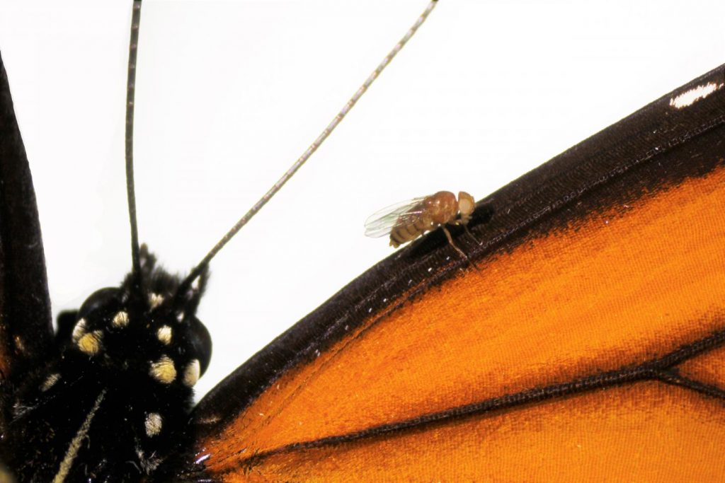
Caption: A Drosophila melanogaster “monarch fly” with mutations introduced by CRISPR-Cas9 genome editing (V111, S119 and H122) to the sodium potassium pump, on a wing of a monarch butterfly (Danaus plexippus). Credit & Ccpyright: Julianne Pelaez

Here’s a link to and a citation for the paper,

Genome editing retraces the evolution of toxin resistance in the monarch butterfly by Marianthi Karageorgi, Simon C. Groen, Fidan Sumbul, Julianne N. Pelaez, Kirsten I. Verster, Jessica M. Aguilar, Amy P. Hastings, Susan L. Bernstein, Teruyuki Matsunaga, Michael Astourian, Geno Guerra, Felix Rico, Susanne Dobler, Anurag A. Agrawal & Noah K. Whiteman. Nature (2019) DOI: https://doi.org/10.1038/s41586-019-1610-8 Published 02 October 2019

This paper is behind a paywall.

Words about a word

I’m glad they changed the headline and substituted vomit for puke. I think we need vulgar and/or taboo words to release anger or disgust or other difficult emotions. Incorporating those words into standard language deprives them of that power.

The last word: Genetivision

The company mentioned in the new release, Genetivision, is the place to go for transgenic flies. Here’s a sampling from the their Testimonials webpage,

GenetiVision‘s service has been excellent in the quality and price. The timeliness of its international service has been a big plus. We are very happy with its consistent service and the flies it generates.”
Kwang-Wook Choi, Ph.D.
Department of Biological Sciences
Korea Advanced Institute of Science and Technology


“We couldn’t be happier with GenetiVision. Great prices on both standard P and PhiC31 transgenics, quick turnaround time, and we’re still batting 1000 with transformant success. We used to do our own injections but your service makes it both faster and more cost-effective. Thanks for your service!”
Thomas Neufeld, Ph.D.
Department of Genetics, Cell Biology and Development
University of Minnesota

You can find out more here at the Genetivision website.
The latest and greatest in gene drives (for flies)

This is a CRISPR (clustered regularly interspaced short palindromic repeats) story where the researchers are working on flies. If successful, this has much wider implications. From an April 10, 2019 news item on phys.org,

New CRISPR-based gene drives and broader active genetics technologies are revolutionizing the way scientists engineer the transfer of specific traits from one generation to another.

Scientists at the University of California San Diego have now developed a new version of a gene drive that opens the door to the spread of specific, favorable subtle genetic variants, also known as “alleles,” throughout a population.

The new “allelic drive,” described April 9 [2019] in Nature Communications, is equipped with a guide RNA (gRNA) that directs the CRISPR system to cut undesired variants of a gene and replace it with a preferred version of the gene. The new drive extends scientists’ ability to modify populations of organisms with precision editing. Using word processing as an analogy, CRISPR-based gene drives allow scientists to edit sentences of genetic information, while the new allelic drive offers letter-by-letter editing.

An April 9, 2019 University of California at San Diego (UCSD) news release (also on EurekAlert) by Mario Aguilera, which originated the news item, delves into this technique’s potential uses while further explaining the work


In one example of its potential applications, specific genes in agricultural pests that have become resistant to insecticides could be replaced by original natural genetic variants conferring sensitivity to insecticides using allelic drives that selectively swap the identities of a single protein residue (amino acid).

In addition to agricultural applications, disease-carrying insects could be a target for allelic drives.

“If we incorporate such a normalizing gRNA on a gene-drive element, for example, one designed to immunize mosquitoes against malaria, the resulting allelic gene drive will spread through a population. When this dual action drive encounters an insecticide-resistant allele, it will cut and repair it using the wild-type susceptible allele,” said Ethan Bier, the new paper’s senior author. “The result being that nearly all emerging progeny will be sensitive to insecticides as well as refractory to malaria transmission.”

“Forcing these species to return to their natural sensitive state using allelic drives would help break a downward cycle of ever-increasing and environmentally damaging pesticide over-use,” said Annabel Guichard, the paper’s first author.

The researchers describe two versions of the allelic drive, including “copy-cutting,” in which researchers use the CRISPR system to selectively cut the undesired version of a gene, and a more broadly applicable version referred to as “copy-grafting” that promotes transmission of a favored allele next to the site that is selectively protected from gRNA cleavage.

“An unexpected finding from this study is that mistakes created by such allelic drives do not get transmitted to the next generation,” said Guichard. “These mutations instead produce an unusual form of lethality referred to as ‘lethal mosaicism.’ This process helps make allelic drives more efficient by immediately eliminating unwanted mutations created by CRISPR-based drives.”

Although demonstrated in fruit flies, the new technology also has potential for broad application in insects, mammals and plants. According to the researchers, several variations of the allelic drive technology could be developed with combinations of favorable traits in crops that, for example, thrive in poor soil and arid environments to help feed the ever-growing world population.

Beyond environmental applications, allelic drives should enable next-generation engineering of animal models to study human disease as well as answer important questions in basic science. As a member of the Tata Institute for Genetics and Society (TIGS), Bier says allelic drives could be used to aid in environmental conservation efforts to protect vulnerable endemic species or stop the spread of invasive species.

Gene drives and active genetics systems are now being developed for use in mammals. The scientists say allelic drives could accelerate new laboratory strains of animal models of human disease that aid in the development of new cures.

Here’s a link to and a citation for the paper,

Efficient allelic-drive in Drosophila by Annabel Guichard, Tisha Haque, Marketta Bobik, Xiang-Ru S. Xu, Carissa Klanseck, Raja Babu Singh Kushwah, Mateus Berni, Bhagyashree Kaduskar, Valentino M. Gantz & Ethan Bier. Nature Communicationsvolume 10, Article number: 1640 (2019) DOI: https://doi.org/10.1038/s41467-019-09694-w Published 09 April 2019

This paper is open access.

For anyone new to gene drives, I have a February 8, 2018 posting that highlights a report from the UK on the latest in genetic engineering, which provides a definition for [synthetic] gene drives, and if you scroll down about 75% of the way, you’ll also find excerpts from an article for The Atlantic by Ed Yong on gene drives as proposed for a project in New Zealand.

Cell-to-cell communication via nanotubes

It turns out that the cells communicating with each other are located in fruit flies. So, it’s perhaps not quite as exciting as one might have imagined, nonetheless, a July 1, 2015 news item on ScienceDaily provides some intriguing insights into cell communication,

When it comes to communicating with each other, some cells may be more “old school” than was previously thought.

Certain types of stem cells use microscopic, threadlike nanotubes to communicate with neighboring cells, like a landline phone connection, rather than sending a broadcast signal, researchers at University of Michigan Life Sciences Institute and University of Texas Southwestern Medical Center have discovered.

The findings, which are scheduled for online publication July 1 in Nature, offer new insights on how stem cells retain their identities when they divide to split off a new, specialized cell.

The fruit-fly research also suggests that short-range, cell-to-cell communication may rely on this type of direct connection more than was previously understood, said co-senior author Yukiko Yamashita, a U-M developmental biologist whose lab is located at the Life Sciences Institute.

A July 1, 2015 University of Michigan news release (also on EurekAlert), which originated the news item, expands on the theme,

“There are trillions of cells in the human body, but nowhere near that number of signaling pathways,” she said. “There’s a lot we don’t know about how the right cells get just the right messages to the right recipients at the right time.”

The nanotubes had actually been hiding in plain sight.

The investigation began when a postdoctoral researcher in Yamashita’s lab, Mayu Inaba, approached her mentor with questions about tiny threads of connection she noticed in an image of fruit fly reproductive stem cells, which are also known as germ line cells. The projections linked individual stem cells back to a central hub in the stem cell “niche.” Niches create a supportive environment for stem cells and help direct their activity.

Yamashita, a Howard Hughes Medical Institute investigator, MacArthur Fellow and an associate professor at the U-M Medical School, looked through her old image files and discovered that the connections appeared in numerous images.

“I had seen them, but I wasn’t seeing them,” Yamashita said. “They were like a little piece of dust on an otherwise normal picture. After we presented our findings at meetings, other scientists who work with the same cells would say, ‘We see them now, too.'”

It’s not surprising that the minute structures went overlooked for so long. Each one is about 3 micrometers long; by comparison, a piece of paper is 100 micrometers thick.

While the study looked specifically at reproductive cells in male Drosophila fruit flies, there have been indications of similar structures in other contexts, including mammalian cells, Yamashita said.

Fruit flies are an important model for this type of investigation, she added. If one was to start instead with human cells, one might find something, but the system’s greater complexity would make it far more difficult to tease apart the underlying mechanisms.

The findings shed new light on a key attribute of stem cells: their ability to make new specialized cells while still retaining their identity as stem cells.

Germ line stem cells typically divide asymmetrically. In the male fruit fly, when a stem cell divides, one part stays attached to the hub and remains a stem cell. The other part moves away from the hub and begins differentiation into a fly sperm cell.

Until the discovery of the nanotubes, scientists had been puzzled as to how cellular signals guiding identity could act on one of the cells but not the other, said collaborator Michael Buszczak, an associate professor of molecular biology at UT Southwestern, who shares corresponding authorship of the paper and currently co-mentors Inaba with Yamashita.

The researchers conducted experiments that showed disruption of nanotube formation compromised the ability of the germ line stem cells to renew themselves.

I gather the fruit fly research offers the basis for more extensive investigations into other species and their cell-to-cell communication.

Here’s a link to and a citation for the paper,

Nanotubes mediate niche–stem-cell signalling in the Drosophila testis by Mayu Inaba, Michael Buszczak, & Yukiko M. Yamashita. Nature (2015) doi:10.1038/nature14602 Published online 01 July 2015

This paper is behind a paywall.

Could engineered nanoparticles be behind rise in obesity and metabolic disorders?

The researchers haven’t published a study and they have used fruit flies as their testing mechanism (animal models) so, it’s a little difficult (futile) to analyze the work at this stage but it is intriguing. A June 9, 2015 news item on Azonano announces a research collaboration  designed to examine the impact engineered nanoparticles have on the gut and the gut microbiome,

Researchers at Binghamton University believe understanding nano particles’ ability to influence our metabolic processing may be integral to mediating metabolic disorders and obesity, both of which are on the rise and have been linked to processed foods.

Anthony Fiumera, associate professor of biological sciences, and Gretchen Mahler, assistant professor of biomedical engineering, are collaborating on a research project funded by a Binghamton University Transdisciplinary Areas of Excellence (TAE) grant to discover the role ingested nanoparticles play in the physiology and function of the gut and gut microbiome.

A June 8, 2015 Binghamton University news release, which originated the news item, describes the reasoning behind the research,

The gut microbiome is the population of microbes living within the human intestine, consisting of tens of trillions of microorganisms (including at least 1,000 different species of known bacteria). Nanoparticles, which are often added to processed foods to enhance texture and color, have been linked to changes in gut function. As processed foods become more common elements of our diet, there has been a significant increase in concentrations of these particles found in the human body.

Fiumera works in vivo with fruit flies while Mahler works in vitro using a 3-D cell-culture model of the gastrointestinal (GI) tract to understand how ingesting nanoparticles influences glucose processing and the gut microbiome. By using complementary research methods, the researchers have helped advance each other’s understanding of nanoparticles.

Using fruit flies, Fiumera looks at the effects of nanoparticles on development, physiology and biochemical composition, as well as the microbial community in the GI tract of the fly. The fly model offers two advantages: 1) research can be done on a wide range of traits that might be altered by changes in metabolism and 2) the metabolic processes within the fly are similar to those in humans. Fiumera also aims to investigate which genes are associated with responses to the nanoparticles, which ultimately may help us understand why individuals react differently to nanoparticles.

For this project, Mahler expanded her GI tract model to include a commensal intestinal bacterial species and used the model to determine a more detailed mechanism of the role of nanoparticle exposure on gut bacteria and intestinal function. Early results have shown that nanoparticle ingestion alters glucose absorption, and that the presence of beneficial gut bacteria eliminates these effects.

Mahler was already investigating nanoparticles when she reached out to Fiumera and proposed they combine their respective expertise. With the help of undergraduate students Gabriella Shull and John Fountain and graduate student Jonathan Richter, Fiumera and Mahler have begun to uncover some effects of ingesting nanoparticles. Since they are using realistic, low concentrations of nanoparticles, the effects are slight, but eventually may be additive.

The most interesting aspect of this research (to me) is the notion that the impact may be additive. In short, you might be able to tolerate a few more nanoparticles in your gut but as more engineered nanoparticles become part of our food and drink (including water) and your gut receives more and more that tolerance may no longer possible.

There is increasing concern about engineered nanoparticles as they cycle through environment and the US Environmental Protection Agency (EPA) funded a programed by Arizona State University (ASU), LCnano Network (part of the EPA’s larger Life Cycle of Nanomaterials project). You can find out more about the ASU program in my April 8, 2014 post (scroll down about 50% of the way).

Getting back to Binghamton, I look forward to hearing more about the research as it progresses.

Wound healing is nature’s way of zipping up your skin

Scientists have been able to observe the healing process at the molecular scale—in fruit flies. From an April 21, 2015 news item on ScienceDaily,

Scientists from the Goethe University (GU) Frankfurt, the European Molecular Biology Laboratory (EMBL) Heidelberg and the University of Zurich explain skin fusion at a molecular level and pinpoint the specific molecules that do the job in their latest publication in the journal Nature Cell Biology.

An April 21, 2015 Goethe University Frankfurt press release on EurekAlert, which originated the news item, describes similarities between humans and fruit flies allowing scientists to infer the wound healing process for human skin,

In order to prevent death by bleeding or infection, every wound (skin opening) must close at some point. The events leading to skin closure had been unclear for many years. Mikhail Eltsov (GU) and colleagues used fruit fly embryos as a model system to understand this process. Similarly to humans, fruit fly embryos at some point in their development have a large opening in the skin on their back that must fuse. This process is called zipping, because two sides of the skin are fastened in a way that resembles a zipper that joins two sides of a jacket.

The scientists have used a top-of-the-range electron microscope to study exactly how this zipping of the skin works. “Our electron microscope allows us to distinguish the molecular components in the cell that act like small machines to fuse the skin. When we look at it from a distance, it appears as if skin cells simply fuse to each other, but if we zoom in, it becomes clear that membranes, molecular machines, and other cellular components are involved”, explains Eltsov.

“In order to visualize this orchestra of healing, a very high-resolution picture of the process is needed. For this purpose we have recorded an enormous amount of data that surpasses all previous studies of this kind”, says Mikhail Eltsov.

As a first step, as the scientists discovered, cells find their opposing partner by “sniffing” each other out. As a next step, they develop adherens junctions which act like a molecular Velcro. This way they become strongly attached to their opposing partner cell. The biggest revelation of this study was that small tubes in the cell, called microtubules, attach to this molecular Velcro and then deploy a self-catastrophe, which results in the skin being pulled towards the opening, as if one pulls a blanket over.

Damian Brunner who led the team at the University of Zurich has performed many genetic manipulations to identify the correct components. The scientists were astonished to find that microtubules involved in cell-division are the primary scaffold used for zipping, indicating a mechanism conserved during evolution.

“What was also amazing was the tremendous plasticity of the membranes in this process which managed to close the skin opening in a very short space of time. When five to ten cells have found their respective neighbors, the skin already appears normal”, says Achilleas Frangakis from the Goethe University Frankfurt, who led the study.

The scientists hope that their results will open new avenues into the understanding of epithelial plasticity and wound healing. They are also investigating the detailed structural organization of the adherens junctions, work for which they were awarded a starting grant from European Research Council (ERC).

Here’s a link to and a citation for the paper,

Quantitative analysis of cytoskeletal reorganization during epithelial tissue sealing by large-volume electron tomography by Mikhail Eltsov, Nadia Dubé, Zhou Yu, Laurynas Pasakarnis, Uta Haselmann-Weiss, Damian Brunner, & Achilleas S. Frangakis. Nature Cell Biology (2015) doi:10.1038/ncb3159 Published online 20 April 2015

This paper is behind a paywall but there is a free preview available via ReadCube Access.

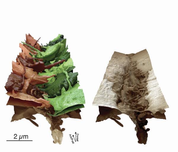
The researchers have provided an image illustrating ‘wound zipping’.

Caption: This is a perspective view of the zipping area with 17 skin cells. Credit: GU

Caption: This is a perspective view of the zipping area with 17 skin cells.
Credit: GU