Tag Archives: Geelsu Hwang

Smart dental implant resists bacterial growth and generates own electricity

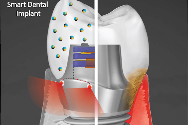
A “smart” dental implant could improve upon current devices by employing biofilm-resisting nanoparticles and a light powered by biomechanical forces to promote health of the surrounding gum tissue. (Image: Courtesy of Albert Kim)

A September 9, 2021 news item on ScienceDaily announces research into ‘smart’ dental implants,

More than 3 million people in America have dental implants, used to replace a tooth lost to decay, gum disease, or injury. Implants represent a leap of progress over dentures or bridges, fitting much more securely and designed to last 20 years or more.

But often implants fall short of that expectation, instead needing replacement in five to 10 years due to local inflammation or gum disease, necessitating a repeat of a costly and invasive procedure for patients.

“We wanted to address this issue, and so we came up with an innovative new implant,” says Geelsu Hwang, an assistant professor in the University of Pennsylvania School of Dental Medicine, who has a background in engineering that he brings to his research on oral health issues.

The novel implant would implement two key technologies, Hwang says. One is a nanoparticle-infused material that resists bacterial colonization. And the second is an embedded light source to conduct phototherapy, powered by the natural motions of the mouth, such as chewing or toothbrushing. In a paper in the journal ACS Applied Materials & Interfaces and a 2020 paper in the journal Advanced Healthcare Materials, Hwang and colleagues lay out their platform, which could one day be integrated not only into dental implants but other technologies, such as joint replacements, as well.

A September 9, 2021 University of Pennsylvania news release (also on EurekAlert), which originated the news item, provides more technical details about the proposed technology,

“Phototherapy can address a diverse set of health issues,” says Hwang. “But once a biomaterial is implanted, it’s not practical to replace or recharge a battery. We are using a piezoelectric material, which can generate electrical power from natural oral motions to supply a light that can conduct phototherapy, and we find that it can successfully protect gingival tissue from bacterial challenge.”

In the paper, the material the researchers explored was barium titanate (BTO), which has piezoelectric properties that are leveraged in applications such as capacitators and transistors, but has not yet been explored as a foundation for anti-infectious implantable biomaterials. To test its potential as the foundation for a dental implant, the team first used discs embedded with nanoparticles of BTO and exposed them to Streptococcus mutans, a primary component of the bacterial biofilm responsible for tooth decay commonly known as dental plaque. They found that the discs resisted biofilm formation in a dose-dependent manner. Discs with higher concentrations of BTO were better at preventing biofilms from binding.

While earlier studies had suggested that BTO might kill bacteria outright using reactive oxygen species generated by light-catalyzed or electric polarization reactions, Hwang and colleagues did not find this to be the case due to the short-lived efficacy and off-target effects of these approaches. Instead, the material generates enhanced negative surface charge that repels the negatively charged cell walls of bacteria. It’s likely that this repulsion effect would be long-lasting, the researchers say.

“We wanted an implant material that could resist bacterial growth for a long time because bacterial challenges are not a one-time threat,” Hwang says.

The power-generating property of the material was sustained and in tests over time the material did not leach. It also demonstrated a level of mechanical strength comparable to other materials used in dental applications.

Finally, the material did not harm normal gingival tissue in the researchers’ experiments, supporting the idea that this could be used without ill effect in the mouth.

The technology is a finalist in the Science Center’s research accelerator program, the QED Proof-of-Concept program. As one of 12 finalists, Hwang and colleagues will receive guidance from experts in commercialization. If the project advances to be one of three finalists, the group has the potential to receive up to $200,000 in funding.

In future work, the team hopes to continue to refine the “smart” dental implant system, testing new material types and perhaps even using assymetric properties on each side of the implant components, one that encourages tissue integration on the side facing the gums and one that resists bacterial formation on the side facing the rest of the mouth.

“We hope to further develop the implant system and eventually see it commercialized so it can be used in the dental field,” Hwang says.

Here’s a link to and a citation for the paper,

Bimodal Nanocomposite Platform with Antibiofilm and Self-Powering Functionalities for Biomedical Applications by Atul Dhall, Sayemul Islam, Moonchul Park, Yu Zhang, Albert Kim, and Geelsu Hwang. ACS Appl. Mater. Interfaces 2021, 13, 34, 40379–40391 DOI: https://doi.org/10.1021/acsami.1c11791 Publication Date:August 18, 2021 Copyright © 2021 American Chemical Society

This paper is behind a paywall.

The work from 2020, mentioned in the news release, laid groundwork for the latest paper.

Human Oral Motion-Powered Smart Dental Implant (SDI) for In Situ Ambulatory Photo-biomodulation Therapy by Moonchul Park, Sayemul Islam, Hye-Eun Kim, Jonathan Korosto, Markus B. Blatz, Geelsu Hwang, and Albert Kim. Adv. Healthcare Mater. 2020, 9, 2000658 DOI: 10.1002/adhm.202000658 First published: 01 July 2020 © 2020 WILEY-VCH Verlag GmbH & Co. KGaA, WeinheimHuman

This paper is behind a paywall.

Nanoparticles for breaking up plaque and preventing cavities

There may be iron in your tooth care future if a team of researchers at the University of Pennsylvania have their way. From a July 26, 2016 news item on ScienceDaily,

The bacteria that live in dental plaque and contribute to tooth decay often resist traditional antimicrobial treatment, as they can “hide” within a sticky biofilm matrix, a glue-like polymer scaffold.

A new strategy conceived by University of Pennsylvania researchers took a more sophisticated approach. Instead of simply applying an antibiotic to the teeth, they took advantage of the pH-sensitive and enzyme-like properties of iron-containing nanoparticles to catalyze the activity of hydrogen peroxide, a commonly used natural antiseptic. The activated hydrogen peroxide produced free radicals that were able to simultaneously degrade the biofilm matrix and kill the bacteria within, significantly reducing plaque and preventing the tooth decay, or cavities, in an animal model.

“Even using a very low concentration of hydrogen peroxide, the process was incredibly effective at disrupting the biofilm,” said Hyun (Michel) Koo, a professor in the Penn School of Dental Medicine’s Department of Orthodontics and divisions of Pediatric Dentistry and Community and Oral Health and the senior author of the study, which was published in the journal Biomaterials. “Adding nanoparticles increased the efficiency of bacterial killing more than 5,000-fold.”

A July 25, 2016 University of Pennsylvania news release, which originated the news item, describes the genesis of the work and provides more details about the current research (Note: A link has been removed),

The work built off a seminal finding by Gao [Lizeng Gao, a postdoctoral researcher in Koo’s lab] and colleagues, published in 2007 in Nature Nanotechnology, showing that nanoparticles, long believed to be biologically and chemically inert, could in fact possess enzyme-like properties. In that study, Gao showed that an iron oxide nanoparticle behaved similarly to a peroxidase, an enzyme found naturally that catalyzes oxidative reactions, often using hydrogen peroxide.

When Gao joined Koo’s lab in 2013, he proposed using these nanoparticles in an oral setting, as the oxidation of hydrogen peroxide produces free radicals that can kill bacteria.

“When he first presented it to me, I was very skeptical,” Koo said, “because these free radicals can also damage healthy tissue. But then he refuted that and told me this is different because the nanoparticles’ activity is dependent on pH.”

Gao had found that the nanoparticles had no catalytic activity at neutral or near-neutral pH of 6.5 or 7, physiological values typically found in blood or in a healthy mouth. But when pH was acidic, closer to 5, they become highly active and can rapidly produce free radicals.

The scenario was ideal for targeting plaque, which can produce an acidic microenvironment when exposed to sugars.

Gao and Koo reached out to Cormode [David Cormode, an assistant professor of radiology and bioengineering], who had experience working with iron oxide nanoparticles in a radiological imaging context, to help them synthesize, characterize and test the effectiveness of the nanoparticles, several forms of which are already FDA-approved for imaging in humans.

Beginning with in vitro studies, which involved growing a biofilm containing the cavity-causing bacteria Streptococcus mutans on a tooth-enamel-like surface and then exposing it to sugar, the researchers confirmed that the nanoparticles adhered to the biofilm, were retained even after treatment stopped and could effectively catalyze hydrogen peroxide in acidic conditions.

They also showed that the nanoparticles’ reaction with a 1 percent or less hydrogen peroxide solution was remarkably effective at killing bacteria, wiping out more than 99.9 percent of the S. mutans in the biofilm within five minutes, an efficacy more than 5,000 times greater than using hydrogen peroxide alone. Even more promising, they demonstrated that the treatment regimen, involving a 30-second topical treatment of the nanoparticles followed by a 30-second treatment with hydrogen peroxide, could break down the biofilm matrix components, essentially removing the protective sticky scaffold.

Moving to an animal model, they applied the nanoparticles and hydrogen peroxide topically to the teeth of rats, which can develop tooth decay when infected with S. mutans just as humans do. Twice-a-day, one-minute treatments for three weeks significantly reduced the onset and severity of carious lesions, the clinical term for tooth decay, compared to the control or treatment with hydrogen peroxide alone. The researchers observed no adverse effects on the gum or oral soft tissues from the treatment.

“It’s very promising,” said Koo. “The efficacy and toxicity need to be validated in clinical studies, but I think the potential is there.”

Among the attractive features of the platform is the fact that the components are relatively inexpensive.

“If you look at the amount you would need for a dose, you’re looking at something like 5 milligrams,” Cormode said. “It’s a tiny amount of material, and the nanoparticles are fairly easily synthesize, so we’re talking about a cost of cents per dose.”

In addition, the platform uses a concentration of hydrogen peroxide, 1 percent, which is lower than many currently available tooth-whitening systems that use 3 to 10 percent concentrations, minimizing the chance of negative side effects.

Looking ahead, Gao, Koo, Cormode and colleagues hope to continue refining and improving upon the effectiveness of the nanoparticle platform to fight biofilms.

“We’re studying the role of nanoparticle coatings, composition, size and so forth so we can engineer the particles for even better performance,” Cormode said.

The funding agencies provide a note of interest (Note: Links have been removed),

The study was funded by the International Association for Dental Research/GlaxoSmithKline Innovation in Oral Health Award, National Science Foundation and University of Pennsylvania Research Foundation.

Presumably the industry as represented by the GlaxoSmithKline Innovation in Oral Health Award is keeping a close eye on this work.

Here’s a link to and a citation for the paper,

Nanocatalysts promote Streptococcus mutans biofilm matrix degradation and enhance bacterial killing to suppress dental caries in vivo by Lizeng Gao, Yuan Liu, Dongyeop Kim, Yong Li, Geelsu Hwang, Pratap C. Naha, David P. Cormode, & Hyun Koo. Biomaterials Volume 101, September 2016, Pages 272–284 doi:10.1016/j.biomaterials.2016.05.051

This paper is behind a paywall.