Tag Archives: gelatin

Optical fibers made from marine algae

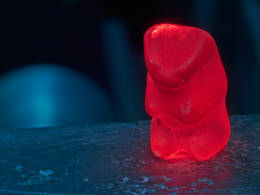
Apparently after you’ve finished imaging with your marine algae-based optical fibers, you can eat them. A July 24, 2020 news item on Nanowerk announces the new research,

An optical fiber made of agar has been produced at the University of Campinas (UNICAMP) in the state of São Paulo, Brazil. This device is edible, biocompatible and biodegradable. It can be used in vivo for body structure imaging, localized light delivery in phototherapy or optogenetics (e.g., stimulating neurons with light to study neural circuits in a living brain), and localized drug delivery.

Another possible application is the detection of microorganisms in specific organs, in which case the probe would be completely absorbed by the body after performing its function.

Caption: Edible, biocompatible and biodegradable, these fibers have potential for various medical applications. Credit: Eric Fujiwara

A July 24, 2020 Fundação de Amparo à Pesquisa dFundação de Amparo à Pesquisa do Estado de São Paulo (FAPESP) do Estado de São Paulo press release on EurekAlert, which originated the news item, provides a few more details about the researches and the work,

The research project, which was supported by São Paulo Research Foundation – FAPESP, was led by Eric Fujiwara, a professor in UNICAMP’s School of Mechanical Engineering, and Cristiano Cordeiro, a professor in UNICAMP’s Gleb Wataghin Institute of Physics, in collaboration with Hiromasa Oku, a professor at Gunma University in Japan.

An article on the study is published) in Scientific Reports, an online journal owned by Springer Nature.

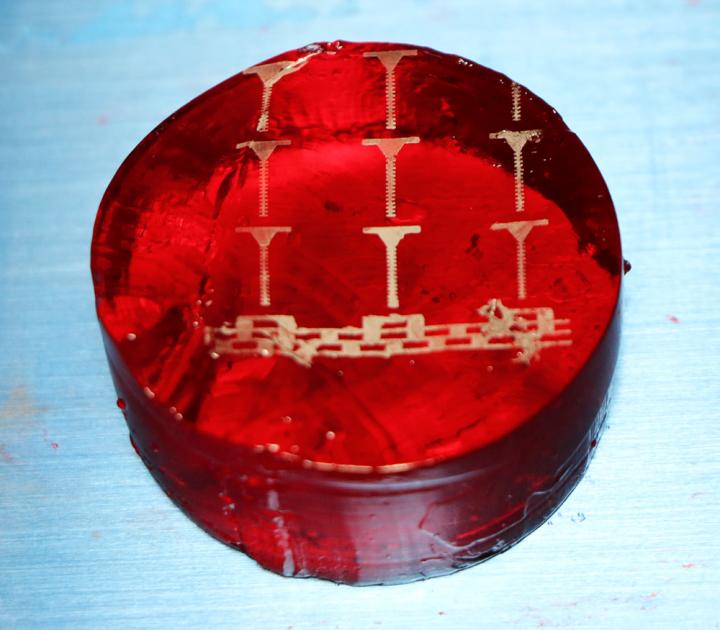
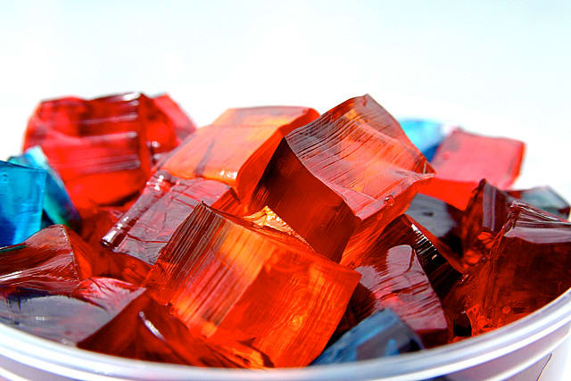
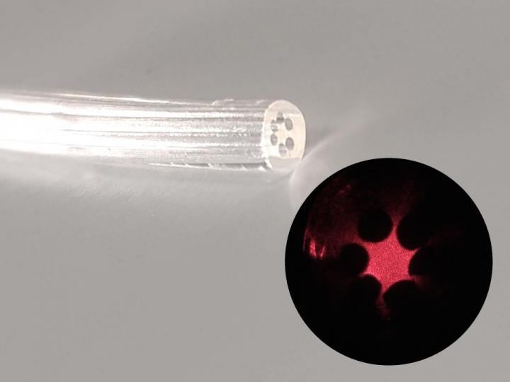
Agar, also called agar-agar, is a natural gelatin obtained from marine algae. Its composition consists of a mixture of two polysaccharides, agarose and agaropectin. “Our optical fiber is an agar cylinder with an external diameter of 2.5 millimeters [mm] and a regular inner arrangement of six 0.5 mm cylindrical airholes around a solid core. Light is confined owing to the difference between the refraction indices of the agar core and the airholes,” Fujiwara told.

“To produce the fiber, we poured food-grade agar into a mold with six internal rods placed lengthwise around the main axis,” he continued. “The gel distributes itself to fill the available space. After cooling, the rods are removed to form airholes, and the solidified waveguide is released from the mold. The refraction index and geometry of the fiber can be adapted by varying the composition of the agar solution and mold design, respectively.”

The researchers tested the fiber in different media, from air and water to ethanol and acetone, concluding that it is context-sensitive. “The fact that the gel undergoes structural changes in response to variations in temperature, humidity and pH makes the fiber suitable for optical sensing,” Fujiwara said.

Another promising application is its simultaneous use as an optical sensor and a growth medium for microorganisms. “In this case, the waveguide can be designed as a disposable sample unit containing the necessary nutrients. The immobilized cells in the device would be optically sensed, and the signal would be analyzed using a camera or spectrometer,” he said.

###

About São Paulo Research Foundation (FAPESP)

The São Paulo Research Foundation (FAPESP) is a public institution with the mission of supporting scientific research in all fields of knowledge by awarding scholarships, fellowships and grants to investigators linked with higher education and research institutions in the State of São Paulo, Brazil. FAPESP is aware that the very best research can only be done by working with the best researchers internationally. Therefore, it has established partnerships with funding agencies, higher education, private companies, and research organizations in other countries known for the quality of their research and has been encouraging scientists funded by its grants to further develop their international collaboration. You can learn more about FAPESP at http://www.fapesp.br/en and visit FAPESP news agency at http://www.agencia.fapesp.br/en to keep updated with the latest scientific breakthroughs FAPESP helps achieve through its many programs, awards and research centers. You may also subscribe to FAPESP news agency at http://agencia.fapesp.br/subscribe.

As per my usual practice, here’s a link to and a citation for the paper,

Agarose-based structured optical fibre by Eric Fujiwara, Thiago D. Cabral, Miko Sato, Hiromasa Oku & Cristiano M. B. Cordeiro. Scientific Reports volume 10, Article number: 7035 (2020) DOI: https://doi.org/10.1038/s41598-020-64103-3 Published: 27 April 2020

This paper is open access.

Should you have a problem accessing the English language version of the FAPESP website, the Portuguese language version of the site seems more accessible (assuming you have the language skills).

Printing metal on flowers or gelatin

Martin Thuo and his research group have developed heat-free technology that can print conductive, metallic lines and traces on just about anything, including a rose petal. Photo courtesy of Martin Thuo.

I’m not sure how I feel about an electrified rose but it is strangely fascinating. Here’s more from a July 29, 2019 news item on Nanowerk,

Martin Thuo of Iowa State University and the Ames Laboratory clicked through the photo gallery for one of his research projects.

How about this one? There was a rose with metal traces printed on a delicate petal.

Or this? A curled sheet of paper with a flexible, programmable LED display.

Maybe this? A gelatin cylinder with metal traces printed across the top.

Caption: Martin Thuo and his research group have printed electronic traces on gelatin. Credit: Martin Thuo/Iowa State University

A July 26, 2019 Iowa State University news release (also on EurekAlert but published on July 29, 2019), which originated the news item,

All those photos showed the latest application of undercooled metal technology developed by Thuo and his research group. The technology features liquid metal (in this case Field’s metal, an alloy of bismuth, indium and tin) trapped below its melting point in polished, oxide shells, creating particles about 10 millionths of a meter across.

When the shells are broken – with mechanical pressure or chemical dissolving – the metal inside flows and solidifies, creating a heat-free weld or, in this case, printing conductive, metallic lines and traces on all kinds of materials, everything from a concrete wall to a leaf.

That could have all kinds of applications, including sensors to measure the structural integrity of a building or the growth of crops. The technology was also tested in paper-based remote controls that read changes in electrical currents when the paper is curved. Engineers also tested the technology by making electrical contacts for solar cells and by screen printing conductive lines on gelatin, a model for soft biological tissues, including the brain.

“This work reports heat-free, ambient fabrication of metallic conductive interconnects and traces on all types of substrates,” Thuo and a team of researchers wrote in a paper describing the technology recently published online by the journal Advanced Functional Materials.

Thuo – an assistant professor of materials science and engineering at Iowa State, an associate of the U.S. Department of Energy’s Ames Laboratory and a co-founder of the Ames startup SAFI-Tech Inc. that’s commercializing the liquid-metal particles – is the lead author. Co-authors are Andrew Martin, a former undergraduate in Thuo’s lab and now an Iowa State doctoral student in materials science and engineering; Boyce Chang, a postdoctoral fellow at the University of California, Berkeley, who earned his doctoral degree at Iowa State Zachariah Martin, Dipak Paramanik and Ian Tevis, of SAFI-Tech; Christophe Frankiewicz, a co-founder of Sep-All in Ames and a former Iowa State postdoctoral research associate; and Souvik Kundu, an Iowa State graduate student in electrical and computer engineering.
The project was supported by university startup funds to establish Thuo’s research lab at Iowa State, Thuo’s Black & Veatch faculty fellowship and a National Science Foundation Small Business Innovation Research grant.

Thuo said he launched the project three years ago as a teaching exercise.

“I started this with undergraduate students,” he said. “I thought it would be fun to get students to make something like this. It’s a really beneficial teaching tool because you don’t need to solve 2 million equations to do sophisticated science.”

And once students learned to use a few metal-processing tools, they started solving some of the technical challenges of flexible, metal electronics.

“The students discovered ways of dealing with metal and that blossomed into a million ideas,” Thuo said. “And now we can’t stop.”

And so the researchers have learned how to effectively bond metal traces to everything from water-repelling rose petals to watery gelatin. Based on what they now know, Thuo said it would be easy for them to print metallic traces on ice cubes or biological tissue.

All the experiments “highlight the versatility of this approach,” the researchers wrote in their paper, “allowing a multitude of conductive products to be fabricated without damaging the base material.”

Here’s a link to and a citation for the paper,

Heat‐Free Fabrication of Metallic Interconnects for Flexible/Wearable Devices by Andrew Martin, Boyce S. Chang, Zachariah Martin, Dipak Paramanik, Christophe Frankiewicz, Souvik Kundu, Ian D. Tevis, Martin Thuo. Advanced Functional Materials Online Version of Record before inclusion in an issue 1903687 DOI: https://doi.org/10.1002/adfm.201903687 First published online: 15 July 2019

This paper is behind a paywall.

Jiggly jell-o as a new hydrogen fuel catalyst

Jello [uploaded from https://www.organicauthority.com/eco-chic-table/new-jell-o-mold-jiggle-chic-holidays]

I’m quite intrigued by this ‘jell-o’ story. It’s hard to believe a childhood dessert might prove to have an application as a catalyst for producing hydrogen fuel. From a December 14, 2018 news item on Nanowerk,

A cheap and effective new catalyst developed by researchers at the University of California, Berkeley, can generate hydrogen fuel from water just as efficiently as platinum, currently the best — but also most expensive — water-splitting catalyst out there.

The catalyst, which is composed of nanometer-thin sheets of metal carbide, is manufactured using a self-assembly process that relies on a surprising ingredient: gelatin, the material that gives Jell-O its jiggle.

Two-dimensional metal carbides spark a reaction that splits water into oxygen and valuable hydrogen gas. Berkeley researchers have discovered an easy new recipe for cooking up these nanometer-thin sheets that is nearly as simple as making Jell-O from a box. (Xining Zang graphic, copyright Wiley)

A December 13, 2018 University of California at Berkeley (UC Berkeley) news release by Kara Manke (also on EurekAlert but published on Dec. 14, 2018), which originated the news item, provides more technical detail,

“Platinum is expensive, so it would be desirable to find other alternative materials to replace it,” said senior author Liwei Lin, professor of mechanical engineering at UC Berkeley. “We are actually using something similar to the Jell-O that you can eat as the foundation, and mixing it with some of the abundant earth elements to create an inexpensive new material for important catalytic reactions.”

The work appears in the Dec. 13 [2018] print edition of the journal Advanced Materials.

A zap of electricity can break apart the strong bonds that tie water molecules together, creating oxygen and hydrogen gas, the latter of which is an extremely valuable source of energy for powering hydrogen fuel cells. Hydrogen gas can also be used to help store energy from renewable yet intermittent energy sources like solar and wind power, which produce excess electricity when the sun shines or when the wind blows, but which go dormant on rainy or calm days.

A black and white image of metal carbide under high magnification.

When magnified, the two-dimensional metal carbides resemble sheets of cell[o]phane. (Xining Zang photo, copyright Wiley)

But simply sticking an electrode in a glass of water is an extremely inefficient method of generating hydrogen gas. For the past 20 years, scientists have been searching for catalysts that can speed up this reaction, making it practical for large-scale use.

“The traditional way of using water gas to generate hydrogen still dominates in industry. However, this method produces carbon dioxide as byproduct,” said first author Xining Zang, who conducted the research as a graduate student in mechanical engineering at UC Berkeley. “Electrocatalytic hydrogen generation is growing in the past decade, following the global demand to lower emissions. Developing a highly efficient and low-cost catalyst for electrohydrolysis will bring profound technical, economical and societal benefit.”

To create the catalyst, the researchers followed a recipe nearly as simple as making Jell-O from a box. They mixed gelatin and a metal ion — either molybdenum, tungsten or cobalt — with water, and then let the mixture dry.

“We believe that as gelatin dries, it self-assembles layer by layer,” Lin said. “The metal ion is carried by the gelatin, so when the gelatin self-assembles, your metal ion is also arranged into these flat layers, and these flat sheets are what give Jell-O its characteristic mirror-like surface.”

Heating the mixture to 600 degrees Celsius triggers the metal ion to react with the carbon atoms in the gelatin, forming large, nanometer-thin sheets of metal carbide. The unreacted gelatin burns away.

The researchers tested the efficiency of the catalysts by placing them in water and running an electric current through them. When stacked up against each other, molybdenum carbide split water the most efficiently, followed by tungsten carbide and then cobalt carbide, which didn’t form thin layers as well as the other two. Mixing molybdenum ions with a small amount of cobalt boosted the performance even more.

“It is possible that other forms of carbide may provide even better performance,” Lin said.

On the left, an illustration of blue spheres, representing gelatin molecules, arranged in a lattice shape. On the right, an illustration of thin sheets of metal carbide.

Molecules in gelatin naturally self-assemble in flat sheets, carrying the metal ions with them (left). Heating the mixture to 600 degrees Celsius burns off the gelatin, leaving nanometer-thin sheets of metal carbide. (Xining Zang illustration, copyright Wiley)

The two-dimensional shape of the catalyst is one of the reasons why it is so successful. That is because the water has to be in contact with the surface of the catalyst in order to do its job, and the large surface area of the sheets mean that the metal carbides are extremely efficient for their weight.

Because the recipe is so simple, it could easily be scaled up to produce large quantities of the catalyst, the researchers say.

“We found that the performance is very close to the best catalyst made of platinum and carbon, which is the gold standard in this area,” Lin said. “This means that we can replace the very expensive platinum with our material, which is made in a very scalable manufacturing process.”

Co-authors on the study are Lujie Yang, Buxuan Li and Minsong Wei of UC Berkeley, J. Nathan Hohman and Chenhui Zhu of Lawrence Berkeley National Lab; Wenshu Chen and Jiajun Gu of Shanghai Jiao Tong University; Xiaolong Zou and Jiaming Liang of the Shenzhen Institute; and Mohan Sanghasadasa of the U.S. Army RDECOM AMRDEC.

Here’s a link to and a citation for the paper,

Self‐Assembly of Large‐Area 2D Polycrystalline Transition Metal Carbides for Hydrogen Electrocatalysis by Xining Zang, Wenshu Chen, Xiaolong Zou, J. Nathan Hohman, Lujie Yang
Buxuan Li, Minsong Wei, Chenhui Zhu, Jiaming Liang, Mohan Sanghadasa, Jiajun Gu, Liwei Lin. Advanced Materials Volume30, Issue 50 December 13, 2018 1805188 DOI: https://doi.org/10.1002/adma.201805188 First published [online]: 09 October 2018

This paper is behind a paywall.

Accelerated healing of the tissue in the blood-brain barrier with gelatin

It’s been a few years since my last brain and gelatin story (Dec. 24, 2014 posting: Gelatin nanoparticles for drug delivery after stroke) and this time they’re trying to make brain surgery easier and to reduce any attendant brain damage according to a Nov. 6, 2017 news item on ScienceDaily,

Researchers already know that gelatin-covered electrode implants cause less damage to brain tissue than electrodes with no gelatin coating. Researchers at the Neuronano Research Centre (NRC) at Lund University in Sweden have now shown that microglia, the brain’s cleansing cells, and the enzymes that the cells use in the cleaning process, change in the presence of gelatin.

“Knowledge about the beneficial effects of gelatin could be significant for brain surgery, but also in the development of brain implants,” say the researchers behind the study.

Our brains are surrounded by a blood brain barrier which protects the brain from harmful substances that could enter it via the bloodstream. When the barrier is penetrated, as in the case of biopsy or brain surgery for example, leaks can occur and cause serious inflammation. Researchers at the NRC have previously shown that gelatin accelerates brain tissue healing and reduces damage to nerve cells in the case of electrode implants, but only now are they starting to understand how.

A November 6, 2017 Lund University press release, which originated the news item, provides more details,

The researchers used sedated rats to investigate how the brain is repaired after an injury. Gelatin-coated needles were used in one group, and needles without gelatin in the other.

“The use of gelatin-coated needles reduced or eliminated the leakage of molecules (which normally don’t get through) through the blood brain barrier within twenty-four hours. Without gelatin, the leakage continued for up to three days”, says Lucas Kumosa, one of the researchers behind the study, which was recently published in the research journal Acta Biomaterialia.

Gelatin

The images in the left-hand column show the healing of an injury caused by a stainless steel needle. The images in the right-hand column show what the process looked like when the researchers used a gelatin-coated needle. Gelatin accelerated the healing process and reduced the leakage of blood-borne molecules capable of passing through the blood brain barrier into the brain and causing inflammation.

FEWER INFLAMMATORY CLEANING CELLS

When there is an injury to the brain, microglial cells – the brain’s cleaning cells – gather at the site. They clean up, but can also damage the nerve cell tissue through enzymes they release. In their study, the researchers observed a change in which cleaning cells moved towards the injury site.

“When we used gelatin, we saw only a small number of the inflammatory microglial cells. Instead, we observed cells of a different kind, that are anti-inflammatory, which we believe could be significant in accelerating healing”, explains Lucas Kumosa.

The hypothesis is that the potentially damaging enzymes are occupied with the gelatin instead.

“Gelatin is a protein and its decomposition releases amino-acids that we believe could promote the reconstruction of blood vessels and tissue”, explains Jens Schouenborg, professor of neurophysiology at Lund University.

SURGICAL SIGNIFICANCE

Research is currently underway on how electrodes implanted in the brain could be used in the treatment of various diseases, such as epilepsy or Parkinson’s. A major challenge has been to find ways of reducing damage to the area when using such implants.

“Although the research field of brain electrodes is promising, it has been a challenge to find solutions that don’t damage the brain tissue. Knowledge of how injuries heal faster with gelatin could therefore be significant for the development of surgical treatment as well,” says Jens Schouenborg.

The research is funded by the Knut and Alice Wallenberg Foundation, the Swedish Research Council, Lund University and the Sven-Olof Jansons livsverk Foundation.

Here’s a link to and a citation for the paper,

Gelatin promotes rapid restoration of the blood brain barrier after acute brain injury by Lucas S. Kumosa, Valdemar Zetterberg, Jens Schouenborg. Acta Biomaterialia https://doi.org/10.1016/j.actbio.2017.10.020 Available online 14 October 2017

This paper is open access.

Slaughterhouse yarn (scientists looking for business investment)

Not everyone is going to feel comfortable with the idea of using gelatine to create fibres for yarn. Nonetheless, here’s a July 29, 2015 ETH Zurich (Swiss Federal Institute of Technology in Zurich, [Eidgenössische Technische Hochschule Zürich]) press release (also on EurekAlert) describes the research (a plea for business investment follows),

Some 70 million tonnes of fibres are traded worldwide every year. Man-made fibres manufactured from products of petroleum or natural gas account for almost two-thirds of this total. The most commonly used natural fibres are wool and cotton, but they have lost ground against synthetic fibres.

Despite their environmental friendliness, fibres made of biopolymers from plant or animal origin remain very much a niche product. At the end of the 19th century, there were already attempts to refine proteins into textiles. For example, a patent for textiles made of gelatine was filed in 1894. After the Second World War, however, the emerging synthetic fibres drove biological protein fibres swiftly and thoroughly from the market.

Over the past few years, there has been increased demand for natural fibres produced from renewable resources using environmentally friendly methods. Wool fibre in particular has experienced a renaissance in performance sportswear made of merino wool. And a few years ago, a young entrepreneur in Germany started making high-quality textiles from the milk protein casein.

New use for waste product

Now Philipp Stössel, a 28-year-old PhD student in Professor Wendelin Stark’s Functional Materials Laboratory (FML), is presenting a new method for obtaining high-quality fibres from gelatine. The method was developed in cooperation with the Advanced Fibers Laboratory at Empa St. Gallen. Stössel was able to spin the fibres into a yarn from which textiles can be manufactured.

Gelatine consists chiefly of collagen, a main component of skin, bone and tendons. Large quantities of collagen are found in slaughterhouse waste and can be easily made into gelatine. For these reasons, Stark and Stössel decided to use this biomaterial for their experiments.

Coincidence helps provide a solution

In his experiments, Stössel noticed that when he added an organic solvent (isopropyl) to a heated, aqueous gelatine solution, the protein precipitated at the bottom of the vessel. He removed the formless mass using a pipette and was able to effortlessly press an elastic, endless thread from it. This was the starting point for his unusual research work.

As part of his dissertation, Stössel developed and refined the method, which he has just recently presented in an article for the journal Biomacromolecules.

The refined method replaces the pipette with several syringe drivers in a parallel arrangement. Using an even application of pressure, the syringes push out fine endless filaments, which are guided over two Teflon-coated rolls. The rolls are kept constantly moist in an ethanol bath; this prevents the filaments from sticking together and allows them to harden quickly before they are rolled onto a conveyor belt. Using the spinning machine he developed, Stössel was able to produce 200 metres of filaments a minute. He then twisted around 1,000 individual filaments into a yarn with a hand spindle and had a glove knitted from the yarn as a showpiece.

Attractive luster

Extremely fine, the individual fibres have a diameter of only 25 micrometres, roughly half the thickness of a human hair. With his first laboratory spinning machines, the fibre thickness was 100 micrometres, Stössel recalls. That was too thick for yarn production.

Whereas natural wool fibres have tiny scales, the surface of the gelatine fibres is smooth. “As a result, they have an attractive luster,” Stössel says. Moreover, the interior of the fibres is filled with cavities, as shown by the researchers’ electron microscope images. This might also be the reason for the gelatine yarn’s good insulation, which Stössel was able to measure in comparison with a glove made of merino wool.

Water-resistant fibres

Gelatine’s major drawback is that it its water-solubility. Stössel had to greatly improve the water resistance of the gelatine yarn through various chemical processing stages. First he treated the glove with an epoxy in order to bond the gelatine components more firmly together. Next, he treated the material with formaldehyde so that it would harden better. Finally, he impregnated the yarn with lanolin, a natural wool grease, to make it supple.

As he completes his dissertation over the coming months, Stössel will research how to make the gelatine fibres even more water-resistant. Sheep’s wool is still superior to the gelatine yarn in this respect. However, Stössel is convinced that he is very close to his ultimate goal: making a biopolymer fibre from a waste product.

It’s been a few months since I’ve seen one of these pleas for commercial interest/partnership (from the press release),

Three years ago, the researchers applied for a patent on their invention. Stössel explains that they have reached the point where their capacity in the laboratory is at its limit, but commercial production will only be possible if they can find partners and funding.

Here’s a link to and a citation for the researchers’ latest published paper (there are also two previous paper listed in the press release),

Porous, Water-Resistant Multifilament Yarn Spun from Gelatin by Philipp R. Stoessel, Urs Krebs, Rudolf Hufenus, Marcel Halbeisen, Martin Zeltner, Robert N. Grass, and Wendelin J. Stark. Biomacromolecules, 2015, 16 (7), pp 1997–2005 DOI: 10.1021/acs.biomac.5b00424 Publication Date (Web): June 2, 2015

Copyright © 2015 American Chemical Society

This paper is behind a paywall.

Gummy bears and an antiparticle story

Gummy bear on the experimental set-up – To avoid influences of the colour, the scientists only examined red gummy bears using positrons. Photo: Wenzel Schürmann / TUM

Gummy bear on the experimental set-up – To avoid influences of the colour, the scientists only examined red gummy bears using positrons. Photo: Wenzel Schürmann / TUM

Gelatin is commonly used as a delivery system for drugs. It’s particularly effective for timed release of medications, in part, due to tiny pores. According to a Dec. 29, 2014 news item on Nanowerk, researchers at the Technische Universität München (TUM) have found a way to measure these pores using gummy bears in a bid to improve gelatin’s effectiveness as a delivery system (Note: A link has been removed),

Gelatin is used in the pharmaceutical industry to encapsulate active agents. It protects against oxidation and overly quick release. Nanopores in the material have a significant influence on this, yet they are difficult to investigate. In experiments on gummy bears, researchers at Technische Universität München (TUM) have now transferred a methodology to determine the free volume of gelatin preparations (“The Free Volume in Dried and H2O-Loaded Biopolymers Studied by Positron Lifetime Measurements”).

A Dec. ??, 2014 TUM press release, which originated the news item, describes the research in more detail,

Custom-tailored gelatin preparations are widely used in the pharmaceutical industry. Medications that do not taste good can be packed into gelatin capsules, making them easier to swallow. Gelatin also protects sensitive active agents from oxidation. Often the goal is to release the medication gradually. In these cases slowly dissolving gelatin is used.

Nanopores in the material play a significant role in all of these applications. “The larger the free volume, the easier it is for oxygen to penetrate it and harm the medication, but also the less brittle the gelatin,” says PD Dr. Christoph Hugenschmidt, a physicist at TU München.

However, characterizing the size and distribution of these free spaces in the unordered biopolymer is difficult. A methodology adapted by the Garching physicists Christoph Hugenschmidt and Hubert Ceeh provides relief. “Using positrons as highly mobile probes, the volume of the nanopores can be determined, especially also in unordered systems like netted gelatins,” says Christoph Hugenschmidt.

Positrons are the antiparticles corresponding to electrons. They can be produced in the laboratory in small quantities, as in this experiment, or in large volumes at the Heinz Maier Leibnitz Research Neutron Source (FRM II) of the TU München. If a positron encounters an electron they briefly form an exotic particle, the so-called positronium. Shortly after it annihilates to a flash of light.

To model gelatin capsules that slowly dissolve in the stomach, the scientists bombarded red gummy bears in various drying stages with positrons. Their measurements showed, that in dry gummy bears the positroniums survive only 1.2 nanoseconds on average while in soaked gummy bears it takes 1.9 nanoseconds before they are annihilated. From the lifetime of the positroniums the scientists can deduce the number and size of nanopores in the material.

Here’s a link to and a citation for the paper,

The Free Volume in Dried and H2O-Loaded Biopolymers Studied by Positron Lifetime Measurements by Christoph Hugenschmidt and Hubert Ceeh. J. Phys. Chem. B, 2014, 118 (31), pp 9356–9360 DOI: 10.1021/jp504504p Publication Date (Web): July 21, 2014
Copyright © 2014 American Chemical Society

This paper is behind a paywall but there is another, freely available, undated paper on the topic (Note: the July 2014 published paper is cited there).

Drying Gummi Bears Reduce Anti-Matter Lifetime by Christoph Hugenschmidt und Hubert Ceeh.

Enjoy!