Tag Archives: gene-editing tool

Off-target CRISPR-Cas9 gene-editing changes closer to home than originally believed according to three studies

Heidi Ledford’s June 25, 2020 article (Note: Links have been removed) for Nature focuses on three studies (not yet peer-reviewed) that viewed together suggest CRISPR (clustered regularly interspaced short palindromic repeats) gene-editing is less like using a pair of scissors to cut out unwanted mutations and more like using a catalyst (a chemical agent which increases chemical reactions) and getting unanticipated and unwatned reactions. Except, it’s an unpredictable catalyst.

A suite of experiments that use the gene-editing tool CRISPR–Cas9 to modify human embryos have revealed how the process can make large, unwanted changes to the genome at or near the target site. [emphasis mine]

The studies were published this month on the preprint server bioRxiv, and have not yet been peer-reviewed1,2,3. But taken together, they give scientists a good look at what some say is an underappreciated risk of CRISPR–Cas9 editing. Previous experiments have revealed that the tool can make ‘off target’ gene mutations far from the target site, but the nearby changes identified in the latest studies can be missed by standard assessment methods.

These safety concerns are likely to inform the ongoing debate over whether scientists should edit human embryos to prevent genetic diseases — a process that is controversial because it creates a permanent change to the genome that can be passed down for generations. “If human embryo editing for reproductive purposes or germline editing were space flight, the new data are the equivalent of having the rocket explode at the launch pad before take-off,” says Fyodor Urnov, who studies genome editing at the University of California, Berkeley, but was not involved in any of the latest research.

These studies,if borne out, offer new concerns (from Ledford’s June 25, 2020 article),

The changes are the result of DNA-repair processes harnessed by genome-editing tools. CRISPR–Cas9 uses a small strand of RNA to direct the Cas9 enzyme to a site in the genome with a similar sequence. The enzyme then cuts both strands of DNA at that site, and the cell’s repair systems heal the gap.

The edits occur during that repair: most often, the cell seals up the cut using an error-prone mechanism that can insert or delete a small number of DNA letters. If researchers provide a DNA template, the cell might sometimes use that sequence to mend the cut, resulting in a true rewrite. But broken DNA can also cause shuffling or loss of a large region of the chromosome.

Previous work using CRISPR in mouse embryos and other kinds of human cell had already demonstrated that editing chromosomes can cause large, unwanted effects4,5. But it was important to demonstrate the work in human embryos as well, says Urnov, because different cell types might respond to genome editing differently.

Such rearrangements could be missed in many experiments, which typically look for other unwanted edits, such as single DNA-letter changes or small insertions or deletions of only a few letters. The latest studies, however, looked specifically for large deletions and chromosomal rearrangements near the target site. [emphasis mine] “This is something that all of us in the scientific community will, starting immediately, take more seriously than we already have,” says Urnov. “This is not a one-time fluke.”

Ledford’s article offers some description and analysis of each of the three papers.Note: All of the research was done with nonviable embryos. For anyone who wants to read the papers for themselves here are links and citations for each of the three,

Frequent loss-of-heterozygosity in CRISPR-Cas9-edited early human embryos by Gregorio Alanis-Lobato, Jasmin Zohren, Afshan McCarthy, Norah M.E. Fogarty, Nada Kubikova, Emily Hardman, Maria Greco, Dagan Wells, James M.A. Turner, Kathy K. Niakan. bioRxiv DOI: https://doi.org/10.1101/2020.06.05.135913 Posted: June 5, 2020

Reading frame restoration at the EYS locus, and allele-specific chromosome removal after Cas9 cleavage in human embryos by Michael V. Zuccaro, Jia Xu, Carl Mitchell, Diego Marin, Raymond Zimmerman, Bhavini Rana, Everett Weinstein, Rebeca T. King, Morgan Smith, Stephen H. Tsang, Robin Goland, Maria Jasin, Rogerio Lobo, Nathan Treff, Dieter Egli. bioRxiv DOI: https://doi.org/10.1101/2020.06.17.149237 Posted June 18, 2020

FREQUENT GENE CONVERSION IN HUMAN EMBRYOS INDUCED BY DOUBLE STRAND BREAKS by Dan Liang, Nuria Marti Gutierrez, Tailai Chen, Yeonmi Lee, Sang-Wook Park, Hong Ma, Amy Koski, Riffat Ahmed, Hayley Darby, Ying Li, Crystal Van Dyken, Aleksei Mikhalchenko, Thanasup Gonmanee, Tomonari Hayama, Han Zhao, Keliang Wu, Jingye Zhang, Zhenzhen Hou, Jumi Park, Chong-Jai Kim, Jianhui Gong, Yilin Yuan, Ying Gu, Yue Shen, Susan B. Olson, Hui Yang, David Battaglia, Thomas O’Leary, Sacha A. Krieg, David M. Lee, Diana H. Wu, P. Barton Duell, Sanjiv Kaul, Jin-Soo Kim, Stephen B. Heitner, Eunju Kang, Zi-Jiang Chen, Paula Amato, Shoukhrat Mitalipov. bioRxiv DOI: https://doi.org/10.1101/2020.06.19.162214 Posted June 20, 2020

These papers are open access.

A July 17, 2018 posting is probably the first time I featured work showing that CRISPR gene-editing can result in off-target effects; it was followed up by a September 20, 2019 posting on the topic.

Acoustic nanomotors deliver Cas9-sgRNA complex to the cell

The gene editing tool .CRISPR (clustered regularly interspaced short palindromic repeats) does feature in this story but only as a minor character; the real focus is on the delivery system. From a February 9, 2018 news item on Nanowerk ()Note: A link has been removed),

In cancer research, the “Cas-9–sgRNA” complex is an effective genomic editing tool, but its delivery across the cell membrane to the target (tumor) genome has not yet been satisfactorily solved.

American and Danish scientists have now developed an active nanomotor for the efficient transport, delivery, and release of this gene scissoring system. As detailed in their paper in the journal Angewandte Chemie (“Active Intracellular Delivery of a Cas9/sgRNA Complex Using Ultrasound-Propelled Nanomotors”), their nanovehicle is propelled towards its target by ultrasound.

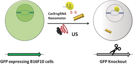
The publisher (Wiley) has made this image illustrating the work available,

Courtesy: Wiley

A February 9, 2018 Wiley Publications news release (also on EurekAlert), which originated the news item, provides more information,

Genomic engineering as a promising cancer therapeutic approach has experienced a tremendous surge since the discovery of the adaptive bacterial immune defense system “CRISPR” and its potential as a gene editing tool over a decade ago. Engineered CRISPR systems for gene editing now contain two main components, a single guide RNA or sgRNA and Cas-9 nuclease. While the sgRNA guides the nuclease to the specified gene sequence, Cas-9 nuclease performs its editing with surgical efficiency. However, the delivery of the large machinery to the target genome is still problematic. The authors of the Angewandte Chemie study, Liangfang Zhang and Joseph Wang from the University of California San Diego, and their colleagues now propose ultrasound-propelled gold nanowires as an active transport/release vehicle for the Cas9-sgRNA complex over the membrane.

Gold nanowires may cross a membrane passively, but thanks to their rod- or wirelike asymmetric shape, active motion can be triggered by ultrasound. “The asymmetric shape of the gold nanowire motor, given by the fabrication process, is essential for the acoustic propulsion,” the authors remarked. They assembled the vehicle by attaching the Cas-9 protein/RNA complex to the gold nanowire through sulfide bridges. These reduceable linkages have the advantage that inside the tumor cell, the bonds would be broken by glutathione, a natural reducing compound enriched in tumor cells. The Cas9-sgRNA would be released and sent to the nucleus to do its editing work, for, example, the knockout of a gene.

As a test system, the scientists monitored the suppression of fluorescence emitted by green fluorescence protein expressing melanoma B16F10 cells. Ultrasound was applied for five minutes, which accelerated the nanomotor carrying the Cas9-sgRNA complex across the membrane, accelerating it even inside the cell, as the authors noted. Moreover, they observed their Cas9-sgRNA complex effectively suppressing fluorescence with only tiny concentrations of the complex needed.

Thus, both the effective use of an acoustic nanomotor as an active transporter and the small payload needed for efficient gene knockout are intriguing results of the study. The simplicity of the system, which uses only few and readily available components, is another remarkable achievement.

Here’s a link to and a citation for the paper,

Active Intracellular Delivery of a Cas9/sgRNA Complex Using Ultrasound-Propelled Nanomotors by Malthe Hansen-Bruhn, Dr. Berta Esteban-Fernández de Ávila, Dr. Mara Beltrán-Gastélum, Prof. Jing Zhao, Dr. Doris E. Ramírez-Herrera, Pavimol Angsantikul, Prof. Kurt Vesterager Gothelf, Prof. Liangfang Zhang, and Prof. Joseph Wang. Angewandte Chemie International Edition Vol. 57 Issue 7 DOI: 10.1002/anie.201713082 Version of Record online: 6 FEB 2018

© 2018 Wiley-VCH Verlag GmbH & Co. KGaA, Weinheim

This paper is behind a paywall.

Café Scientifique Vancouver talk on January 30, 2018 and a couple of February 2018 art/sci events in Toronto

Vancouver

This could be a first for Café Scientifique Vancouver. From a January 28, 2018 Café Scientifique Vancouver announcement (received via email)

This is a reminder that our next café with biotech entrepreneur Dr.Andrew Tait (TUESDAY, JANUARY 30TH [2018] at 7:30PM) in the back room of YAGGER'S DOWNTOWN (433 W Pender).

COMBINING TRADITIONAL NATURAL MEDICINES WITH SCIENTIFIC RESEARCH: UNVEILING THE POTENTIAL OF THE MANDARIN ORANGE PEEL

The orange peel is something most of us may think of as a throw-away compost item, but it is so much more. Travel back in time 9,000 years to China, where orange peel was found in the first fermented alcoholic beverage, and return to today, where mandarin orange peel remains one of China’s top selling herbs that promotes digestion. Now meet Tait Laboratories Inc., a company that was founded based on one chemistry Ph.D. student’s idea, that mandarin orange peel has the potential to reverse incurable neurodegenerative diseases like multiple sclerosis. You will learn about the company’s journey through a scientific lens, from its early days to the present, having developed a mandarin orange peel product sold across Canada in over 1,000 stores including 400 Rexall pharmacies. You will leave with a basic understanding of how herbal products like the company’s mandarin orange peel-based product are developed and brought to market in Canada, and about the science that is required to substantiate health claims on this and other exciting new botanical products.

Bio:

Dr. Andrew Tait is the founder of Tait Laboratories Inc., a company devoted to developing natural medicines from agricultural bi-products. After a B.Sc. in Biochemistry and M.Sc. in Chemistry from Concordia University (Montreal), he completed a Ph.D. in Chemistry at the
University of British Columbia [UBC].

Inspired by his thesis work on multiple sclerosis, he subsequently identified Traditional Chinese Medicines as having potential to treat a wide range of chronic diseases; he founded the company while finishing his graduate studies.

In 2012, he was invited to Ottawa to be awarded the NSERC [{Canada} Natural Sciences and Engineering Research Council] Innovation Challenge Award, for successfully translating his Ph.D. research to an entrepreneurial venture. In 2014, he was awarded the BC Food Processors Association “Rising Star” award.

Dr. Tait is a regularly invited speaker on the topics of entrepreneurship and the science supporting natural health products; he was keynote speaker in 2012 at the Annual Symposium of the Boucher Institute of Naturopathic Medicine (Vancouver) and in 2016 at the
Functional Foods and Natural Health Products Graduate Research Symposium (Winnipeg).

Supported by the Futurpreneur Canada, the Bank of Development of Canada, the UBC’s Entrepreneurship@UBC program, and the NSERC  and NRC  [{Canada} National Research Council] Industry Research Assistance Program (IRAP), he works with industrial and academic researchers developing safe, affordable, and clinically proven medicines. He successfully launched MS+ Mandarin Skin PlusÒ, a patent-pending digestive product now on shelf in over 1000 pharmacies and health food stores across Canada, including 400 Rexall pharmacies.

Dr. Tait mentors young companies as an Entrepreneur in Residence at both SFU [Simon Fraser University] Coast Capital Savings Venture Connection and also the Health Tech Innovation Hub and he also volunteers his time to mentor students of the Student Biotechnology Network.

Lest it be forgotten, many drugs and therapeutic agents are based on natural remedies; a fact often ignored in the discussion about drugs and natural remedies. In any event, I am surprised this talk is being hosted by Café Scientifique Vancouver which has tended to more ‘traditional’ (i.e., university academic) presentations without any hint of ‘alternative’ or ‘entrepreneurial’ aspects. I wonder if this is the harbinger of new things to come from the Café Scientifique Vancouver community.

Meanwhile, interested parties can find out more about Tait Laboratories on their company website. They are selling one product at this time (from the MS+ [Mandarin Skin Plus] product webpage,

MS+™ (Mandarin Skin Plus) is a revolutionary natural health product that aids with digestion and promotes gastrointestinal health. It is a patent-pending proprietary extract based on dry-aged mandarin orange peel, an ancient Traditional Chinese Medicine. This remedy has been safely used for centuries to relieve bloating, indigestion, diarrhea, nausea, upset stomach, cough with phlegm. Experience ULTIMATE DIGESTIVE RELIEF and top gastrointestinal health for only about a dollar a day!

Directions: take one capsule twice a day, up to six capsules per day. Swallow capsule directly OR dissolve powder in water.
60 vegan capsules for ~ 1 month supply

I would have liked to have seen a list of research papers and discussion of human clinical trials regarding their ‘digestive’ product. Will Tait be discussing his research and results into what seems to be a new direction (i.e., the use of mandarin skin peel-derived therapeutics for neurodegenerative diseases)?

I don’t think I’m going to make it to the talk but should anyone who attends care to answer the question, please feel free to add a comment.

ArtSci Salon in Toronto

2018 is proving to be an active year for the ArtSci Salon folks in Toronto. They’ve just finished hosting a January 24-25, 2018 workshop and January 26, 2018 panel discussion on the gene-editing tool CRISPR/CAS9 (see my January 10, 2018 posting for a description).

Now they’ve announced another workshop and panel discussion on successive nights in February, the topic being: cells. From a January 29, 2018 ArtSci Salon announcement (received via email), Note: The panel discussion is listed first, then the workshop, then the artists’ biographies,

FROM CELL TO CANVAS: CREATIVE EXPLORATIONS OF THE MICROSCOPIC [panel discussion]

From the complex forms of the cell to the colonies created by the microbiota; from the undetectable chemical reactions activated by enzymes and natural processes to the environmental information captured through data visualization, the five local and international artists presenting tonight have developed a range of very diverse practices all inspired by the invisible, the undetectable and the microscopic.

We invite you to an evening of artist talks and discussion on the creative process of exploring the microscopic and using living organisms in art, on its potentials and implication for science and its popular dissemination, as well as on its ethics.

WITH:
Robyn Crouch
Mellissa Fisher
JULIA KROLIK
SHAVON MADDEN
TOSCA TERAN

FRIDAY, FEB 9, 2018
6:00-8:00 PM
THE FIELDS INSTITUTE
222 COLLEGE STREET,
RM 230

[Go to this page for access to registration]

FROM CELL TO CANVAS: CREATIVE EXPLORATIONS OF THE MICROSCOPIC [workshop]

THE EVENT WILL BE FOLLOWED BY A WORKSHOP BY: MELLISSA FISHER, SHAVON MADDEN AND JULIA KROLIK
FEB. 10, 2018
11:00AM-5:00PM
AT HACKLAB,
1266 Queen St West

[Go to this page for access to registration]

Workshop:

Design My Microbiome

Artist Mellissa Fisher invites participants to mould parts of her body in agar to create their own microbial version of her, alongside producing their own microbial portrait with painting techniques.

Cooking with the Invasive

Artist Shavon Madden invites participants to discuss invasive species like garlic mustard and cook invasive species whilst exploring, do species which we define and brand as invasive simply have no benefits?

Intoduction to Biological Staining

Artist & Scientist Julia Krolik invites participants to learn about 3 different types of biological staining and have a chance to try staining procedures.

BIOS:

ROBYN CROUCH
The symbolic imagery that comes through Robyn’s work invites one’s gaze inward to the cellular realms. There, one discovers playful depictions of chemical processes; the unseen lattice upon which our macro­cosmic world is constructed. Technological advancements create windows into this molecular realm, and human consciousness acts as the interface between the seen and the unseen worlds. In her functional ceramic work, the influence of Chinese and Japanese tea ceremony encourages contem­plation and appreciation of a quiet
moment. The viewer-participant can lose their train of thought while meandering through geometry and biota, con­nected by strands of double-helical DNA. A flash of recognition, a momentary mirror.

MELLISSA FISHER
Mellissa Fisher is a British Bio Artist based in Kent. Her practice explores the invisible world on our skin by using living organisms and by creating sculptures made with agar to show the public what the surface of our skin really looks like. She is best known for her work with bacteria and works extensively with collaborators in microbiology and immunology. She has exhibited an installation _ “Microbial Me”_with Professor Mark Clements and Dr Richard Harvey at The Eden Project for their permanent exhibition _“The Invisible You: The Human
Microbiome”._The installation included a living portrait in bacteria of the artists face as well as a time-lapse film of the sculpture growing.

JULIA KROLIK
Julia Krolik is a creative director, entrepreneur, scientist and award-winning artist. Her diverse background enables a rare cross-disciplinary empathy, and she continuously advocates for both art and science through several initiatives. Julia is the founder of Art the Science, a non-profit organization dedicated to facilitating artist residencies in scientific research laboratories to foster Canadian science-art culture and expand scientific knowledge communication to benefit the public. Through her consulting agency Pixels and Plans, Julia works with private and public organizations, helping them with strategy, data visualization and knowledge mobilization, often utilizing creative technology and skills-transfer workshops.

SHAVON MADDEN
Shavon Madden is a Brampton based artist, specializing in sculptural, performance and instillation based work exploring the social injustices inflicted on the environment and its creatures. Her work focuses on challenging social-environmental and political ethics, through the embodied experience and feelings of self. She graduated from the University of Toronto Specializing in Art and Art History, along with studies in Environmental Science and will be on her way to Edinburgh for her MFA. Shavon has had works shown at Shelly Peterson, the Burlington Art Gallery and the Art Gallery of Mississauga, among many others. Website: www.greenheartartistry.com [4]

TOSCA TERAN
Working with metal for over 30+ years, Tosca was introduced to glass as an artistic medium in 2004. Through developing bodies of work incorporating metal + glass Tosca has been awarded scholarships at The Corning Museum of Glass, Pilchuck Glass School and The Penland school of Crafts. Her work has been featured at SOFA New York, Culture Canada,
Metalsmith Magazine, The Toronto Design Exchange, and the Memphis Metal Museum. She has been awarded residencies at Gullkistan, Nes, and the Ayatana Research Program. A long-term guest artist instructor at the Ontario Science Centre, Tosca continues to explore materials, code, BioArt, SciArt and teach Metal + Glass courses out of her studio in Toronto.

It seems that these February events and the two events with Marta de Menezes are part of the FACTT (transdisciplinary and transnational festival of art and science) Toronto, from the FACTT Toronto webpage,

FACTT Toronto – Festival of Art & Science posted in: blog, events

The Arte Institute, in partnership with Cultivamos Cultura and ArtSi Salon, has the pleasure to announce FACTT – Festival of Art & Science in Toronto.

The Festival took place in Lisbon, New York, Mexico, Berlin and will continue in Toronto.
Exhibition: The Cabinet Project/ Art Sci Salon / FACTT

Artists:

Andrew Carnie
Elaine Whittaker
Erich Berger
Joana Ricou
Ken Rinaldo
Laura Beloff and Maria Antonia Gonzalez Valerio
Marta de Menezes and Luís Graça
Pedro Cruz

Dates: Jan 26- feb 15 [2018 {sic}]

Where: Meet us on Jan 26 [2018] in the Lobby of the Physics Department, 255 Huron Street
University of Toronto
When: 4:45 PM

You may want to keep an eye on the ArtSci Salon website although I find their posting schedule a bit erratic. Sometimes, I get email notices for events that aren’t yet listed on their website.
CRISPR-CAS9 and gold

As so often happens in the sciences, now that the initial euphoria has expended itself problems (and solutions) with CRISPR ((clustered regularly interspaced short palindromic repeats))-CAAS9 are being disclosed to those of us who are not experts. From an Oct. 3, 2017 article by Bob Yirka for phys.org,

A team of researchers from the University of California and the University of Tokyo has found a way to use the CRISPR gene editing technique that does not rely on a virus for delivery. In their paper published in the journal Nature Biomedical Engineering, the group describes the new technique, how well it works and improvements that need to be made to make it a viable gene editing tool.

CRISPR-Cas9 has been in the news a lot lately because it allows researchers to directly edit genes—either disabling unwanted parts or replacing them altogether. But despite many success stories, the technique still suffers from a major deficit that prevents it from being used as a true medical tool—it sometimes makes mistakes. Those mistakes can cause small or big problems for a host depending on what goes wrong. Prior research has suggested that the majority of mistakes are due to delivery problems, which means that a replacement for the virus part of the technique is required. In this new effort, the researchers report that they have discovered just a such a replacement, and it worked so well that it was able to repair a gene mutation in a Duchenne muscular dystrophy mouse model. The team has named the new technique CRISPR-Gold, because a gold nanoparticle was used to deliver the gene editing molecules instead of a virus.

An Oct. 2, 2017 article by Abby Olena for The Scientist lays out the CRISPR-CAS9 problems the scientists are trying to solve (Note: Links have been removed),

While promising, applications of CRISPR-Cas9 gene editing have so far been limited by the challenges of delivery—namely, how to get all the CRISPR parts to every cell that needs them. In a study published today (October 2) in Nature Biomedical Engineering, researchers have successfully repaired a mutation in the gene for dystrophin in a mouse model of Duchenne muscular dystrophy by injecting a vehicle they call CRISPR-Gold, which contains the Cas9 protein, guide RNA, and donor DNA, all wrapped around a tiny gold ball.

The authors have made “great progress in the gene editing area,” says Tufts University biomedical engineer Qiaobing Xu, who did not participate in the work but penned an accompanying commentary. Because their approach is nonviral, Xu explains, it will minimize the potential off-target effects that result from constant Cas9 activity, which occurs when users deliver the Cas9 template with a viral vector.

Duchenne muscular dystrophy is a degenerative disease of the muscles caused by a lack of the protein dystrophin. In about a third of patients, the gene for dystrophin has small deletions or single base mutations that render it nonfunctional, which makes this gene an excellent candidate for gene editing. Researchers have previously used viral delivery of CRISPR-Cas9 components to delete the mutated exon and achieve clinical improvements in mouse models of the disease.

“In this paper, we were actually able to correct [the gene for] dystrophin back to the wild-type sequence” via homology-directed repair (HDR), coauthor Niren Murthy, a drug delivery researcher at the University of California, Berkeley, tells The Scientist. “The other way of treating this is to do something called exon skipping, which is where you delete some of the exons and you can get dystrophin to be produced, but it’s not [as functional as] the wild-type protein.”

The research team created CRISPR-Gold by covering a central gold nanoparticle with DNA that they modified so it would stick to the particle. This gold-conjugated DNA bound the donor DNA needed for HDR, which the Cas9 protein and guide RNA bound to in turn. They coated the entire complex with a polymer that seems to trigger endocytosis and then facilitate escape of the Cas9 protein, guide RNA, and template DNA from endosomes within cells.

In order to do HDR, “you have to provide the cell [with] the Cas9 enzyme, guide RNA by which you target Cas9 to a particular part of the genome, and a big chunk of DNA, which will be used as a template to edit the mutant sequence to wild-type,” explains coauthor Irina Conboy, who studies tissue repair at the University of California, Berkeley. “They all have to be present at the same time and at the same place, so in our system you have a nanoparticle which simultaneously delivers all of those three key components in their active state.”

Olena’s article carries on to describe how the team created CRISPR-Gold and more.

Additional technical details are available in an Oct. 3, 2017 University of California at Berkeley news release by Brett Israel (also on EurekAlert), which originated the news item (Note: A link has been removed) ,

Scientists at the University of California, Berkeley, have engineered a new way to deliver CRISPR-Cas9 gene-editing technology inside cells and have demonstrated in mice that the technology can repair the mutation that causes Duchenne muscular dystrophy, a severe muscle-wasting disease. A new study shows that a single injection of CRISPR-Gold, as the new delivery system is called, into mice with Duchenne muscular dystrophy led to an 18-times-higher correction rate and a two-fold increase in a strength and agility test compared to control groups.

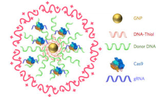
Diagram of CRISPR-Gold

CRISPR–Gold is composed of 15 nanometer gold nanoparticles that are conjugated to thiol-modified oligonucleotides (DNA-Thiol), which are hybridized with single-stranded donor DNA and subsequently complexed with Cas9 and encapsulated by a polymer that disrupts the endosome of the cell.

Since 2012, when study co-author Jennifer Doudna, a professor of molecular and cell biology and of chemistry at UC Berkeley, and colleague Emmanuelle Charpentier, of the Max Planck Institute for Infection Biology, repurposed the Cas9 protein to create a cheap, precise and easy-to-use gene editor, researchers have hoped that therapies based on CRISPR-Cas9 would one day revolutionize the treatment of genetic diseases. Yet developing treatments for genetic diseases remains a big challenge in medicine. This is because most genetic diseases can be cured only if the disease-causing gene mutation is corrected back to the normal sequence, and this is impossible to do with conventional therapeutics.

CRISPR/Cas9, however, can correct gene mutations by cutting the mutated DNA and triggering homology-directed DNA repair. However, strategies for safely delivering the necessary components (Cas9, guide RNA that directs Cas9 to a specific gene, and donor DNA) into cells need to be developed before the potential of CRISPR-Cas9-based therapeutics can be realized. A common technique to deliver CRISPR-Cas9 into cells employs viruses, but that technique has a number of complications. CRISPR-Gold does not need viruses.

In the new study, research lead by the laboratories of Berkeley bioengineering professors Niren Murthy and Irina Conboy demonstrated that their novel approach, called CRISPR-Gold because gold nanoparticles are a key component, can deliver Cas9 – the protein that binds and cuts DNA – along with guide RNA and donor DNA into the cells of a living organism to fix a gene mutation.

“CRISPR-Gold is the first example of a delivery vehicle that can deliver all of the CRISPR components needed to correct gene mutations, without the use of viruses,” Murthy said.

The study was published October 2 [2017] in the journal Nature Biomedical Engineering.

CRISPR-Gold repairs DNA mutations through a process called homology-directed repair. Scientists have struggled to develop homology-directed repair-based therapeutics because they require activity at the same place and time as Cas9 protein, an RNA guide that recognizes the mutation and donor DNA to correct the mutation.

To overcome these challenges, the Berkeley scientists invented a delivery vessel that binds all of these components together, and then releases them when the vessel is inside a wide variety of cell types, triggering homology directed repair. CRISPR-Gold’s gold nanoparticles coat the donor DNA and also bind Cas9. When injected into mice, their cells recognize a marker in CRISPR-Gold and then import the delivery vessel. Then, through a series of cellular mechanisms, CRISPR-Gold is released into the cells’ cytoplasm and breaks apart, rapidly releasing Cas9 and donor DNA.

Schematic of CRISPR-Gold's method of action

CRISPR-Gold’s method of action (Click to enlarge).

A single injection of CRISPR-Gold into muscle tissue of mice that model Duchenne muscular dystrophy restored 5.4 percent of the dystrophin gene, which causes the disease, to the wild- type, or normal, sequence. This correction rate was approximately 18 times higher than in mice treated with Cas9 and donor DNA by themselves, which experienced only a 0.3 percent correction rate.

Importantly, the study authors note, CRISPR-Gold faithfully restored the normal sequence of dystrophin, which is a significant improvement over previously published approaches that only removed the faulty part of the gene, making it shorter and converting one disease into another, milder disease.

CRISPR-Gold was also able to reduce tissue fibrosis – the hallmark of diseases where muscles do not function properly – and enhanced strength and agility in mice with Duchenne muscular dystrophy. CRISPR-Gold-treated mice showed a two-fold increase in hanging time in a common test for mouse strength and agility, compared to mice injected with a control.

“These experiments suggest that it will be possible to develop non-viral CRISPR therapeutics that can safely correct gene mutations, via the process of homology-directed repair, by simply developing nanoparticles that can simultaneously encapsulate all of the CRISPR components,” Murthy said.

CRISPR-Cas9

CRISPR in action: A model of the Cas9 protein cutting a double-stranded piece of DNA

The study found that CRISPR-Gold’s approach to Cas9 protein delivery is safer than viral delivery of CRISPR, which, in addition to toxicity, amplifies the side effects of Cas9 through continuous expression of this DNA-cutting enzyme. When the research team tested CRISPR-Gold’s gene-editing capability in mice, they found that CRISPR-Gold efficiently corrected the DNA mutation that causes Duchenne muscular dystrophy, with minimal collateral DNA damage.

The researchers quantified CRISPR-Gold’s off-target DNA damage and found damage levels similar to the that of a typical DNA sequencing error in a typical cell that was not exposed to CRISPR (0.005 – 0.2 percent). To test for possible immunogenicity, the blood stream cytokine profiles of mice were analyzed at 24 hours and two weeks after the CRISPR-Gold injection. CRISPR-Gold did not cause an acute up-regulation of inflammatory cytokines in plasma, after multiple injections, or weight loss, suggesting that CRISPR-Gold can be used multiple times safely, and that it has a high therapeutic window for gene editing in muscle tissue.

“CRISPR-Gold and, more broadly, CRISPR-nanoparticles open a new way for safer, accurately controlled delivery of gene-editing tools,” Conboy said. “Ultimately, these techniques could be developed into a new medicine for Duchenne muscular dystrophy and a number of other genetic diseases.”

A clinical trial will be needed to discern whether CRISPR-Gold is an effective treatment for genetic diseases in humans. Study co-authors Kunwoo Lee and Hyo Min Park have formed a start-up company, GenEdit (Murthy has an ownership stake in GenEdit), which is focused on translating the CRISPR-Gold technology into humans. The labs of Murthy and Conboy are also working on the next generation of particles that can deliver CRISPR into tissues from the blood stream and would preferentially target adult stem cells, which are considered the best targets for gene correction because stem and progenitor cells are capable of gene editing, self-renewal and differentiation.

“Genetic diseases cause devastating levels of mortality and morbidity, and new strategies for treating them are greatly needed,” Murthy said. “CRISPR-Gold was able to correct disease-causing gene mutations in vivo, via the non-viral delivery of Cas9 protein, guide RNA and donor DNA, and therefore has the potential to develop into a therapeutic for treating genetic diseases.”

The study was funded by the National Institutes of Health, the W.M. Keck Foundation, the Moore Foundation, the Li Ka Shing Foundation, Calico, Packer, Roger’s and SENS, and the Center of Innovation (COI) Program of the Japan Science and Technology Agency.

Here’s a link to and a citation for the paper,

Nanoparticle delivery of Cas9 ribonucleoprotein and donor DNA in vivo induces homology-directed DNA repair by Kunwoo Lee, Michael Conboy, Hyo Min Park, Fuguo Jiang, Hyun Jin Kim, Mark A. Dewitt, Vanessa A. Mackley, Kevin Chang, Anirudh Rao, Colin Skinner, Tamanna Shobha, Melod Mehdipour, Hui Liu, Wen-chin Huang, Freeman Lan, Nicolas L. Bray, Song Li, Jacob E. Corn, Kazunori Kataoka, Jennifer A. Doudna, Irina Conboy, & Niren Murthy. Nature Biomedical Engineering (2017) doi:10.1038/s41551-017-0137-2 Published online: 02 October 2017

This paper is behind a paywall.

Using CRISPR to reverse retinosa pigmentosa (eye disease)

Years ago I worked as a publicist for the BC (British Columbia) Motorcycle Federation’s Ride for Sight; they were raising funds for research into retinitis pigmentosa (RP). I hadn’t thought about that in years but it all came back when I saw this April 21, 2017 news item on ScienceDaily,

Using the gene-editing tool CRISPR/Cas9, researchers at University of California San Diego [UCSD] School of Medicine and Shiley Eye Institute at UC San Diego Health, with colleagues in China, have reprogrammed mutated rod photoreceptors to become functioning cone photoreceptors, reversing cellular degeneration and restoring visual function in two mouse models of retinitis pigmentosa.

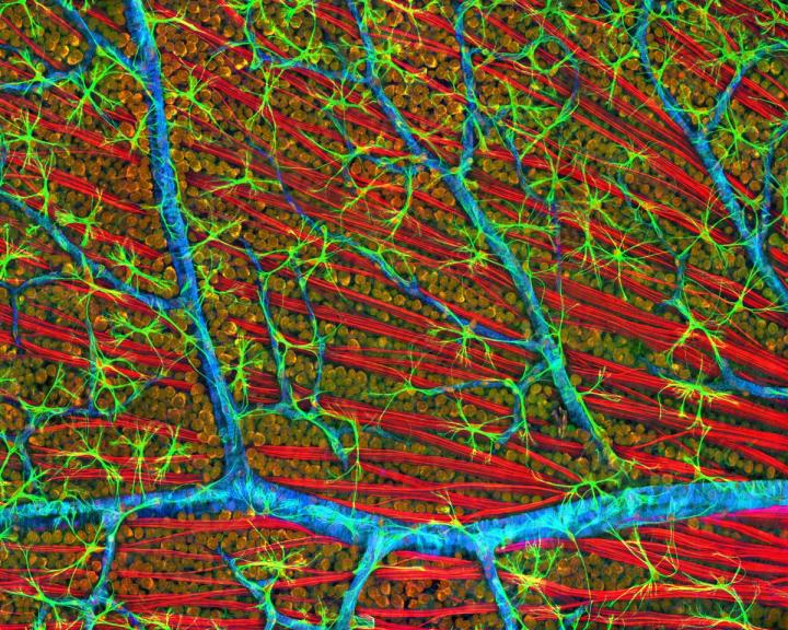
Caption: This is a confocal micrograph of mouse retina depicting optic fiber layer. Credit: Image courtesy of National Center for Microscopy and Imaging Research, UC San Diego.

An April 21, 2017 UCSD news release by Scott LaFee (also on EurekAlert), which originated the news item, delves further into retinitis pigmentosa and this CRISPR research,

Retinitis pigmentosa (RP) is a group of inherited vision disorders caused by numerous mutations in more than 60 genes. The mutations affect the eyes’ photoreceptors, specialized cells in the retina that sense and convert light images into electrical signals sent to the brain. There are two types: rod cells that function for night vision and peripheral vision, and cone cells that provide central vision (visual acuity) and discern color. The human retina typically contains 120 million rod cells and 6 million cone cells.

In RP, which affects approximately 100,000 Americans and 1 in 4,000 persons worldwide, rod-specific genetic mutations cause rod photoreceptor cells to dysfunction and degenerate over time. Initial symptoms are loss of peripheral and night vision, followed by diminished visual acuity and color perception as cone cells also begin to fail and die. There is no treatment for RP. The eventual result may be legal blindness.

In their published research, a team led by senior author Kang Zhang, MD, PhD, chief of ophthalmic genetics, founding director of the Institute for Genomic Medicine and co-director of biomaterials and tissue engineering at the Institute of Engineering in Medicine, both at UC San Diego School of Medicine, used CRISPR/Cas9 to deactivate a master switch gene called Nrl and a downstream transcription factor called Nr2e3.

CRISPR, which stands for Clustered Regularly Interspaced Short Palindromic Repeats, allows researchers to target specific stretches of genetic code and edit DNA at precise locations, modifying select gene functions. Deactivating either Nrl or Nr2e3 reprogrammed rod cells to become cone cells.

“Cone cells are less vulnerable to the genetic mutations that cause RP,” said Zhang. “Our strategy was to use gene therapy to make the underlying mutations irrelevant, resulting in the preservation of tissue and vision.”

The scientists tested their approach in two different mouse models of RP. In both cases, they found an abundance of reprogrammed cone cells and preserved cellular architecture in the retinas. Electroretinography testing of rod and cone receptors in live mice show improved function.

Zhang said a recent independent study led by Zhijian Wu, PhD, at National Eye Institute, part of the National Institutes of Health, also reached similar conclusions.

The researchers used adeno-associated virus (AAV) to perform the gene therapy, which they said should help advance their work to human clinical trials quicker. “AAV is a common cold virus and has been used in many successful gene therapy treatments with a relatively good safely profile,” said Zhang. “Human clinical trials could be planned soon after completion of preclinical study. There is no treatment for RP so the need is great and pressing. In addition, our approach of reprogramming mutation-sensitive cells to mutation-resistant cells may have broader application to other human diseases, including cancer.”

Here’s a link to and a citation for the paper,

Gene and mutation independent therapy via CRISPR-Cas9 mediated cellular reprogramming in rod photoreceptors by Jie Zhu, Chang Ming, Xin Fu, Yaou Duan, Duc Anh Hoang, Jeffrey Rutgard, Runze Zhang, Wenqiu Wang, Rui Hou, Daniel Zhang, Edward Zhang, Charlotte Zhang, Xiaoke Hao, Wenjun Xiong, and Kang Zhang. Cell Research advance online publication 21 April 2017; doi: 10.1038/cr.2017.57

This paper (it’s in the form of a letter to the editor) is open access.