Tag Archives: gold Nanoparticles (AU NPs)

“Injectable tissue prosthesis coupled with closed-loop bioelectronic system” for damaged muscle/nerve regeneration and robot-assisted rehabilitation

A fascinating new use for hyaluronic acid (usually discussed in relation to cosmetic wrinkle-reduction) has been found according to a November 1, 2023 news item on ScienceDaily.

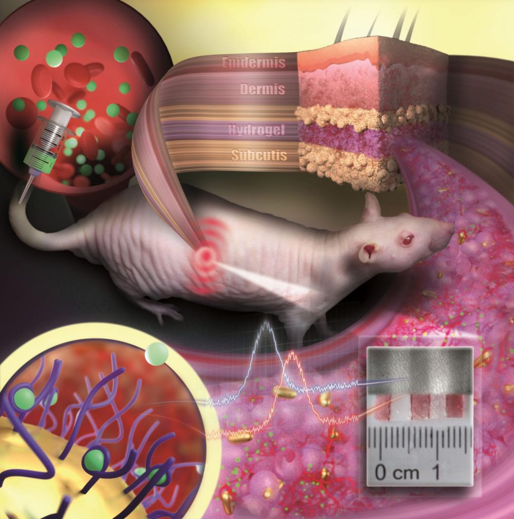
In a recent publication in the journal Nature, researchers from the Institute of Basic Science (IBS) in South Korea have made significant strides in biomaterial technology and rehabilitation medicine. They’ve developed a novel approach to healing muscle injury by employing “injectable tissue prosthesis” in the form of conductive hydrogels and combining it with a robot-assisted rehabilitation system.

Let’s imagine you are swimming in the ocean. A giant shark approaches and bites a huge chunk of meat out of your thigh, resulting in a complete loss of motor/sensor function in your leg.

If left untreated, such severe muscle damage would result in permanent loss of function and disability.

How on Earth will you be able to recover from this kind of injury?

Traditional rehabilitation methods for these kinds of muscle injuries have long sought an efficient closed-loop gait rehabilitation system that merges lightweight exoskeletons and wearable/implantable devices.

Such assistive prosthetic system is required to aid the patients through the process of recovering sensory and motor functions linked to nerve and muscle damage.

Unfortunately, the mechanical properties and rigid nature of existing electronic materials render them incompatible with soft tissues.

This leads to friction and potential inflammation, stalling patient rehabilitation.

To overcome these limitations, the IBS researchers turned to a material commonly used as a wrinkle-smoothing filler, called hyaluronic acid.

A November 2, 2023 Institute of Basic Science (IBS) press release (also on EurekAlert but published November 1, 2023), which originated the news item, explains how hyaluronic acid helps in tissue rehabilitation and regeneration,

Using this substance [hyaluronic acid], an injectable hydrogel was developed for “tissue prostheses”, which can temporarily fill the gap of the missing muscle/nerve tissues while it regenerates. The injectable nature of this material gives it a significant advantage over traditional bioelectronic devices, which are unsuitable for narrow, deep, or small areas, and necessitate invasive surgeries.

Thanks to its highly “tissue-like” properties, this hydrogel seamlessly interfaces with biological tissues and can be easily administered to hard-to-reach body areas without surgery. The reversible and irreversible crosslinks within the hydrogel adapt to high shear stress during injection, ensuring excellent mechanical stability. This hydrogel also incorporates gold nanoparticles, which gives it decent electrical properties. Its conductive nature allows for the effective transmission of electrophysiological signals between the two ends of injured tissues. In addition, the hydrogel is biodegrdable, meaning that the patients do not need to get surgery again.

With mechanical properties akin to natural tissues, exceptional tissue adhesion, and injectable characteristics, researchers believe this material offers a novel approach to rehabilitation.

Next, the researchers put this novel idea to the test in rodent models. To simulate volumetric muscle loss injury, a large chunk of muscle has been removed from the hind legs of these animals. By injecting the hydrogel and implanting the two kinds of stretchable tissue-interfacing devices for electrical sensing and stimulation, the researchers were able to improve the gait in the “injured” rodents. The hydrogel prosthetics were combined with robot assistance, guided by muscle electromyography signals. Together, the two helped enhance the animal’s gait without nerve stimulation. Furthermore, muscle tissue regeneration was effectively improved over the long term after the conductive hydrogel was used to fill muscle damage.

The injectable conductive hydrogel developed in this study excels in electrophysiological signal recording and stimulation performance, offering the potential to expand its applications. It presents a fresh approach to the field of bioelectronic devices and holds promise as a soft tissue prosthesis for rehabilitation support.

Emphasizing the significance of the research, Professor SHIN Mikyung notes, “We’ve created an injectable, mechanically tough, and electrically conductive soft tissue prosthesis ideal for addressing severe muscle damage requiring neuromusculoskeletal rehabilitation. The development of this injectable hydrogel, utilizing a novel cross-linking method, is a notable achievement. We believe it will be applicable not only in muscles and peripheral nerves but also in various organs like the brain and heart.”

Professor SON Donghee added, “In this study, the closed-loop gait rehabilitation system entailing tough injectable hydrogel and stretchable and self-healing sensors could significantly enhance the rehabilitation prospects for patients with neurological and musculoskeletal challenges. It could also play a vital role in precise diagnosis and treatment across various organs in the human body.”

The research team is currently pursuing further studies to develop new materials for nerve and muscle tissue regeneration that can be implanted in a minimally invasive manner. They are also exploring the potential for recovery in various tissue damages through the injection of the conductive hydrogel, eliminating the need for open surgery.

Here’s a link to and a citation for the paper,

Injectable tissue prosthesis for instantaneous closed-loop rehabilitation by Subin Jin, Heewon Choi, Duhwan Seong, Chang-Lim You, Jong-Sun Kang, Seunghyok Rho, Won Bo Lee, Donghee Son & Mikyung Shin. Nature volume 623, pages 58–65 (2023) DOI: https://doi.org/10.1038/s41586-023-06628-x Published: 01 November 2023 Issue Date: 02 November 2023

This paper is behind a paywall.

A roly-poly (woodlouse) gold rush

This environmental monitoring story focused on the roly-poly was announced in an April 18, 2023 news item on Statnano,

The woodlouse goes by many names: roly-poly, pill bug, potato bug, tomato bug, butchy boy, cheesy bob, and chiggy pig, to name just a few. It is best known for contracting into a ball when agitated. This crustacean (yes, it’s a crustacean, not an insect) thrives in heavily metal-contaminated areas due to its specialized digestive organ, called a hepatopancreas, that stores and expels unwanted metals.

Metal nanoparticles are common in industrial and research plants. However, they can leach into the surrounding environment. Currently, little is known about the toxicity of metal nanoparticles for nearby organisms because detecting metal nanoparticles, particularly gold, requires microscopic, 3D imaging that cannot be done in the field

….

Caption: (a) Cartoon of a woodlouse depicting the hepatopancreas (HP) and the hind gut (HG). (b) Transmission overview of a single HP tubule, showing the helical structure. (c) Section from a HP tubule with the nuclei fluorescently labeled in blue. Credit: Iestyn Pope, Nuno G.C. Ferreira, Peter Kille, Wolfgang Langbein, and Paola Borri

An April 11, 2023 American Institute of Physics (AIP) news release (also on EurekAlert), which originated the news item, describes a new approach to detecting gold nanoparticles in roly-polys,

In Applied Physics Letters, by AIP Publishing, researchers from Cardiff University in the U.K. introduce a novel imaging method to detect gold nanoparticles in woodlice. With information about the quantity, location, and impact of gold nanoparticles within the organism, scientists can better understand the potential harm other metals may have on nature.

“Gold nanoparticles are used extensively for biological research applications owing to their biocompatibility and photostability and are available in a large range of shapes and sizes,” said author Wolfgang Langbein. “By using gold nanoparticles, which would not normally be present in the woodlice diet, we can study the journey of nanoparticles inside complex biological systems.”

The researchers developed an imaging method known as four-wave mixing microscopy, which flashes light that the gold nanoparticles absorb. The light flashes again and the subsequent scattering reveals the nanoparticles’ locations. Using this state-of-the-art technique, they locate the individual gold nanoparticles in the 3D cellular environment.

“By precisely pinpointing the fate of individual gold nanoparticles in the hepatopancreas of woodlice, we can gain a better understanding of how these organisms sequester and respond to metals ingested from the environment,” said Langbein. “Tracking this metal within these organisms is the first step enabling further study to determine, for example, if gold is collected within specific cells, or if it can interfere with the metabolisms in high doses.”

The use of gold nanoparticles in medical devices is increasing and with it, their abundance in the environment. This imaging technique will provide clarity into the little-understood mechanisms in the woodlice hepatopancreas and will also provide helpful environmental monitoring.

In the future, background-free four-wave mixing microscopy could be used to detect other metal nanoparticles and may be applied to organisms like fish larvae and even human cell cultures.

Here’s a link to and a citation for the paper,

Background-free four-wave mixing microscopy of small gold nanoparticles inside a multi-cellular organ by Iestyn Pope, Nuno G.C. Ferreira, Peter Kille, Wolfgang Langbein, and Paola Borri. Appl. Phys. Lett. 122, 153701 (2023) DOI: https://doi.org/10.1063/5.0140651Published online April 11, 2023

This paper is open access.

Gold nanoparticle tattoo changes medical diagnostics?

The tattoos are in fact implantable sensors. Here’s more from an April 6, 2021 news item on ScienceDaily,

The idea of implantable sensors that continuously transmit information on vital values and concentrations of substances or drugs in the body has fascinated physicians and scientists for a long time. Such sensors enable the constant monitoring of disease progression and therapeutic success. However, until now implantable sensors have not been suitable to remain in the body permanently but had to be replaced after a few days or weeks. On the one hand, there is the problem of implant rejection because the body recognizes the sensor as a foreign object. On the other hand, the sensor’s color which indicates concentration changes has been unstable so far and faded over time. Scientists at Johannes Gutenberg University Mainz (JGU) have developed a novel type of implantable sensor which can be operated in the body for several months. The sensor is based on color-stable gold nanoparticles that are modified with receptors for specific molecules. Embedded into an artificial polymeric tissue, the nanogold is implanted under the skin where it reports changes in drug concentrations by changing its color.

An April 6, 2021 Johannes Gutenberg Universitaet Mainz press release (also on EurekAlert), which originated the news item, provides more detail about the proposed tattoo/implantable sensors,

Implant reports information as an “invisible tattoo”

Professor Carsten Sönnichsen’s research group at JGU has been using gold nanoparticles as sensors to detect tiny amounts of proteins in microscopic flow cells for many years. Gold nanoparticles act as small antennas for light: They strongly absorb and scatter it and, therefore, appear colorful. They react to alterations in their surrounding by changing color. Sönnichsen’s team has exploited this concept for implanted medical sensing.

To prevent the tiny particles from swimming away or being degraded by immune cells, they are embedded in a porous hydrogel with a tissue-like consistency. Once implanted under the skin, small blood vessels and cells grow into the pores. The sensor is integrated in the tissue and is not rejected as a foreign body. “Our sensor is like an invisible tattoo, not much bigger than a penny and thinner than one millimeter,” said Professor Carsten Sönnichsen, head of the Nanobiotechnology Group at JGU. Since the gold nanoparticles are infrared, they are not visible to the eye. However, a special kind of measurement device can detect their color noninvasively through the skin.

In their study published in Nano Letters, the JGU researchers implanted their gold nanoparticle sensors under the skin of hairless rats. Color changes in these sensors were monitored following the administration of various doses of an antibiotic. The drug molecules are transported to the sensor via the bloodstream. By binding to specific receptors on the surface of the gold nanoparticles, they induce color change that is dependent on drug concentration. Thanks to the color-stable gold nanoparticles and the tissue-integrating hydrogel, the sensor was found to remain mechanically and optically stable over several months.

Huge potential of gold nanoparticles as long-lasting implantable medical sensors

“We are used to colored objects bleaching over time. Gold nanoparticles, however, do not bleach but keep their color permanently. As they can be easily coated with various different receptors, they are an ideal platform for implantable sensors,” explained Dr. Katharina Kaefer, first author of the study.

The novel concept is generalizable and has the potential to extend the lifetime of implantable sensors. In future, gold nanoparticle-based implantable sensors could be used to observe concentrations of different biomarkers or drugs in the body simultaneously. Such sensors could find application in drug development, medical research, or personalized medicine, such as the management of chronic diseases.

Interdisciplinary team work brought success

Sönnichsen had the idea of using gold nanoparticles as implanted sensors already in 2004 when he started his research in biophysical chemistry as a junior professor in Mainz. However, the project was not realized until ten years later in cooperation with Dr. Thies Schroeder and Dr. Katharina Kaefer, both scientists at JGU. Schroeder was experienced in biological research and laboratory animal science and had already completed several years of research work in the USA. Kaefer was looking for an exciting topic for her doctorate and was particularly interested in the complex and interdisciplinary nature of the project. Initial results led to a stipend awarded to Kaefer by the Max Planck Graduate Center (MPGC) as well as financial support from Stiftung Rheinland-Pfalz für Innovation. “Such a project requires many people with different scientific backgrounds. Step by step we were able to convince more and more people of our idea,” said Sönnichsen happily. Ultimately, it was interdisciplinary teamwork that resulted in the successful development of the first functional implanted sensor with gold nanoparticles.

The researchers have provided an image which illustrates several elements described in the press release,

Caption: Gold nanoparticles embedded in a porous hydrogel can be implanted under the skin and used as medical sensors. The sensor is like an invisible tattoo revealing concentration changes of substances in the blood by color change. Credit: ill./©: Nanobiotechnology Group, JGU Department of Chemistry

Here’s a link to and a citation for the paper,

Implantable Sensors Based on Gold Nanoparticles for Continuous Long-Term Concentration Monitoring in the Body by Katharina Kaefer, Katja Krüger, Felix Schlapp, Hüseyin Uzun, Sirin Celiksoy, Bastian Flietel, Axel Heimann, Thies Schroeder, Oliver Kempski, and Carsten Sönnichsen. Nano Lett. 2021, XXXX, XXX, XXX-XXX DOI: https://doi.org/10.1021/acs.nanolett.1c00887 Publication Date:March 30, 2021 © 2021 The Authors. Published by American Chemical Society

This paper is behind a paywall.

Detecting COVID-19 in under five minutes with paper-based sensor made of graphene

A Dec. 7, 2020 news item on Nanowerk announced a new technology for rapid COVID-19 testing (Note: A link has been removed),

As the COVID-19 pandemic continues to spread across the world, testing remains a key strategy for tracking and containing the virus. Bioengineering graduate student, Maha Alafeef, has co-developed a rapid, ultrasensitive test using a paper-based electrochemical sensor that can detect the presence of the virus in less than five minutes.

The team led by professor Dipanjan Pan reported their findings in ACS Nano (“Rapid, Ultrasensitive, and Quantitative Detection of SARS-CoV-2 Using Antisense Oligonucleotides Directed Electrochemical Biosensor Chip”).

“Currently, we are experiencing a once-in-a-century life-changing event,” said Alafeef. “We are responding to this global need from a holistic approach by developing multidisciplinary tools for early detection and diagnosis and treatment for SARS-CoV-2.”

I wonder why they didn’t think to provide a caption for the graphene substrate (the square surface) underlying the gold electrode (the round thing) or provide a caption for the electrode. Maybe they assumed anyone knowledgeable about graphene would be able to identify it?

Caption: COVID-19 electrochemical sensing platform. Credit: University of Illinois

A Dec. 7, 2020 University of Illinois Grainger College of Engineering news release (also on EurekAlert) by Huan Song, which originated the news item, provides more technical detail including a description of the graphene substrate and the gold electrode, which make up the cheaper, faster COVID-19 sensing platform,

There are two broad categories of COVID-19 tests on the market. The first category uses reverse transcriptase real-time polymerase chain reaction (RT-PCR) and nucleic acid hybridization strategies to identify viral RNA. Current FDA [US Food and Drug Administration]-approved diagnostic tests use this technique. Some drawbacks include the amount of time it takes to complete the test, the need for specialized personnel and the availability of equipment and reagents.

The second category of tests focuses on the detection of antibodies. However, there could be a delay of a few days to a few weeks after a person has been exposed to the virus for them to produce detectable antibodies.

In recent years, researchers have had some success with creating point-of-care biosensors using 2D nanomaterials such as graphene to detect diseases. The main advantages of graphene-based biosensors are their sensitivity, low cost of production and rapid detection turnaround. “The discovery of graphene opened up a new era of sensor development due to its properties. Graphene exhibits unique mechanical and electrochemical properties that make it ideal for the development of sensitive electrochemical sensors,” said Alafeef. The team created a graphene-based electrochemical biosensor with an electrical read-out setup to selectively detect the presence of SARS-CoV-2 genetic material.

There are two components [emphasis mine] to this biosensor: a platform to measure an electrical read-out and probes to detect the presence of viral RNA. To create the platform, researchers first coated filter paper with a layer of graphene nanoplatelets to create a conductive film [emphasis mine]. Then, they placed a gold electrode with a predefined design on top of the graphene [emphasis mine] as a contact pad for electrical readout. Both gold and graphene have high sensitivity and conductivity which makes this platform ultrasensitive to detect changes in electrical signals.

Current RNA-based COVID-19 tests screen for the presence of the N-gene (nucleocapsid phosphoprotein) on the SARS-CoV-2 virus. In this research, the team designed antisense oligonucleotide (ASOs) probes to target two regions of the N-gene. Targeting two regions ensures the reliability of the senor in case one region undergoes gene mutation. Furthermore, gold nanoparticles (AuNP) are capped with these single-stranded nucleic acids (ssDNA), which represents an ultra-sensitive sensing probe for the SARS-CoV-2 RNA.

The researchers previously showed the sensitivity of the developed sensing probes in their earlier work published in ACS Nano. The hybridization of the viral RNA with these probes causes a change in the sensor electrical response. The AuNP caps accelerate the electron transfer and when broadcasted over the sensing platform, results in an increase in the output signal and indicates the presence of the virus.

The team tested the performance of this sensor by using COVID-19 positive and negative samples. The sensor showed a significant increase in the voltage of positive samples compared to the negative ones and confirmed the presence of viral genetic material in less than five minutes. Furthermore, the sensor was able to differentiate viral RNA loads in these samples. Viral load is an important quantitative indicator of the progress of infection and a challenge to measure using existing diagnostic methods.

This platform has far-reaching applications due to its portability and low cost. The sensor, when integrated with microcontrollers and LED screens or with a smartphone via Bluetooth or wifi, could be used at the point-of-care in a doctor’s office or even at home. Beyond COVID-19, the research team also foresees the system to be adaptable for the detection of many different diseases.

“The unlimited potential of bioengineering has always sparked my utmost interest with its innovative translational applications,” Alafeef said. “I am happy to see my research project has an impact on solving a real-world problem. Finally, I would like to thank my Ph.D. advisor professor Dipanjan Pan for his endless support, research scientist Dr. Parikshit Moitra, and research assistant Ketan Dighe for their help and contribution toward the success of this study.”

Here’s a link to and a citation for the paper,

Rapid, Ultrasensitive, and Quantitative Detection of SARS-CoV-2 Using Antisense Oligonucleotides Directed Electrochemical Biosensor Chip by Maha Alafeef, Ketan Dighe, Parikshit Moitra, and Dipanjan Pan. ACS Nano 2020, 14, 12, 17028–17045 DOI: https://doi.org/10.1021/acsnano.0c06392 Publication Date:October 20, 2020 Copyright © 2020 American Chemical Society

I’m not sure where I found this notice but it is most definitely from the American Chemical Society: “This paper is freely accessible, at this time, for unrestricted RESEARCH re-use and analyses in any form or by any means with acknowledgement of the original source. These permissions are granted for the duration of the World Health Organization (WHO) declaration of COVID-19 as a global pandemic.”

Climate change and black gold

A July 3, 2019 news item on Nanowerk describes research coming from India and South Korea where nano gold is turned into black nanogold (Note: A link has been removed),

One of the main cause of global warming is the increase in the atmospheric CO2 level. The main source of this CO2 is from the burning of fossil fuels (electricity, vehicles, industry and many more).

Researchers at TIFR [Tata Institute of Fundamental Research] have developed the solution phase synthesis of Dendritic Plasmonic Colloidosomes (DPCs) with varying interparticle distances between the gold Nanoparticles (AU NPs) using a cycle-by-cycle growth approach by optimizing the nucleation-growth step. These DPCs absorb the entire visible and near-infrared region of solar light, due to interparticle plasmonic coupling as well as the heterogeneity in the Au NP [gold nanoparticle] sizes, which transformed golden gold material to black gold (Chemical Science, “Plasmonic colloidosomes of black gold for solar energy harvesting and hotspots directed catalysis for CO2 to fuel conversion”).

A July 3, 2019 Tata Institute of Fundamental Research (TIFR) press release on EurekAlert, which originated the news item, provides more technical detail,

Black (nano)gold was able to catalyze CO2 to methane (fuel) conversion at atmospheric pressure and temperature, using solar energy. They also observed the significant effect of the plasmonic hotspots on the performance of these DPCs for the purification of seawater to drinkable water via steam generation, temperature jump assisted protein unfolding, oxidation of cinnamyl alcohol using pure oxygen as the oxidant, and hydrosilylation of aldehydes.

This was attributed to varying interparticle distances and particle sizes in these DPCs. The results indicate the synergistic effects of EM and thermal hotspots as well as hot electrons on DPCs performance. Thus, DPCs catalysts can effectively be utilized as Vis-NIR light photo-catalysts, and the design of new plasmonic nanocatalysts for a wide range of other chemical reactions may be possible using the concept of plasmonic coupling.

Raman thermometry and SERS (Surface-enhanced Raman Spectroscopy) provided information about the thermal and electromagnetic hotspots and local temperatures which was found to be dependent on the interparticle plasmonic coupling. The spatial distribution of the localized surface plasmon modes by STEM-EELS plasmon mapping confirmed the role of the interparticle distances in the SPR (Surface Plasmon Resonance) of the material.

Thus, in this work, by using the techniques of nanotechnology, the researchers transformed golden gold to black gold, by changing the size and gaps between gold nanoparticles. Similar to the real trees, which use CO2, sunlight and water to produce food, the developed black gold acts like an artificial tree that uses CO2, sunlight and water to produce fuel, which can be used to run our cars. Notably, black gold can also be used to convert sea water into drinkable water using the heat that black gold generates after it captures sunlight.

This work is a way forward to develop “Artificial Trees” which capture and convert CO2 to fuel and useful chemicals. Although at this stage, the production rate of fuel is low, in coming years, these challenges can be resolved. We may be able to convert CO2 to fuel using sunlight at atmospheric condition, at a commercially viable scale and CO2 may then become our main source of clean energy.

Here’s an image illustrating the work

Caption: Use of black gold can get us one step closer to combat climate change. Credit: Royal Society of Chemistry, Chemical Science

A July 3, 2019 Royal Society of Chemistry Highlight features more information about the research,

A “black” gold material has been developed to harvest sunlight, and then use the energy to turn carbon dioxide (CO2) into useful chemicals and fuel.

In addition to this, the material can also be used for applications including water purification, heating – and could help further research into new, efficient catalysts.

“In this work, by using the techniques of nanotechnology, we transformed golden gold to black gold, by simply changing the size and gaps between gold nanoparticles,” said Professor Vivek Polshettiwar from Tata Institute of Fundamental Research (TIFR) in India.

Tuning the size and gaps between gold nanoparticles created thermal and electromagnetic hotspots, which allowed the material to absorb the entire visible and near-infrared region of sunlight’s wavelength – making the gold “black”.

The team of researchers, from TIFR and Seoul National University in South Korea, then demonstrated that this captured energy could be used to combat climate change.

Professor Polshettiwar said: “It not only harvests solar energy but also captures and converts CO2 to methane (fuel). Synthesis and use of black gold for CO2-to-fuel conversion, which is reported for the first time, has the potential to resolve the global CO2 challenge.

“Now, like real trees which use CO2, sunlight and water to produce food, our developed black gold acts like an artificial tree to produce fuel – which we can use to run our cars,” he added.
Although production is low at this stage, Professor Polshettiwar (who was included in the RSC’s 175 Faces of Chemistry) believes that the commercially-viable conversion of CO2 to fuel at atmospheric conditions is possible in the coming years.

He said: “It’s the only goal of my life – to develop technology to capture and convert CO2 and combat climate change, by using the concepts of nanotechnology.”

Other experiments described in the Chemical Science paper demonstrate using black gold to efficiently convert sea water into drinkable water via steam generation.

It was also used for protein unfolding, alcohol oxidation, and aldehyde hydrosilylation: and the team believe their methodology could lead to novel and efficient catalysts for a range of chemical transformations.

Here’s a link to and a citation for the paper,

Plasmonic colloidosomes of black gold for solar energy harvesting and hotspots directed catalysis for CO2 to fuel conversion by Mahak Dhiman, Ayan Maity, Anirban Das, Rajesh Belgamwar, Bhagyashree Chalke, Yeonhee Lee, Kyunjong Sim, Jwa-Min Nam and Vivek Polshettiwar. Chem. Sci., 2019, Advance Article. DOI: 10.1039/C9SC02369K First published on July 3, 2019

This paper is freely available in the open access journal Chemical Science.