Tag Archives: gold nanostructures

Create gold nanoparticles and nanowires with water droplets.

For some reason it took a lot longer than usual to find this research paper despite having the journal (Nature Communications), the title (Spontaneous formation …), and the authors’ names. Thankfully, success was wrested from the jaws of defeat (I don’t care if that is trite; it’s how I felt) and links, etc. follow at the end as usual.

An April 19, 2018 Stanford University news release (also on EurekAlert) spins fascinating tale,

An experiment that, by design, was not supposed to turn up anything of note instead produced a “bewildering” surprise, according to the Stanford scientists who made the discovery: a new way of creating gold nanoparticles and nanowires using water droplets.

The technique, detailed April 19 [2018] in the journal Nature Communications, is the latest discovery in the new field of on-droplet chemistry and could lead to more environmentally friendly ways to produce nanoparticles of gold and other metals, said study leader Richard Zare, a chemist in the School of Humanities and Sciences and a co-founder of Stanford Bio-X.

“Being able to do reactions in water means you don’t have to worry about contamination. It’s green chemistry,” said Zare, who is the Marguerite Blake Wilbur Professor in Natural Science at Stanford.

Noble metal

Gold is known as a noble metal because it is relatively unreactive. Unlike base metals such as nickel and copper, gold is resistant to corrosion and oxidation, which is one reason it is such a popular metal for jewelry.

Around the mid-1980s, however, scientists discovered that gold’s chemical aloofness only manifests at large, or macroscopic, scales. At the nanometer scale, gold particles are very chemically reactive and make excellent catalysts. Today, gold nanostructures have found a role in a wide variety of applications, including bio-imaging, drug delivery, toxic gas detection and biosensors.

Until now, however, the only reliable way to make gold nanoparticles was to combine the gold precursor chloroauric acid with a reducing agent such as sodium borohydride.

The reaction transfers electrons from the reducing agent to the chloroauric acid, liberating gold atoms in the process. Depending on how the gold atoms then clump together, they can form nano-size beads, wires, rods, prisms and more.

A spritz of gold

Recently, Zare and his colleagues wondered whether this gold-producing reaction would proceed any differently with tiny, micron-size droplets of chloroauric acid and sodium borohydide. How large is a microdroplet? “It is like squeezing a perfume bottle and out spritzes a mist of microdroplets,” Zare said.

From previous experiments, the scientists knew that some chemical reactions proceed much faster in microdroplets than in larger solution volumes.

Indeed, the team observed that gold nanoparticle grew over 100,000 times faster in microdroplets. However, the most striking observation came while running a control experiment in which they replaced the reducing agent – which ordinarily releases the gold particles – with microdroplets of water.

“Much to our bewilderment, we found that gold nanostructures could be made without any added reducing agents,” said study first author Jae Kyoo Lee, a research associate.

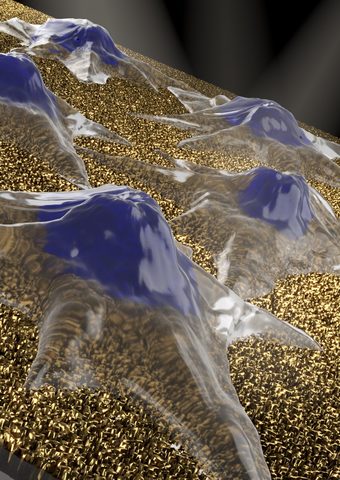
Viewed under an electron microscope, the gold nanoparticles and nanowires appear fused together like berry clusters on a branch.

The surprise finding means that pure water microdroplets can serve as microreactors for the production of gold nanostructures. “This is yet more evidence that reactions in water droplets can be fundamentally different from those in bulk water,” said study coauthor Devleena Samanta, a former graduate student in Zare’s lab and co-author on the paper.

If the process can be scaled up, it could eliminate the need for potentially toxic reducing agents that have harmful health side effects or that can pollute waterways, Zare said.

It’s still unclear why water microdroplets are able to replace a reducing agent in this reaction. One possibility is that transforming the water into microdroplets greatly increases its surface area, creating the opportunity for a strong electric field to form at the air-water interface, which may promote the formation of gold nanoparticles and nanowires.

“The surface area atop a one-liter beaker of water is less than one square meter. But if you turn the water in that beaker into microdroplets, you will get about 3,000 square meters of surface area – about the size of half a football field,” Zare said.

The team is exploring ways to utilize the nanostructures for various catalytic and biomedical applications and to refine their technique to create gold films.

“We observed a network of nanowires that may allow the formation of a thin layer of nanowires,” Samanta said.

Here’s a link and a citation for the paper,

Spontaneous formation of gold nanostructures in aqueous microdroplets by Jae Kyoo Lee, Devleena Samanta, Hong Gil Nam, & Richard N. Zare. Nature Communicationsvolume 9, Article number: 1562 (2018) doi:10.1038/s41467-018-04023-z Published online: 19 April 2018

Not unsurprisingly given Zare’s history as recounted in the news release, this paper is open access.

Nanomushroom sensors

Schematic illustration of cells (blue mountain-like shapes) on top of nanoscale mushroom-like structures with silicone dioxide stems and gold caps, which have the potential to detect cell proliferation in real-time. Courtesy: OIST

The nanomushroom sensors depicted in the above illustration and announced in a February 23, 2018 news item on Nanowerk probably aren’t edible but they certainly make up for that deficiency with other properties,

A small rectangle of pink glass, about the size of a postage stamp, sits on Professor Amy Shen’s desk. Despite its outwardly modest appearance, this little glass slide has the potential to revolutionize a wide range of processes, from monitoring food quality to diagnosing diseases.

The slide is made of a ‘nanoplasmonic’ material — its surface is coated in millions of gold nanostructures, each just a few billionths of a square meter in size. Plasmonic materials absorb and scatter light in interesting ways, giving them unique sensing properties. Nanoplasmonic materials have attracted the attention of biologists, chemists, physicists and material scientists, with possible uses in a diverse array of fields, such as biosensing, data storage, light generation and solar cells.

A February 23, 2018 Okinawa Institute of Science and Technology Graduate University (OIST) press release (also on EurekAlert), which originated the news item, provides more detail,

In several recent papers, Prof. Shen and colleagues at the Micro/Bio/Nanofluidics Unit at the Okinawa Institute of Science and Technology (OIST), described their creation of a new biosensing material that can be used to monitor processes in living cells.

“One of the major goals of nanoplasmonics is to search for better ways to monitor processes in living cells in real time,” says Prof. Shen. Capturing such information can reveal clues about cell behavior, but creating nanomaterials on which cells can survive for long periods of time yet don’t interfere with the cellular processes being measured is a challenge, she explains.

Counting Dividing Cells

One of the team’s new biosensors is made from a nanoplasmonic material that is able to accommodate a large number of cells on a single substrate and to monitor cell proliferation, a fundamental process involving cell growth and division, in real time. Seeing this process in action can reveal important insights into the health and functions of cells and tissues.

Researchers in OIST’s Micro/Bio/Nanofluidics Unit described the sensor in a study recently published in the journal Advanced Biosystems [citation and link follow this press release].

The most attractive feature of the material is that it allows cells to survive over long time periods. “Usually, when you put live cells on a nanomaterial, that material is toxic and it kills the cells,” says Dr. Nikhil Bhalla, a postdoctoral researcher at OIST and first author of the paper. “However, using our material, cells survived for over seven days.” The nanoplasmonic material is also highly sensitive: It can detect an increase in cells as small as 16 in 1000 cells.

The material looks just like an ordinary pieces of glass. However, the surface is coated in tiny nanoplasmonic mushroom-like structures, known as nanomushrooms, with stems of silicon dioxide and caps of gold. Together, these form a biosensor capable of detecting interactions at the molecular level.

The biosensor works by using the nanomushroom caps as optical antennae. When white light passes through the nanoplasmonic slide, the nanomushrooms absorb and scatter some of the light, changing its properties. The absorbance and scattering of light is determined by the size, shape and material of the nanomaterial and, more importantly, it is also affected by any medium in close proximity to the nanomushroom, such as cells that have been placed on the slide. By measuring how the light has changed once it emerges through the other side of the slide, the researchers can detect and monitor processes occurring on the sensor surface, such as cell division.

“Normally, you have to add labels, such as dyes or molecules, to cells, to be able to count dividing cells,” says Dr. Bhalla. “However, with our method, the nanomushrooms can sense them directly.”

Scaling Up

This work builds on a new method, developed by scientists at the Micro/Bio/Nanofluidics Unit at OIST, for fabricating nanomushroom biosensors. The technique was published in the journal ACS Applied Materials and Interfaces in December 2017.

Producing large-scale nanoplasmonic materials is challenging because it is difficult to ensure uniformity across the entire material surface. For this reason, biosensors for routine clinical examinations, such as disease testing, are still lacking.

In response to this problem, the OIST researchers developed a novel printing technique to create large-scale nanomushroom biosensors. With their method, they were able to develop a material consisting of approximately one million mushroom-like structures on a 2.5cm by 7.5cm silicon dioxide substrate.

“Our technique is like taking a stamp, covering it with ink made from biological molecules, and printing onto the nanoplasmonic slide,” says Shivani Sathish, a PhD student at OIST and co-author of the paper. The biological molecules increase the sensitivity of the material, meaning it can sense extremely low concentrations of substances, such as antibodies, and thus potentially detect diseases in their earliest stages.

“Using our method, it is possible to create a highly sensitive biosensor that can detect even single molecules,” says Dr. Bhalla, first author of the paper.

Plasmonic and nanoplasmonic sensors offer important tools for many fields, from electronics to food production to medicine. For example, in December 2017, second year Ph.D student Ainash Garifullina from the Unit developed a new plasmonic material for monitoring the quality of food products during the manufacturing process. The results were published in the journal Analytical Methods.

Prof. Shen and her unit say that, in the future, nanoplasmonic materials may even be integrated with emerging technologies, such as wireless systems in microfluidic devices, allowing users to take readings remotely and thereby minimizing the risk of contamination.

Here’s a link to and a citation for the paper,

Large-Scale Nanophotonic Structures for Long-Term Monitoring of Cell Proliferation by Nikhil Bhalla, Shivani Sathish, Abhishek Sinha, and Amy Q. Shen. Advanced Biosystems Vol. 2 Issue 2 DOI: 10.1002/adbi.201700258 Version of Record online: 19 JAN 2018

© 2018 WILEY-VCH Verlag GmbH & Co. KGaA, Weinheim

This paper is behind a paywall.

Here’s a link to and a citation for the earlier paper,

Probing specific gravity in real-time with graphene oxide plasmonics by Ainash Garifullina, Nikhil Bhalla, and Amy Q. Shen. Anal. Methods 2018, 10, 290-297 DOI: 10.1039/C7AY02423A first published [online] on 06 Dec 2017

This paper is open access provided you have registered for a free account.