Tag Archives: Graz University of Technology

Save energy with neuromorphic (brainlike) hardware

It seems the appetite for computing power is bottomless, which presents a problem in a world where energy resources are increasingly constrained. A May 24, 2022 news item on ScienceDaily announces research into neuromorphic computing which hints the energy efficiency long promised by the technology may be realized in the foreseeable future,

For the first time TU Graz’s [Graz University of Technology; Austria] Institute of Theoretical Computer Science and Intel Labs demonstrated experimentally that a large neural network can process sequences such as sentences while consuming four to sixteen times less energy while running on neuromorphic hardware than non-neuromorphic hardware. The new research based on Intel Labs’ Loihi neuromorphic research chip that draws on insights from neuroscience to create chips that function similar to those in the biological brain.

Rich Uhlig, managing director of Intel Labs, holds one of Intel’s Nahuku boards, each of which contains 8 to 32 Intel Loihi neuromorphic chips. Intel’s latest neuromorphic system, Pohoiki Beach, is made up of multiple Nahuku boards and contains 64 Loihi chips. Pohoiki Beach was introduced in July 2019. (Credit: Tim Herman/Intel Corporation)

A May 24, 2022 Graz University of Technology (TU Graz) press release (also on EurekAlert), which originated the news item, delves further into the research, Note: Links have been removed,

The research was funded by The Human Brain Project (HBP), one of the largest research projects in the world with more than 500 scientists and engineers across Europe studying the human brain. The results of the research are published in the research paper “Memory for AI Applications in Spike-based Neuromorphic Hardware” [sic] (DOI 10.1038/s42256-022-00480-w) which in published in Nature Machine Intelligence.  

Human brain as a role model

Smart machines and intelligent computers that can autonomously recognize and infer objects and relationships between different objects are the subjects of worldwide artificial intelligence (AI) research. Energy consumption is a major obstacle on the path to a broader application of such AI methods. It is hoped that neuromorphic technology will provide a push in the right direction. Neuromorphic technology is modelled after the human brain, which is highly efficient in using energy. To process information, its hundred billion neurons consume only about 20 watts, not much more energy than an average energy-saving light bulb.

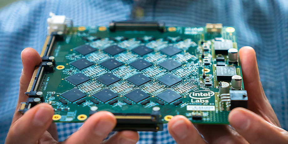
In the research, the group focused on algorithms that work with temporal processes. For example, the system had to answer questions about a previously told story and grasp the relationships between objects or people from the context. The hardware tested consisted of 32 Loihi chips.

Loihi research chip: up to sixteen times more energy-efficient than non-neuromorphic hardware

“Our system is four to sixteen times more energy-efficient than other AI models on conventional hardware,” says Philipp Plank, a doctoral student at TU Graz’s Institute of Theoretical Computer Science. Plank expects further efficiency gains as these models are migrated to the next generation of Loihi hardware, which significantly improves the performance of chip-to-chip communication.

“Intel’s Loihi research chips promise to bring gains in AI, especially by lowering their high energy cost,“ said Mike Davies, director of Intel’s Neuromorphic Computing Lab. “Our work with TU Graz provides more evidence that neuromorphic technology can improve the energy efficiency of today’s deep learning workloads by re-thinking their implementation from the perspective of biology.”

Mimicking human short-term memory

In their neuromorphic network, the group reproduced a presumed memory mechanism of the brain, as Wolfgang Maass, Philipp Plank’s doctoral supervisor at the Institute of Theoretical Computer Science, explains: “Experimental studies have shown that the human brain can store information for a short period of time even without neural activity, namely in so-called ‘internal variables’ of neurons. Simulations suggest that a fatigue mechanism of a subset of neurons is essential for this short-term memory.”

Direct proof is lacking because these internal variables cannot yet be measured, but it does mean that the network only needs to test which neurons are currently fatigued to reconstruct what information it has previously processed. In other words, previous information is stored in the non-activity of neurons, and non-activity consumes the least energy.

Symbiosis of recurrent and feed-forward network

The researchers link two types of deep learning networks for this purpose. Feedback neural networks are responsible for “short-term memory.” Many such so-called recurrent modules filter out possible relevant information from the input signal and store it. A feed-forward network then determines which of the relationships found are very important for solving the task at hand. Meaningless relationships are screened out, the neurons only fire in those modules where relevant information has been found. This process ultimately leads to energy savings.

“Recurrent neural structures are expected to provide the greatest gains for applications running on neuromorphic hardware in the future,” said Davies. “Neuromorphic hardware like Loihi is uniquely suited to facilitate the fast, sparse and unpredictable patterns of network activity that we observe in the brain and need for the most energy efficient AI applications.”

This research was financially supported by Intel and the European Human Brain Project, which connects neuroscience, medicine, and brain-inspired technologies in the EU. For this purpose, the project is creating a permanent digital research infrastructure, EBRAINS. This research work is anchored in the Fields of Expertise Human and Biotechnology and Information, Communication & Computing, two of the five Fields of Expertise of TU Graz.

Here’s a link to and a citation for the paper,

A Long Short-Term Memory for AI Applications in Spike-based Neuromorphic Hardware by Arjun Rao, Philipp Plank, Andreas Wild & Wolfgang Maass. Nature Machine Intelligence (2022) DOI: https://doi.org/10.1038/s42256-022-00480-w Published: 19 May 2022

This paper is behind a paywall.

For anyone interested in the EBRAINS project, here’s a description from their About page,

EBRAINS provides digital tools and services which can be used to address challenges in brain research and brain-inspired technology development. Its components are designed with, by, and for researchers. The tools assist scientists to collect, analyse, share, and integrate brain data, and to perform modelling and simulation of brain function.

EBRAINS’ goal is to accelerate the effort to understand human brain function and disease.

This EBRAINS research infrastructure is the entry point for researchers to discover EBRAINS services. The services are being developed and powered by the EU-funded Human Brain Project.

You can register to use the EBRAINS research infrastructure HERE

One last note, the Human Brain Project is a major European Union (EU)-funded science initiative (1B Euros) announced in 2013 and to be paid out over 10 years.

An easier way to make highly ordered porous films for commercial sensors

An April 3, 2017 news item on Nanowerk describes Japanese research into a new technique for producing MOF’s (metallic organic frameworks),

Osaka-based researchers developed a new method to create films of porous metal–organic frameworks fully aligned on inorganic substrates. The method is simple, requiring only that the substrate and an organic linker are mixed under mild conditions, and fast, producing perfectly aligned films within minutes. The films oriented fluorescent dye molecules within their pores, and the fluorescence response of these dyes was switched on or off simply by rotating the material in polarized light.

An April 3, 2017 Osaka University press release on the Alpha Gallileo news service, which originated the news item, explains more about MOFs and gives some details about the new technique,,

Metal–organic frameworks, or MOFs, are highly ordered crystalline structures made of metal ion nodes and organic molecule linkers. Many MOFs can take up and store gases, such as carbon dioxide or hydrogen, thanks to their porous, sponge-like structures.

MOFs are also potential chemical sensors. They can be designed to change color or display another optical signal if a particular molecule is taken up into the framework. However, most studies on MOFs are performed on tiny single crystals, which is not practical for the commercial development of these materials.

Chemists have now come a step closer to making commercially viable sensors that contain highly ordered MOFs, thanks to the collaboration of an international team of researchers at Osaka Prefecture University, Osaka University and Graz University of Technology. The method will allow researchers to fabricate large tailor-made MOF films on any substrate of any size, which will vastly improve their prospects for commercial development.

In a study recently published in Nature Materials and highlighted on the cover and in the ‘News and Views’ section of the journal, the Osaka-based researchers report a one-step method to prepare thin MOF films directly on inorganic copper hydroxide substrates. Using this method, the researchers produced large MOF films with areas of more than 1 cm2 that were, for the first time, fully aligned with the crystal lattice of the underlying substrate.

Noting that microcrystals of copper hydroxide can be converted into MOFs by adding organic linker molecules under mild conditions, the researchers used the same strategy to create a thin MOF layer on larger copper hydroxide substrates. They carefully chose the carboxylic acid-based linker molecule 1,4-dibenzenedicarboxylic acid because it fit exactly to the spacing between the copper atoms on the substrate surface.

A MOF film began to grow on the copper hydroxide substrates within minutes of mixing it with the linker molecule, making this technique much easier and faster than previous step-wise approaches to build up MOF films. Using microscopy and X-ray diffraction techniques, the researchers found that the film was precisely oriented along the copper hydroxide lattice.

To demonstrate the unique optical behavior of their films, the researchers filled the MOF’s ordered pores with fluorescent molecules, which fluoresce when light is shone on them in a particular direction. When they shone polarized light on the ordered material, the researchers found that they could easily switch the fluorescence response on or off simply by rotating the material.

Here’s a link to and a citation for the paper,

Centimetre-scale micropore alignment in oriented polycrystalline metal–organic framework films via heteroepitaxial growth by Paolo Falcaro, Kenji Okada, Takaaki Hara, Ken Ikigaki, Yasuaki Tokudome, Aaron W. Thornton, Anita J. Hill, Timothy Williams, Christian Doonan, & Masahide Takahashi. Nature Materials 16, 342–348  (2017) doi:10.1038/nmat4815 Published online 05 December 2016

This paper is behind a paywall.

Monitoring the life of bacteria in microdroplets

Trying to establish better ways to test the effect of drugs on bacteria has led the Institute of Physical Chemistry of the Polish Academy of Sciences to develop a new monitoring technique. From a Jan.  11, 2017 news item on Nanowerk,

So far, however, there has been no quick or accurate method of assessing the oxygen conditions in individual microdroplets. This key obstacle has been overcome at the Institute of Physical Chemistry of the Polish Academy of Sciences.

Not in rows of large industrial tanks, nor on shelves laden with test tubes and beakers. The future of chemistry and biology is barely visible to the eye: it’s hundreds and thousands of microdroplets, whizzing through thin tubules of microfluidic devices. The race is on to find technologies that will make it possible to carry out controlled chemical and biological experiments in microdroplets. At the Institute of Physical Chemistry of the Polish Academy of Sciences (IPC PAS) in Warsaw a method of remote, yet rapid and accurate assessment of oxygen consumption by micro-organisms living in individual microdroplets has been demonstrated for the first time.

“Devices for the cultivation of bacteria in microdroplets have the chance to revolutionize work on the development of new antibiotics and the study of mechanisms responsible for the acquisition of drug resistance by bacteria. In one small microfluidic system it is possible to accommodate several hundred or even several thousand microdroplets – and to carry out a different experiment in each of them, for example with different types of microorganisms and at different concentrations of antibiotic in each drop,” describes Prof. Piotr Garstecki (IPC PAS), then explains: “For such studies to be possible, one has to provide the bacteria with conditions for development for even a few weeks. Thus, knowledge about the flow of oxygen to the droplets and the rate of its consumption by the microorganisms becomes extremely important. In our latest system we demonstrate how to read this key information.”

A Jan. 11, 2017 IPC PAS press release on EurekAlert, which originated the  news item, describes the work in more detail,

The bioreactors of the future are water droplets with culture medium suspended in a carrier liquid with which they are immiscible (usually this is oil). In the channel of the microfluidic device each droplet is longer than it is wide and it almost completely fills its lumen; sizes matched in this manner ensure that the drops do not swop places in the channel and throughout the duration of the experiment they can be identified without any problems. At the same time, there has to be a thin layer of oil maintained continuously between each microdroplet and the wall of the channel. Without this, the bacteria would be in direct contact with the walls of the channel so they would be able to settle on them and move from drop to drop. Unfortunately, when the microdroplet is stationary, with time it pushes out the oil separating it from the walls, laying it open to contamination. For this reason the drops must be kept in constant motion – even for weeks.

Growing bacteria need culture medium, and waste products need to be removed from their environment at an appropriate rate. Information about the bacterial oxygen consumption in individual droplets is therefore crucial to the operation of microbioreactors.

“It is immediately obvious where the problem lies. In each of the hundreds of moving droplets measurements need to be carried out at a frequency corresponding to the frequency of division of the bacteria or more, in practice at least once every 15 minutes. In addition, the measurement cannot cause any interference in the microdroplets,” says PhD student Michal Horka (IPC PAS), a co-author of the publication in the journal Analytical Chemistry.

Help was at hand for the Warsaw researchers from chemists from the Austrian Institute of Analytical Chemistry and Food Chemistry at the Graz University of Technology. They provided polymer nanoparticles with a phosphorescent dye, which after excitation emit light for longer the higher the concentration of oxygen in the surrounding solution (the nanoparticles underwent tests at the IPC PAS on bacteria in order to determine their possible toxicity – none was found).

Research on monitoring oxygen consumption in the droplets commenced with the preparation of an aqueous solution with the bacteria, the culture medium and a suitable quantity of nanoparticles. The mixture was injected into the microfluidic system constructed of tubing with Teflon connectors with correspondingly shaped channels. The first module formed droplets with a volume of approx. 4 microlitres, which were directed to the incubation tube wound on a spool. In the middle of its length there was another module, with detectors for measuring oxygen and absorbance.

“In the incubation part in one phase of the cycle the droplets flowed in one direction, in the second – in another, electronically controlled by means of suitable solenoid valves. All this looks seemingly simple enough, but in practice one of the biggest challenges was to ensure a smooth transition between the detection module and the tubing, so that bacterial contamination did not occur at the connections,” explains PhD student Horka.

During their passage through the detection module the droplets flowed under an optical sensor which measured the so-called optical density, which is the standard parameter used to evaluate the number of cells (the more bacteria in the droplets, the less light passes through them). In turn, the measurement of the duration of the phosphorescence of the nanoparticles, evaluating the concentration of oxygen in the microdroplets, was carried out using the Piccolo2 optical detector, provided by the Austrian group. This detector, which looks like a big pen drive, was connected directly to the USB port on the control computer. Comparing information from both sensors, IPC PAS researchers showed that the microfluidic device they had constructed made it possible to regularly and quickly monitor the metabolic activity of bacteria in the individual microdroplets.

“We carried out our tests both with bacteria floating in water singly – this is how the common Escherichia coli bacteria behave – as well as with those having a tendency to stick together in clumps – as is the case for tuberculosis bacilli or others belonging to the same family including Mycobacterium smegmatis which we studied. Evaluation of the rate of oxygen consumption by both species of microorganisms proved to be not only possible, but also reliable,” stresses PhD student Artur Ruszczak (IPC PAS).

The results of the research, funded by the European ERC Starting Grant (Polish side) and the Maria Sklodowska-Curie grant (Austrian side) are an important step in the process of building fully functional microfluidic devices for conducting biological experiments lasting many weeks. A system for culturing bacteria in microdroplets was developed at the IPC PAS a few years ago, however it was constructed on a polycarbonate plate. The maximum dimensions of the plate did not exceed 10 cm, which greatly limited the number of droplets; in addition, as a result of interaction with the polycarbonate, after four days the channels were contaminated with bacteria. Devices of Teflon modules and tubing would not have these disadvantages, and would be suitable for practical applications.

Here’s a link to and a citation for the paper,

Lifetime of Phosphorescence from Nanoparticles Yields Accurate Measurement of Concentration of Oxygen in Microdroplets, Allowing One To Monitor the Metabolism of Bacteria by Michał Horka, Shiwen Sun, Artur Ruszczak, Piotr Garstecki, and Torsten Mayr. Anal. Chem., 2016, 88 (24), pp 12006–12012 DOI: 10.1021/acs.analchem.6b03758 Publication Date (Web): November 23, 2016

Copyright © 2016 American Chemical Society

This paper is behind a paywall.

Bioplastics and the Salar de Uyuni (Bolivia)

The longest continuous salt desert in the world, the Salar de Uyuni in Bolivia, is home to a bacterium that stores the polymer, PHB (poly-beta-hydroxybutyrate), a possible substitute for petroleum-based plastics. according to a July 10, 2013 news release on EurekAlert,

In Bolivia, in the largest continuous salt desert in the world, researchers from the Polytechnic University of Catalonia have found a bacterium that stores large amounts of PHB, a prized polymer. This biodegradable plastic is used by the food and pharmaceutical industries, for example to produce nanospheres to transport antibiotics.

In the quest for natural polymers to substitute for petroleum-based plastics, scientists have recently discovered that a microorganism in South America produces poly-beta-hydroxybutyrate (PHB), a biodegradable compound of great utility for the food, pharmaceutical, cosmetic and packaging industries.

The bacterium in question is Bacillus megaterium Uyuni S29, a strain that produces the largest amount of polymer of the genus. It has been found in the water ‘eyes’ of the famous Salar de Uyuni or Uyuni salt flat, in Bolivia.

“These are very extreme environments, which facilitate intracellular accumulation of PHB, a reserve material used by bacteria in times when nutrients are scarce,” Dr Marisol Marqués, microbiologist at the Polytechnic University of Catalonia (UPC, Spain), explains to SINC.

Scientists from the UPC and the Graz University of Technology in Austria have successfully made the bacillus produce significant quantities of the compound in the laboratory in cultivation conditions similar to those used in industry. The technique is published in the journals Food Technology & Biotechnology and Journal of Applied Microbiology.

“The resulting biopolymer has thermal properties different from conventional PHBs, which makes it easier to process, independently of its application,” Marqués goes on.

The researcher recognises that the costs of producing biopolymers are, in general, “still high and not competitive when compared with conventional polymers, although progress is being made in this regard.”

The news release includes citations for the team’s  two recently published papers,

A. Rodríguez-Contreras, M Koller, M. Miranda de Sousa Dias, M. Calafell, G. Braunegg, M. S. Marqués-Calvo. “Novel Poly[(R)-3-Hydroxybutyrate]-Producing Bacterium Isolated from a Bolivian Hypersaline Lake”. Food Technology & Biotechnology 51 (1): 123-130, 2013.

A. Rodríguez-Contreras, M. Koller, M. Miranda-de Sousa Dias, M. Calafell, G. Braunegg, M. S. Marqués-Calvo. “High production of poly(3-hydroxybutyrate) from a wild Bacillus megaterium Bolivian strain. Journal of Applied Microbiology 114 (5):1378-87, 2013.

It’s always a pleasure to feature a country I haven’t had an opportunity to mention before (Bolivia) within the nanotechnology context, so, thank you to whomever ensured that there was an English language version of the news release.

FEI/National Geographic image contest: Explore the Unseen

It’s not unusual to see contests for the best ‘nanoimage’ but this one offers some special prizes including exposure (pun intended)  in a National Geographic project on nanotechnology. From the June 27, 2012 news item on Azonano,

FEI is excited to announce this year’s FEI Image Contest, “Explore the Unseen” and invites owners and users to submit their best nano-scale images online at fei.com. This year FEI are pleased to partner with National Geographic on a film tentatively titled “Invisible Worlds”.

Winning images will be posted on National Geographic’s website and all images will be considered for inclusion in the film’s promotional materials.

Inspired by the upcoming film, the FEI Image Contest offers owners and users an opportunity to explore their creativity and share their images with National Geographic’s worldwide audience.

I was a little curious about FEI and found it’s a microscopy company, from their About FEI page,

FEI  is the world leader in the production and distribution of electron microscopes, including scanning electron microscopes (SEM), transmission electron microscopes (TEM), DualBeam™­ instruments, and focused ion beam tools (FIB), for nanoscale research, serving a broad range of customers worldwide. Nanotechnology is the science of finding, characterizing, analyzing and fabri­cating materials smaller than 100 nano­meters (a nanometer is one billionth of a meter). FEI’s global customer base includes researchers, scientists, engineers, lab managers, and other skilled professionals.

Here’s more about the contest from the FEI’s 2012 contest page,

Contest Benefits

What’s in it for you?

All images submitted will be considered for inclusion in the National Geographic film promotional materials. This may include a companion game, book, education guide and poster.

Monthly Category Prizes

Everyone who enters will have the opportunity to win one of four monthly prizes. Prizes will be awarded in the following categories: The Human Body, Around the House, The Natural World, and Other Relevant Science. Monthly winners will receive a custom 24 x 24 inch bamboo mounted print of their image to put on display.

Plus, the four winning images will be posted to the Nat Geo Movies section of their website and Facebook page.

Grand Prize

At the conclusion of the contest, a grand prize will be awarded for the best image received from the monthly category winners. The grand prize is two coach class tickets to a United States destination of the winners choosing.

In addition, the winning image will be part of a special photo gallery promoting the film “Invisible Worlds”.

Here are more details about the individual categories,

Image Categories

This year, we’ve chosen image categories with broad audience appeal. The following examples, while not an exhaustive list, provide an idea of what we’re looking for:

The Natural World:

  • Insect parts – wings, eyes, etc. (ideal insects include moth, ladybug, fly, dragonfly, butterfly, cicada, cricket, etc.)
  • Spider silk / webs
  • Pollen, allergens, leaves, tree slime, fungus, bacteria & mold
  • Micro-invertebrates seen in water-quality testing
  • Plants, flowers, blades of grass
  • Rock, minerals, sand, etc.
  • Ice/snow/snowflakes, other crystals, raindrops
  • Close-up of animals or animal parts: dog, cat, bird, fish (pets a kid would own)

The Human Body:

  • Insects that live on your body (eyebrows, lashes, etc.) lice, bacteria
  • Body parts: bone (including fractures/breaks), human hair, skin flakes
  • Bodily fluids: snot, sweat, blood, saliva, tears, etc
  • Hands (finger, skin) before and after washing
  • Viruses
  • Endoplasmic reticulum, cell walls, etc
  • What a tattoo looks like under the skin

Around the House:

  • Things you would find in a kids room: t-shirt fibers, stuff on the soles of dirty shoes, dust mites, carpet fibers, hair inside of a baseball cap, sloughed skin, dust, pencil lead, crayons
  • Food: ice cream, candy, bread, french fries, apples, carrots, tomatoes, etc.
  • Creatures that live on the mouthpiece of a phone, in the kitchen sink
  • Tires, cars, bikes, toys
  • Lint from clothing
  • The inside workings of a clock, computer, smartphone or TV
  • Gems and jewels: rubies, diamonds, other gems
  • Sports equipment: baseball, basketball, soccer ball, bathing suit, etc.

Other Relevant Science:

Do your best images not fit into the categories above? Are you interested in sharing what you’re working on today? Whether you are investigating advanced materials, working to understand complex chemical reactions, or researching the 3D architecture of tissues and cells, this is the category for submitting your best work.

Here’s FEI’s 2011 winning image (from FEI”s 2011 Owner Image Contest Winner announcement page),

Microcanyon: a micro-crack in steel after bending tests Credit: Martina Dienstleder of the Institute for Electron Microscopy at the Graz University of Technology

The reasons it was selected as the ‘grand’ prize winner (from FEI”s 2011 Owner Image Contest Winner announcement page) were,

Overall, the entries were judged on their aesthetic appeal, application and scientific relevance, and overall creativity.

Given that there is mention of a micro-crack and the grand prize winner is titled Microcanyon, I’m assuming last year’s theme was less specific than this year’s invitation to submit ‘nanoscale’ inflected images.

Given there are monthly winners I assume there are monthly deadlines but I couldn’t find them on the FEI contest webpage however, the  final deadline for submissions is Sept. 14, 2012.

Good luck to the 2012 entrants.