Tag Archives: green algae

Steering a synthetic nanorobot using light

This news comes from the University of Hong Kong. A Nov. 8, 2016 news item on Nanowerk throws some light on the matter (Note: A link has been removed),

A team of researchers led by Dr Jinyao Tang of the Department of Chemistry, the University of Hong Kong, has developed the world’s first light-seeking synthetic Nano robot. With size comparable to a blood cell, those tiny robots have the potential to be injected into patients’ bodies, helping surgeons to remove tumors and enabling more precise engineering of targeted medications. The findings have been published in October [2016] earlier in leading scientific journal Nature Nanotechnology (“Programmable artificial phototactic microswimmer”).

An Oct. 24, 2016 University of Hong Kong press release (also on EurekAlert), which originated the news item, expands on the theme,

It has been a dream in science fiction for decades that tiny robots can fundamentally change our daily life. The famous science fiction  movie “Fantastic  Voyage” is a very good example, with a group of scientists driving their miniaturized nano-submarine inside human body to repair a damaged brain. In the film “Terminator  2,” billions of nanorobots were assembled into the amazing shapeshifting body: the T-1000. In the real world, it is quite challenging to make and design a sophisticated nanorobot with advanced functions.

The Nobel Prize in Chemistry 2016 was awarded to three scientists for “the design and synthesis of molecular machines.” They developed a set of mechanical components at molecular scale which may be  assembled into  more complicated nanomachines  to  manipulate single  molecule such as DNA or proteins in the future. The development of tiny nanoscale machines for biomedical applications has been a major trend of scientific research in recent years. Any breakthroughs will potentially open the door to new knowledge and treatments of diseases and development of new drugs.

One difficulty in nanorobot design is to make these nanostructures sense and respond to the environment. Given each nanorobot is only a few micrometer in size which is ~50 times smaller than the diameter of a human hair, it  is very difficult  to  squeeze  normal electronic sensors and circuits into  nanorobots with reasonable price. Currently, the only method to remotely control nanorobots is to  incorporate tiny magnetic inside the nanorobot and guide the motion via external magnetic field.

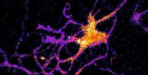
The  nanorobot developed by Dr Tang’s team use light as the propelling  force, and is the first research team globally to explore the light-guided nanorobots and demonstrated its feasibility and effectiveness. In their paper published in Nature  Nanotechnology, Dr Tang’s team  demonstrated  the  unprecedented ability of these light-controlled nanorobots as they are “dancing”  or even spell a word under light control. With a novel  nanotree structure, the nanorobots can respond to the light shining on it like  moths  being drawn to flames. Dr Tang described the motions as if “they can “see” the light and drive itself towards it”.

The team gained inspiration from natural green algae
for the nanorobot design. In nature, some green algae have evolved  with  the  ability  of  sensing  light  around  it.  Even just a single cell, these green  algae can sense the intensity of light and swim  towards the light source for photosynthesis. Dr  Jinyao  Tang’s team successfully developed the nanorobots after over three years’ efforts. With a novel nanotree structure, they are composed of two  common and low-price semiconductor materials: silicon  and titanium oxide. During  the  synthesis, silicon  and titanium oxide are shaped into nanowire and then further arranged into a tiny nanotree heterostructure.

Dr Tang said: “Although the current nanorobot cannot be used for disease treatment yet, we are working on the next generation nanorobotic system which is more efficient and biocompatible.”

“Light is a more effective option to communicate between microscopic world and macroscopic world. We can conceive that more complicated instructions can be sent to nanorobots which provide scientists with a new tool to further develop more functions into nanorobot and get us one step closer to daily life applications,” he added.

Here’s a link to and a citation for the paper,

Programmable artificial phototactic microswimmer by Baohu Dai, Jizhuang Wang, Ze Xiong, Xiaojun Zhan, Wei Dai, Chien-Cheng Li, Shien-Ping Feng, & Jinyao Tang.  Nature Nanotechnology (2016)  doi:10.1038/nnano.2016.187 Published online 17 October 2016

So, this ‘bot’ seems to be a microbot or microrobot with some nanoscale features. In any event, the paper is behind a paywall.

Getting your brain cells to glow in the dark

The extraordinary effort to colonize our brains continues apace with a new sensor from Vanderbilt University. From an Oct. 27, 2016 news item on ScienceDaily,

A new kind of bioluminescent sensor causes individual brain cells to imitate fireflies and glow in the dark.

The probe, which was developed by a team of Vanderbilt scientists, is a genetically modified form of luciferase, the enzyme that a number of other species including fireflies use to produce light. …

The scientists created the technique as a new and improved method for tracking the interactions within large neural networks in the brain.

“For a long time neuroscientists relied on electrical techniques for recording the activity of neurons. These are very good at monitoring individual neurons but are limited to small numbers of neurons. The new wave is to use optical techniques to record the activity of hundreds of neurons at the same time,” said Carl Johnson, Stevenson Professor of Biological Sciences, who headed the effort.

Individual neuron glowing with bioluminescent light produced by a new genetically engineered sensor. (Johnson Lab / Vanderbilt University)

Individual neuron glowing with bioluminescent light produced by a new genetically engineered sensor. (Johnson Lab / Vanderbilt University)

An Oct. 27, 2016 Vanderbilt University news release (also on EurekAlert) by David Salisbury, which originated the news item, explains the work in more detail,

“Most of the efforts in optical recording use fluorescence, but this requires a strong external light source which can cause the tissue to heat up and can interfere with some biological processes, particularly those that are light sensitive,” he [Carl Johnson] said.

Based on their research on bioluminescence in “a scummy little organism, the green alga Chlamydomonas, that nobody cares much about” Johnson and his colleagues realized that if they could combine luminescence with optogenetics – a new biological technique that uses light to control cells, particularly neurons, in living tissue – they could create a powerful new tool for studying brain activity.

“There is an inherent conflict between fluorescent techniques and optogenetics. The light required to produce the fluorescence interferes with the light required to control the cells,” said Johnson. “Luminescence, on the other hand, works in the dark!”

Johnson and his collaborators – Associate Professor Donna Webb, Research Assistant Professor Shuqun Shi, post-doctoral student Jie Yang and doctoral student Derrick Cumberbatch in biological sciences and Professor Danny Winder and postdoctoral student Samuel Centanni in molecular physiology and biophysics – genetically modified a type of luciferase obtained from a luminescent species of shrimp so that it would light up when exposed to calcium ions. Then they hijacked a virus that infects neurons and attached it to their sensor molecule so that the sensors are inserted into the cell interior.

The researchers picked calcium ions because they are involved in neuron activation. Although calcium levels are high in the surrounding area, normally they are very low inside the neurons. However, the internal calcium level spikes briefly when a neuron receives an impulse from one of its neighbors.

They tested their new calcium sensor with one of the optogenetic probes (channelrhodopsin) that causes the calcium ion channels in the neuron’s outer membrane to open, flooding the cell with calcium. Using neurons grown in culture they found that the luminescent enzyme reacted visibly to the influx of calcium produced when the probe was stimulated by brief light flashes of visible light.

To determine how well their sensor works with larger numbers of neurons, they inserted it into brain slices from the mouse hippocampus that contain thousands of neurons. In this case they flooded the slices with an increased concentration of potassium ions, which causes the cell’s ion channels to open. Again, they found that the sensor responded to the variations in calcium concentrations by brightening and dimming.

“We’ve shown that the approach works,” Johnson said. “Now we have to determine how sensitive it is. We have some indications that it is sensitive enough to detect the firing of individual neurons, but we have to run more tests to determine if it actually has this capability.”

Here’s a link to and a citation for the paper,

Coupling optogenetic stimulation with NanoLuc-based luminescence (BRET) Ca++ sensing by Jie Yang, Derrick Cumberbatch, Samuel Centanni, Shu-qun Shi, Danny Winder, Donna Webb, & Carl Hirschie Johnson. Nature Communications 7, Article number: 13268 (2016)  doi:10.1038/ncomms13268 Published online: 27 October 2016

This paper is open access.