Tag Archives: green fluorescent proteins

Lifesaving moths and nanomagnets

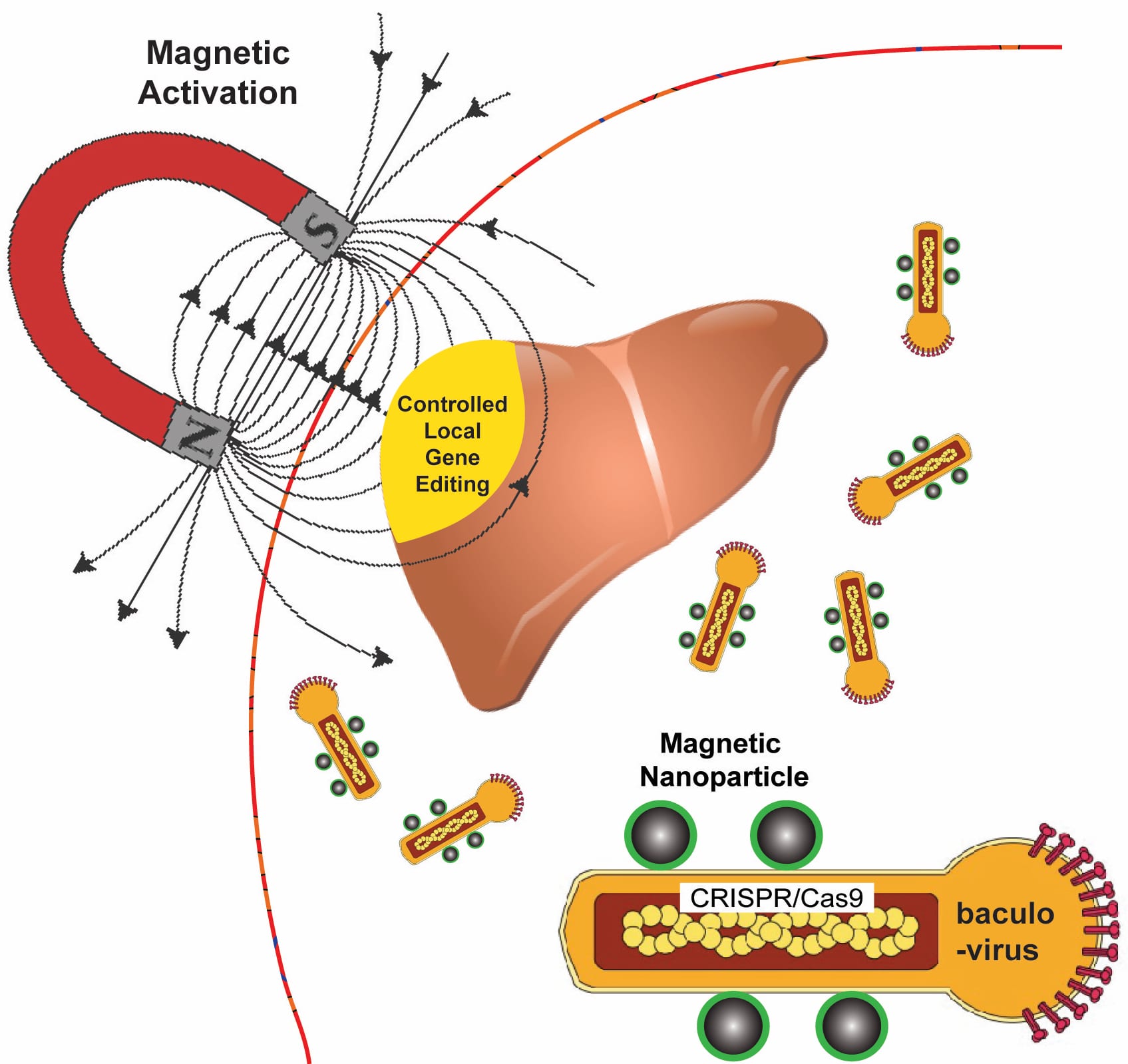
Rice University bioengineers use a magnetic field to activate nanoparticle-attached baculoviruses in a tissue. The viruses, which normally infect alfalfa looper moths, are modified to deliver gene-editing DNA code only to cells that are targeted with magnetic field-induced local transduction. Courtesy of the Laboratory of Biomolecular Engineering and Nanomedicine

Kudos to whomever put that diagram together! That’s a lot of well conveyed information.

Now for the details about how this technology might save lives. From a November 13, 2018 news item on Nanowerk,

A new technology that relies on a moth-infecting virus and nanomagnets could be used to edit defective genes that give rise to diseases like sickle cell, muscular dystrophy and cystic fibrosis.

Rice University bioengineer Gang Bao has combined magnetic nanoparticles with a viral container drawn from a particular species of moth to deliver CRISPR/Cas9 payloads that modify genes in a specific tissue or organ with spatial control.

A November 12, 2018 Rice University news release (also on EurekAlert published on November 13, 2018), which originated the news item, provides detail,

Because magnetic fields are simple to manipulate and, unlike light, pass easily through tissue, Bao and his colleagues want to use them to control the expression of viral payloads in target tissues by activating the virus that is otherwise inactivated in blood.

The research appears in Nature Biomedical Engineering. In nature, CRISPR/Cas9 bolsters microbes’ immune systems by recording the DNA of invaders. That gives microbes the ability to recognize and attack returning invaders, but scientists have been racing to adapt CRISPR/Cas9 to repair mutations that cause genetic diseases and to manipulate DNA in laboratory experiments.

CRISPR/Cas9 has the potential to halt hereditary disease – if scientists can get the genome-editing machinery to the right cells inside the body. But roadblocks remain, especially in delivering the gene-editing payloads with high efficiency.

Bao said it will be necessary to edit cells in the body to treat many diseases. “But efficiently delivering genome-editing machinery into target tissue in the body with spatial control remains a major challenge,” Bao said. “Even if you inject the viral vector locally, it can leak to other tissues and organs, and that could be dangerous.”

The delivery vehicle developed by Bao’s group is based on a virus that infects Autographa californica, aka the alfalfa looper, a moth native to North America. The cylindrical baculovirus vector (BV), the payload-carrying part of the virus, is considered large at up to 60 nanometers in diameter and 200-300 nanometers in length. That’s big enough to transport more than 38,000 base pairs of DNA, which is enough to supply multiple gene-editing units to a target cell, Bao said.

He said the inspiration to combine BV and magnetic nanoparticles came from discussions with Rice postdoctoral researcher and co-lead author Haibao Zhu, who learned about the virus during a postdoctoral stint in Singapore but knew nothing about magnetic nanoparticles until he joined the Bao lab. The Rice team had previous experience using iron oxide nanoparticles and an applied magnetic field to open blood vessel walls just enough to let large-molecule drugs pass through.

“We really didn’t know if this would work for gene editing or not, but we thought, ‘worth a shot,'” Bao said.

The researchers use the magnetic nanoparticles to activate BV and deliver gene-editing payloads only where they’re needed. To do this, they take advantage of an immune-system protein called C3 that normally inactivates baculoviruses.

“If we combine BV with magnetic nanoparticles, we can overcome this deactivation by applying the magnetic field,” Bao said. “The beauty is that when we deliver it, gene editing occurs only at the tissue, or the part of the tissue, where we apply the magnetic field.”

Application of the magnetic field allows BV transduction, the payload-delivery process that introduces gene-editing cargo into the target cell. The payload is also DNA, which encodes both a reporter gene and the CRISPR/Cas9 system.

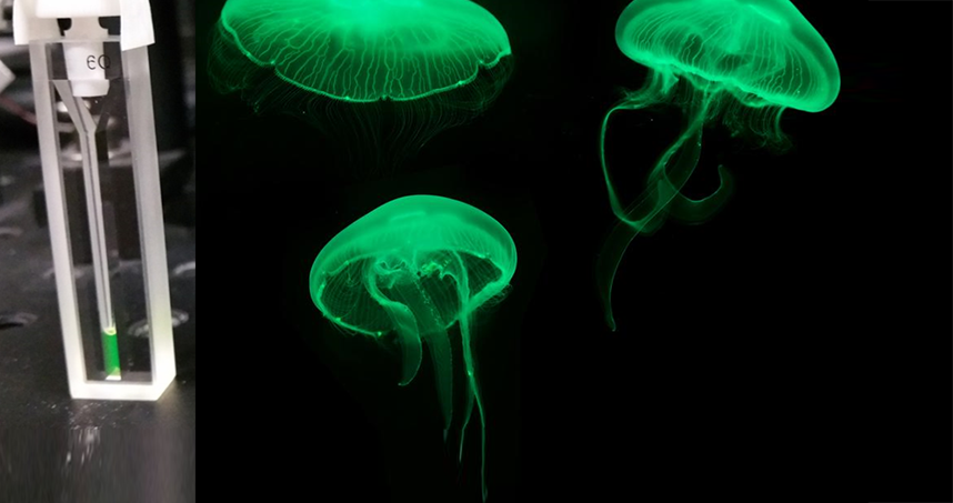
In tests, the BV was loaded with green fluorescent proteins or firefly luciferase. Cells with the protein glowed brightly under a microscope, and experiments showed the magnets were highly effective at targeted delivery of BV cargoes in both cell cultures and lab animals.

Bao noted his and other labs are working on the delivery of CRISPR/Cas9 with adeno-associated viruses (AAV), but he said BV’s capacity for therapeutic cargo is roughly eight times larger. “However, it is necessary to make BV transduction into target cells more efficient,” he said.

Here’s a link to and a citation for the paper,

Spatial control of in vivo CRISPR–Cas9 genome editing via nanomagnets by Haibao Zhu, Linlin Zhang, Sheng Tong, Ciaran M. Lee, Harshavardhan Deshmukh, & Gang Bao. Nature Biomedical Engineering (2018) DOI: https://doi.org/10.1038/s41551-018-0318-7 Published: 12 November 2018

This paper is behind a paywall.

Entanglement and biological systems

I think it was about five years ago thatI wrote a paper on something I called ‘cognitive entanglement’ (mentioned in my July 20,2012 posting) so the latest from Northwestern University (Chicago, Illinois, US) reignited my interest in entanglement. A December 5, 2017 news item on ScienceDaily describes the latest ‘entanglement’ research,

Nearly 75 years ago, Nobel Prize-winning physicist Erwin Schrödinger wondered if the mysterious world of quantum mechanics played a role in biology. A recent finding by Northwestern University’s Prem Kumar adds further evidence that the answer might be yes.

Kumar and his team have, for the first time, created quantum entanglement from a biological system. This finding could advance scientists’ fundamental understanding of biology and potentially open doors to exploit biological tools to enable new functions by harnessing quantum mechanics.

A December 5, 2017 Northwestern University news release (also on EurekAlert), which originated the news item, provides more detail,

“Can we apply quantum tools to learn about biology?” said Kumar, professor of electrical engineering and computer science in Northwestern’s McCormick School of Engineering and of physics and astronomy in the Weinberg College of Arts and Sciences. “People have asked this question for many, many years — dating back to the dawn of quantum mechanics. The reason we are interested in these new quantum states is because they allow applications that are otherwise impossible.”

Partially supported by the [US] Defense Advanced Research Projects Agency [DARPA], the research was published Dec. 5 [2017] in Nature Communications.

Quantum entanglement is one of quantum mechanics’ most mystifying phenomena. When two particles — such as atoms, photons, or electrons — are entangled, they experience an inexplicable link that is maintained even if the particles are on opposite sides of the universe. While entangled, the particles’ behavior is tied one another. If one particle is found spinning in one direction, for example, then the other particle instantaneously changes its spin in a corresponding manner dictated by the entanglement. Researchers, including Kumar, have been interested in harnessing quantum entanglement for several applications, including quantum communications. Because the particles can communicate without wires or cables, they could be used to send secure messages or help build an extremely fast “quantum Internet.”

“Researchers have been trying to entangle a larger and larger set of atoms or photons to develop substrates on which to design and build a quantum machine,” Kumar said. “My laboratory is asking if we can build these machines on a biological substrate.”

In the study, Kumar’s team used green fluorescent proteins, which are responsible for bioluminescence and commonly used in biomedical research. The team attempted to entangle the photons generated from the fluorescing molecules within the algae’s barrel-shaped protein structure by exposing them to spontaneous four-wave mixing, a process in which multiple wavelengths interact with one another to produce new wavelengths.

Through a series of these experiments, Kumar and his team successfully demonstrated a type of entanglement, called polarization entanglement, between photon pairs. The same feature used to make glasses for viewing 3D movies, polarization is the orientation of oscillations in light waves. A wave can oscillate vertically, horizontally, or at different angles. In Kumar’s entangled pairs, the photons’ polarizations are entangled, meaning that the oscillation directions of light waves are linked. Kumar also noticed that the barrel-shaped structure surrounding the fluorescing molecules protected the entanglement from being disrupted.

“When I measured the vertical polarization of one particle, we knew it would be the same in the other,” he said. “If we measured the horizontal polarization of one particle, we could predict the horizontal polarization in the other particle. We created an entangled state that correlated in all possibilities simultaneously.”

Now that they have demonstrated that it’s possible to create quantum entanglement from biological particles, next Kumar and his team plan to make a biological substrate of entangled particles, which could be used to build a quantum machine. Then, they will seek to understand if a biological substrate works more efficiently than a synthetic one.

Here’s an image accompanying the news release,

Featured in the cuvette on the left, green fluorescent proteins responsible for bioluninescence in jellyfish. Courtesy: Northwestern University

Here’s a link to and a citation for the paper,

Generation of photonic entanglement in green fluorescent proteins by Siyuan Shi, Prem Kumar & Kim Fook Lee. Nature Communications 8, Article number: 1934 (2017) doi:10.1038/s41467-017-02027-9 Published online: 05 December 2017

This paper is open access.