Tag Archives: halloysite

Nanoparticles for prolonged anti-lice protection

Caption Graphical abstract [the animal is a capybara, world’s largest rodent] Credit: Kazan Federal University, Louisiana Tech University, Gubkin University

A September 28, 2021Kazan Federal University (Russia) press release (also on EurekAlert; Source text: Larisa Busil Photo: Rawil Fakhrullin) announces news that could lead to relief for anyone who owns animals,

An international researcher team of Louisiana Tech University, Gubkin University [also known as, Gubkin Russian State University of Oil and Gas] and Kazan Federal University reported the fabrication of nanoscale insecticidal hair coating for prolonged anti-lice protection. The study was supported by the Russian Science Foundation.

“Treating agricultural and domestic animals infected with ectoparasites (such as lice, fleas, chewing lice, etc.) is among the primary challenges of veterinary medicine and agriculture. In case of mass infestation, regular measures, such as isolation of infected animals or repeated reapplication of insecticides, are not always effective. These methods are time-limited and provide a short-term therapeutic effect,” explains co-author Rawil Fakhrullin, Head of Kazan University’s Bionanotechnology Lab. “Using an inorganic nanoscale carrier as a component of a therapeutic formulation for topical application of insecticides might be the optimal way to address this challenge.”

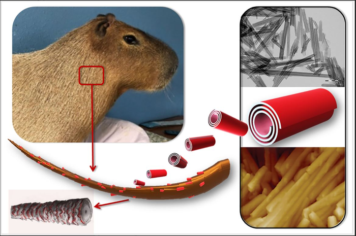
Halloysite, a natural nanosized tubular mineral, was used as a drug carrier capable of forming a durable and uniform coating on the surface of animal hair.

“Loading an insecticidal drug, permethrin, into halloysite nanotubes reduces the release rate, leading to fewer re-treatments and fewer side effects,” continues Dr. Fakhrullin.

The paper shows that after goat hair samples were treated with halloysite-based nanocontainers, a stable 2-3 micron waterproof coating was formed on the surface of the hair, suitable for long-term antiparasitic protection.

“Long-term insecticidal activity is the result of the gradual release of the drug from the nanotubes. A formulation based on halloysite retains its protective antiparasitic properties after washing the animal’s hair with water. This stable and water-resistant composite coating provides a drug dose effective for long-term protection of animals,” says the interviewee.

The authors also examined the hair structure of the capybara, world’s largest rodent native to South America. They found that the wax-like layer present on the hair surface of this semi-aquatic animal facilitates the formation of a denser and more durable coating of halloysite than in terrestrial animals (guinea pigs and goats). The wax helps retaining nanoclay particles on the surface of the animal’s hair.

Dr. Fakhrullin comments about the test subjects, “We studied the suppressive effects of nanocontainers on goat ectoparasites Damalinia caprae from the Trichodectidae family. At the same time, our technique can be effective towards other types of lice, since these parasites live in hair and maintain close contact with hair cuticles, regardless of the animal’s dietary preferences. We believe that this approach can be used for long-term and sustainable antiparasitic protection of farm animals, especially if other insecticidal preparations are encapsulated in addition to permethrin. In addition, similar drugs can be used for the prevention or treatment of head lice in humans.”

Furthermore, the described material can also be helpful in treating fur in zoological collections.

Here’s a link to and a citation for the paper,

Clay Nanotube Immobilization on Animal Hair for Sustained Anti-Lice Protection by Naureen Rahman, Faith Hannah Scott, Yuri Lvov, Anna Stavitskaya, Farida Akhatova, Svetlana Konnova, Gӧlnur Fakhrullina and Rawil Fakhrullin. Pharmaceutics 2021, 13(9), 1477; DOI: https://doi.org/10.3390/pharmaceutics13091477 Published: 15 September 2021

This paper is open access.

Combining chitosan, agarose, and protein gelatine with clay nanotubes to create scaffolds for tissue engineering

Russian scientists have published work on clay nanotube-bipolymer composite scaffolds according to an April 29, 2016 news item on ScienceDaily,

Scientists combined three biopolymers, chitosan and agarose (polysaccharides), and a protein gelatine, as the materials to produce tissue engineering scaffolds and demonstrated the enhancement of mechanical strength (doubled pick load), higher water uptake and thermal properties in chitosan-gelatine-agarose hydrogels doped with halloysite [a clay mineral and a naturally occurring nanotube].

An April 29, 2016 Kazan Federal University (Russia) press release on EurekAlert, which originated the news item, provides more detail and context,

The fabrication of a prototype tissue having functional properties close to the natural ones is crucial for effective transplantation. Tissue engineering scaffolds are typically used as supports which allow cells to form tissue-like structures essentially required for the correct functioning of the cells under the conditions close to the three-dimensional tissue.

Chitosan, a natural biodegradable and chemically versatile biopolymer, has been effectively used in antibacterial, antifungal, anti-tumour and immunostimulating formulations. To overcome the disadvantages of pure chitosan scaffolds such as mechanical fragility and low biological resistance, chitosan scaffolds are typically doped with other supporting compounds which allow for mechanical strengthening, thus yielding ?omposite biologically resistant scaffolds.

Agarose is a galactose-based backbone polysaccharide isolated from red algae, having remarkable mechanical properties which are useful in the design of tissue engineering scaffolds.

Gelatine is formed from collagen by hydrolysis (breaking the triple-helix structure into single-strand molecules) and has a number of advantages over its precursor. It is less immunogenic compared with collagen and it retains informational signal sequences promoting cell adhesion, migration, differentiation and proliferation.

The surface irregularities of the scaffold pores due to the insoluble nanosized components promote the best adhesion of the cells on scaffold materials, while the nanoparticle fillers increase the composites’ strength. Thus, researchers doped halloysite nanotubes into a chitosan-agarose-gelatine matrix to design the implantable 3D cell scaffolds.

The resulting scaffolds demonstrate the shape memory upon deformation and have the porous structure suitable for cell adhesion and proliferation which is essential for artificial tissue fabrication. Macroscopic observations have confirmed that all the samples of scaffolds exhibited the sponge-like behaviour with the shape memory and shape reconstitution after deformation both in wet and dry states.

The swelling experiments indicated that the addition of halloysite can greatly improve the hydrophilicity and wetting of composite scaffolds. The incorporation of halloysite nanotubes into the scaffolds increases the water uptake and subsequently improves the biocompatibility. The intrinsic properties of halloysite nanotubes can be used for further improving the biocompatibility of scaffolds by the loading and sustained release of different bioactive compounds. This opens the prospect for fabrication of scaffolds with defined properties for directed differentiation of cells on matrixes due to gradual release of differentiation factors.

Experiments on two types of human cancer cells (A549 and Hep3B) show that in vitro cell adhesion and proliferation on the nanocomposites occur without changes in viability and cytoskeleton formation.

Further in vivo biocompatibility and biodegradability evaluation in rats has confirmed that the scaffolds promote the formation of novel blood vessels around the implantation sites. The scaffolds show excellent resorption within six weeks after implantation in rats. Neo-vascularization observed in newly formed connective tissue placed near the scaffold allows for the complete restoration of blood flow.

The results obtained indicate that the halloysite doped scaffolds are biocompatible as demonstrated both in vitro and in vivo. In addition, they confirm the great potential of chitosan-agarose-gelatine nanocomposite porous scaffolds doped with halloysite in tissue engineering with potential for sustained nanotube drug delivery.

For anyone interested about drug delivery and nanoparticles, there’s some interesting research profiled in my April 27, 2016 posting which describes how very few nanoparticles are actually delivered to specific sites.

Getting back to the regular program, here’s a link to and a citation for the paper on scaffolds and clay nanotubes,

Clay nanotube–biopolymer composite scaffolds for tissue engineering by Ekaterina A. Naumenko, Ivan D. Guryanov, Raghuvara Yendluri, Yuri M. Lvova, and Rawil F. Fakhrullin. Nanoscale, 2016,8, 7257-7271 DOI: 10.1039/C6NR00641H First published online 01 Mar 2016

This paper is behind a paywall.