Tag Archives: Hanadi F. Sleiman

Disinfectant for backyard pools could be key to new nanomaterials

Research from McGill University (Québec, Canada) focuses on cyanuric acid, one of the chemicals used to disinfect backyard pools. according to a March 1, 2016 McGill University news release (received by email; it can also be found in a March 1, 2016 news item on Nanowerk *and on EurekAlert*),

Cyanuric acid is commonly used to stabilize chlorine in backyard pools; it binds to free chlorine and releases it slowly in the water. But researchers at McGill University have now discovered that this same small, inexpensive molecule can also be used to coax DNA into forming a brand new structure: instead of forming the familiar double helix, DNA’s nucleobases — which normally form rungs in the DNA ladder — associate with cyanuric acid molecules to form a triple helix.

The discovery “demonstrates a fundamentally new way to make DNA assemblies,” says Hanadi Sleiman, Canada Research Chair in DNA Nanoscience at McGill and senior author of the study, published in Nature Chemistry. “This concept may apply to many other molecules, and the resulting DNA assemblies could have applications in a range of technologies.”

The DNA alphabet, composed of the four letters A, T, G and C, is the underlying code that gives rise to the double helix famously discovered by Watson and Crick more than 60 years ago. The letters, or bases, of DNA can also interact in other ways to form a variety of DNA structures used by scientists in nanotechnology applications – quite apart from DNA’s biological role in living cells.

For years, scientists have sought to develop a larger, designer alphabet of DNA bases that would enable the creation of more DNA structures with unique, new properties. For the most part, however, devising these new molecules has involved costly and complex procedures.

The road to the McGill team’s discovery began some eight years ago, when Sleiman mentioned to others in her lab that cyanuric acid might be worth experimenting with because of its properties. The molecule has three faces with the same binding features as thymine (T in the DNA alphabet), the natural complement to adenine (A).  “One of my grad students tried it,” she recalls, “and came back and said he saw fibres” through an atomic force microscope.

The researchers later discovered that these fibres have a unique underlying structure. Cyanuric acid is able to coax strands composed of adenine bases into forming a novel motif in DNA assembly. The adenine and cyanuric acid units associate into flower-like rosettes; these form the cross-section of a triple helix.  The strands then combine to form long fibres.

“The nanofibre material formed in this way is easy to access, abundant and highly structured,” says Nicole Avakyan, a PhD student in Sleiman’s lab and first author of the study. “With further development, we can envisage a variety of applications of this material, from medicinal chemistry to tissue engineering and materials science.”

Here’s a link to and a citation for the paper,

Reprogramming the assembly of unmodified DNA with a small molecule by Nicole Avakyan, Andrea A. Greschner, Faisal Aldaye, Christopher J. Serpell, Violeta Toader,    Anne Petitjean, & Hanadi F. Sleiman. Nature Chemistry (2016) doi:10.1038/nchem.2451 Published online 22 February 2016

This paper is behind a paywall.

*’also on EurekAlert’ added on March 2, 2016.
A nanoparticle ‘printing press’

This research comes from Montréal, Canada via a Jan. 7, 2016 McGill University news release (also on EurekAlert*),

Gold nanoparticles have unusual optical, electronic and chemical properties, which scientists are seeking to put to use in a range of new technologies, from nanoelectronics to cancer treatments.

Some of the most interesting properties of nanoparticles emerge when they are brought close together – either in clusters of just a few particles or in crystals made up of millions of them. Yet particles that are just millionths of an inch in size are too small to be manipulated by conventional lab tools, so a major challenge has been finding ways to assemble these bits of gold while controlling the three-dimensional shape of their arrangement.

One approach that researchers have developed has been to use tiny structures made from synthetic strands of DNA to help organize nanoparticles. Since DNA strands are programmed to pair with other strands in certain patterns, scientists have attached individual strands of DNA to gold particle surfaces to create a variety of assemblies. But these hybrid gold-DNA nanostructures are intricate and expensive to generate, limiting their potential for use in practical materials. The process is similar, in a sense, to producing books by hand.

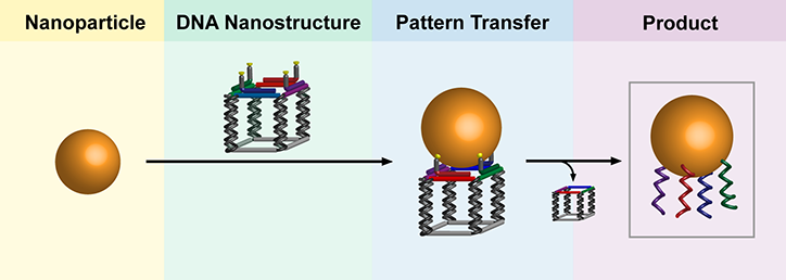
Enter the nanoparticle equivalent of the printing press. It’s efficient, re-usable and carries more information than previously possible. In results reported online in Nature Chemistry, researchers from McGill’s Department of Chemistry outline a procedure for making a DNA [deoxyribonucleic acid] structure with a specific pattern of strands coming out of it; at the end of each strand is a chemical “sticky patch.”  When a gold nanoparticle is brought into contact to the DNA nanostructure, it sticks to the patches. The scientists then dissolve the assembly in distilled water, separating the DNA nanostructure into its component strands and leaving behind the DNA imprint on the gold nanoparticle. …

The researchers have made an illustration of their concept available,

Credit: Thomas Edwardson

Credit: Thomas Edwardson

“These encoded gold nanoparticles are unprecedented in their information content,” says senior author Hanadi Sleiman, who holds the Canada Research Chair in DNA Nanoscience. “The DNA nanostructures, for their part, can be re-used, much like stamps in an old printing press.”

The news release includes suggestions for possible future applications,

From stained glass to optoelectronics

Some of the properties of gold nanoparticles have been recognized for centuries.  Medieval artisans added gold chloride to molten glass to create the ruby-red colour in stained-glass windows – the result, as chemists figured out much later, of the light-scattering properties of tiny gold particles.

Now, the McGill researchers hope their new production technique will help pave the way for use of DNA-encoded nanoparticles in a range of cutting-edge technologies. First author Thomas Edwardson says the next step for the lab will be to investigate the properties of structures made from these new building blocks. “In much the same way that atoms combine to form complex molecules, patterned DNA gold particles can connect to neighbouring particles to form well-defined nanoparticle assemblies.”

These could be put to use in areas including optoelectronic nanodevices and biomedical sciences, the researchers say. The patterns of DNA strands could, for example, be engineered to target specific proteins on cancer cells, and thus serve to detect cancer or to selectively destroy cancer cells.

Here’s a link to and a citation for the paper,

Transfer of molecular recognition information from DNA nanostructures to gold nanoparticles by Thomas G. W. Edwardson, Kai Lin Lau, Danny Bousmail, Christopher J. Serpell, & Hanadi F. Sleiman. Nature Chemistry (2016)  doi:10.1038/nchem.2420 Published online 04 January 2016

This paper is behind a paywall.

*’also on EurekAlert’ added on Jan. 8, 2016.

Customizing DNA nanotubes quickly and cheaply

Building on some work published earlier this year, scientists from McGill University (Montréal, Québec) created a new technique for building DNA nanotubes block by block (my March 2, 2015 posting) and, now, the newest research from the McGill team features a way of making long DNA strands with that technique, as mentioned in a May 7, 2015 news item on Azonano,

Imagine taking strands of DNA – the material in our cells that determines how we look and function – and using it to build tiny structures that can deliver drugs to targets within the body or take electronic miniaturization to a whole new level.

While it may still sound like science fiction to most of us, researchers have been piecing together and experimenting with DNA structures for decades. And, in recent years, work by scientists such as McGill University chemistry professor Hanadi Sleiman has moved the use of man-made DNA structures closer to a variety of real-world applications.

But as these applications continue to develop, they require increasingly large and complex strands of DNA. That has posed a problem, because the automated systems used for making synthetic DNA can’t produce strands containing more than about 100 bases (the chemicals that link up to form the strands). It can take hundreds of these short strands to assemble nanotubes for applications such as smart drug-delivery systems.

Here’s a video featuring one of the researchers taking about this latest work from McGill University,

A May 6, 2015 McGill University news release, which originated the news item, describes the long DNA nanotubes in more detail,

In new research published May 5 in Nature Communications, however, Sleiman’’s team at McGill reports that it has devised a technique to create much longer strands of DNA, including custom-designed sequence patterns. What’s more, this approach also produces large amounts of these longer strands in just a few hours, making the process potentially more economical and commercially viable than existing techniques.

The new method involves piecing together small strands one after the other, so that they attach into a longer DNA strand with the help of an enzyme known as ligase.  A second enzyme, polymerase, is then used to generate many copies of the long DNA strand, yielding larger volumes of the material. The polymerase process has the added advantage of correcting any errors that may have been introduced into the sequence, amplifying only the correctly sequenced, full-length product.

Designer DNA materials

The team used these strands as a scaffold to make DNA nanotubes, demonstrating that the technique allows the length and functions of the tubes to be precisely programmed. “In the end, what we get is a long, synthetic DNA strand with exactly the sequence of bases that we want, and with exactly as many repeat units as we want,” explains Sleiman, who co-authored the study with Graham Hamblin, who recently completed his doctorate, and PhD student Janane Rahbani.

“This work opens the door toward a new design strategy in DNA nanotechnology,” Sleiman says. “This could provide access to designer DNA materials that are economical and can compete with cheaper, but less versatile technologies. In the future, uses could range from customized gene and protein synthesis, to applications in nanoelectronics, nano-optics, and medicine, including diagnosis and therapy.”

Here’s a link to and a citation for the paper,

Sequential growth of long DNA strands with user-defined patterns for nanostructures and scaffolds by Graham D. Hamblin, Janane F. Rahbani, & Hanadi F. Sleiman. Nature Communications 6, Article number: 7065 doi:10.1038/ncomms8065 Published 05 May 2015

This article is behind a paywall.

McGill University (Canada) researchers build DNA nanotubes block by block

McGill University (Montréal, Québec, Canada) researchers have found a new technique for creating DNA (deoxyribonucleic acid) nanotubes according to a Feb. 24, 2015 news item on Azonano,

Researchers at McGill University have developed a new, low-cost method to build DNA nanotubes block by block – a breakthrough that could help pave the way for scaffolds made from DNA strands to be used in applications such as optical and electronic devices or smart drug-delivery systems.

A Feb. 23, 2015 McGill University news release (also on EurekAlert), which originated the news item, describes current practice and the new technique,

Many researchers, including the McGill team, have previously constructed nanotubes using a method that relies on spontaneous assembly of DNA in solution. The new technique, reported today in Nature Chemistry, promises to yield fewer structural flaws than the spontaneous-assembly method. The building-block approach also makes it possible to better control the size and patterns of the DNA structures, the scientists report.

“Just like a Tetris game, where we manipulate the game pieces with the aim of creating a horizontal line of several blocks, we can now build long nanotubes block by block,” said Amani Hariri, a PhD student in McGill’s Department of Chemistry and lead author of the study. “By using a fluorescence microscope we can further visualize the formation of the tubes at each stage of assembly, as each block is tagged with a fluorescent compound that serves as a beacon. We can then count the number of blocks incorporated in each tube as it is constructed.”

This new technique was made possible by the development in recent years of single-molecule microscopy, which enables scientists to peer into the nano-world by turning the fluorescence of individual molecules on and off. (That groundbreaking work won three U.S.- and German-based scientists the 2014 Nobel Prize in Chemistry.)

Hariri’s research is jointly supervised by chemistry professors Gonzalo Cosa and Hanadi Sleiman, who co-authored the new study. Cosa’s research group specializes in single-molecule fluorescence techniques, while Sleiman’s uses DNA chemistry to design new materials for drug delivery and diagnostic tools.

The custom-built assembly technique developed through this collaboration “gives us the ability to monitor the nanotubes as we’re building them, and see their structure, robustness and morphology,” Cosa said.

“We wanted to control the nanotubes’ lengths and features one-by-one,” said Sleiman, who holds the Canada Research Chair in DNA Nanoscience. The resulting “designer nanotubes,” she adds, promise to be far cheaper to produce on a large scale than those created with so-called DNA origami, another innovative technique for using DNA as a nanoscale construction material.

Here’s a link to and a citation for the paper,

Stepwise growth of surface-grafted DNA nanotubes visualized at the single-molecule level by Amani A. Hariri, Graham D. Hamblin, Yasser Gidi, Hanadi F. Sleiman & Gonzalo Cosa. Nature Chemistry (2015) doi:10.1038/nchem.2184 Published online 23 February 2015

This article is behind a paywall.