The European Union’s Human Brain Project was announced in January 2013. It, along with the Graphene Flagship, had won a multi-year competition for the extraordinary sum of one million euros each to be paid out over a 10-year period. (My January 28, 2013 posting gives the details available at the time.)
At a little more than half-way through the project period, Ed Yong, in his July 22, 2019 article for The Atlantic, offers an update (of sorts),
Ten years ago, a neuroscientist said that within a decade he could simulate a human brain. Spoiler: It didn’t happen.
On July 22, 2009, the neuroscientist Henry Markram walked onstage at the TEDGlobal conference in Oxford, England, and told the audience that he was going to simulate the human brain, in all its staggering complexity, in a computer. His goals were lofty: “It’s perhaps to understand perception, to understand reality, and perhaps to even also understand physical reality.” His timeline was ambitious: “We can do it within 10 years, and if we do succeed, we will send to TED, in 10 years, a hologram to talk to you.” …
It’s been exactly 10 years. He did not succeed.
One could argue that the nature of pioneers is to reach far and talk big, and that it’s churlish to single out any one failed prediction when science is so full of them. (Science writers joke that breakthrough medicines and technologies always seem five to 10 years away, on a rolling window.) But Markram’s claims are worth revisiting for two reasons. First, the stakes were huge: In 2013, the European Commission awarded his initiative—the Human Brain Project (HBP)—a staggering 1 billion euro grant (worth about $1.42 billion at the time). Second, the HBP’s efforts, and the intense backlash to them, exposed important divides in how neuroscientists think about the brain and how it should be studied.
Markram’s goal wasn’t to create a simplified version of the brain, but a gloriously complex facsimile, down to the constituent neurons, the electrical activity coursing along them, and even the genes turning on and off within them. From the outset, the criticism to this approach was very widespread, and to many other neuroscientists, its bottom-up strategy seemed implausible to the point of absurdity. The brain’s intricacies—how neurons connect and cooperate, how memories form, how decisions are made—are more unknown than known, and couldn’t possibly be deciphered in enough detail within a mere decade. It is hard enough to map and model the 302 neurons of the roundworm C. elegans, let alone the 86 billion neurons within our skulls. “People thought it was unrealistic and not even reasonable as a goal,” says the neuroscientist Grace Lindsay, who is writing a book about modeling the brain. And what was the point? The HBP wasn’t trying to address any particular research question, or test a specific hypothesis about how the brain works. The simulation seemed like an end in itself—an overengineered answer to a nonexistent question, a tool in search of a use. …
…
Markram seems undeterred. In a recent paper, he and his colleague Xue Fan firmly situated brain simulations within not just neuroscience as a field, but the entire arc of Western philosophy and human civilization. And in an email statement, he told me, “Political resistance (non-scientific) to the project has indeed slowed us down considerably, but it has by no means stopped us nor will it.” He noted the 140 people still working on the Blue Brain Project, a recent set of positive reviews from five external reviewers, and its “exponentially increasing” ability to “build biologically accurate models of larger and larger brain regions.”
No time frame, this time, but there’s no shortage of other people ready to make extravagant claims about the future of neuroscience. In 2014, I attended TED’s main Vancouver conference and watched the opening talk, from the MIT Media Lab founder Nicholas Negroponte. In his closing words, he claimed that in 30 years, “we are going to ingest information. …
I’m happy to see the update. As I recall, there was murmuring almost immediately about the Human Brain Project (HBP). I never got details but it seemed that people were quite actively unhappy about the disbursements. Of course, this kind of uproar is not unusual when great sums of money are involved and the Graphene Flagship also had its rocky moments.
As for Yong’s contribution, I’m glad he’s debunking some of the hype and glory associated with the current drive to colonize the human brain and other efforts (e.g. genetics) which they often claim are the ‘future of medicine’.
To be fair. Yong is focused on the brain simulation aspect of the HBP (and Markram’s efforts in the Blue Brain Project) but there are other HBP efforts, as well, even if brain simulation seems to be the HBP’s main interest.
In 2013, the European Union funded the Human Brain Project, led by Markram, to the tune of $1.3 billion. Markram claimed that the project would create a simulation of the entire human brain on a supercomputer within a decade, revolutionising the treatment of Alzheimer’s disease and other brain disorders. Less than two years into it, the project was recognised to be mismanaged and its claims overblown, and Markram was asked to step down.[7][8]
On 8 October 2015, the Blue Brain Project published the first digital reconstruction and simulation of the micro-circuitry of a neonatal rat somatosensory cortex.[9]
…
I also looked up the Human Brain Project and, talking about their other efforts, was reminded that they have a neuromorphic computing platform, SpiNNaker (mentioned here in a January 24, 2019 posting; scroll down about 50% of the way). For anyone unfamiliar with the term, neuromorphic computing/engineering is what scientists call the effort to replicate the human brain’s ability to synthesize and process information in computing processors.
In fact, there was some discussion in 2013 that the Human Brain Project and the Graphene Flagship would have some crossover projects, e.g., trying to make computers more closely resemble human brains in terms of energy use and processing power.
The Human Brain Project’s (HBP) Silicon Brains webpage notes this about their neuromorphic computing platform,
Neuromorphic computing implements aspects of biological neural networks as analogue or digital copies on electronic circuits. The goal of this approach is twofold: Offering a tool for neuroscience to understand the dynamic processes of learning and development in the brain and applying brain inspiration to generic cognitive computing. Key advantages of neuromorphic computing compared to traditional approaches are energy efficiency, execution speed, robustness against local failures and the ability to learn.
Neuromorphic Computing in the HBP
In the HBP the neuromorphic computing Subproject carries out two major activities: Constructing two large-scale, unique neuromorphic machines and prototyping the next generation neuromorphic chips.
The large-scale neuromorphic machines are based on two complementary principles. The many-core SpiNNaker machine located in Manchester [emphasis mine] (UK) connects 1 million ARM processors with a packet-based network optimized for the exchange of neural action potentials (spikes). The BrainScaleS physical model machine located in Heidelberg (Germany) implements analogue electronic models of 4 Million neurons and 1 Billion synapses on 20 silicon wafers. Both machines are integrated into the HBP collaboratory and offer full software support for their configuration, operation and data analysis.
The most prominent feature of the neuromorphic machines is their execution speed. The SpiNNaker system runs at real-time, BrainScaleS is implemented as an accelerated system and operates at 10,000 times real-time. Simulations at conventional supercomputers typical run factors of 1000 slower than biology and cannot access the vastly different timescales involved in learning and development ranging from milliseconds to years.
Recent research in neuroscience and computing has indicated that learning and development are a key aspect for neuroscience and real world applications of cognitive computing. HBP is the only project worldwide addressing this need with dedicated novel hardware architectures.
I’ve highlighted Manchester because that’s a very important city where graphene is concerned. The UK’s National Graphene Institute is housed at the University of Manchester where graphene was first isolated in 2004 by two scientists, Andre Geim and Konstantin (Kostya) Novoselov. (For their effort, they were awarded the Nobel Prize for physics in 2010.)
Getting back to the HBP (and the Graphene Flagship for that matter), the funding should be drying up sometime around 2023 and I wonder if it will be possible to assess the impact.
I have two brain news bits, one about neural networks and quantum entanglement and another about how the brain operates in* more than three dimensions.
Quantum entanglement and neural networks
A June 13, 2017 news item on phys.org describes how machine learning can be used to solve problems in physics (Note: Links have been removed),
Machine learning, the field that’s driving a revolution in artificial intelligence, has cemented its role in modern technology. Its tools and techniques have led to rapid improvements in everything from self-driving cars and speech recognition to the digital mastery of an ancient board game.
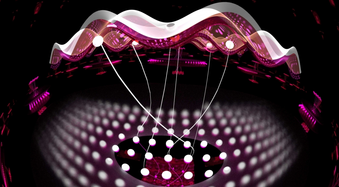
Now, physicists are beginning to use machine learning tools to tackle a different kind of problem, one at the heart of quantum physics. In a paper published recently in Physical Review X, researchers from JQI [Joint Quantum Institute] and the Condensed Matter Theory Center (CMTC) at the University of Maryland showed that certain neural networks—abstract webs that pass information from node to node like neurons in the brain—can succinctly describe wide swathes of quantum systems.
An artist’s rendering of a neural network with two layers. At the top is a real quantum system, like atoms in an optical lattice. Below is a network of hidden neurons that capture their interactions (Credit: E. Edwards/JQI)
A June 12, 2017 JQI news release by Chris Cesare, which originated the news item, describes how neural networks can represent quantum entanglement,
Dongling Deng, a JQI Postdoctoral Fellow who is a member of CMTC and the paper’s first author, says that researchers who use computers to study quantum systems might benefit from the simple descriptions that neural networks provide. “If we want to numerically tackle some quantum problem,” Deng says, “we first need to find an efficient representation.”
On paper and, more importantly, on computers, physicists have many ways of representing quantum systems. Typically these representations comprise lists of numbers describing the likelihood that a system will be found in different quantum states. But it becomes difficult to extract properties or predictions from a digital description as the number of quantum particles grows, and the prevailing wisdom has been that entanglement—an exotic quantum connection between particles—plays a key role in thwarting simple representations.
The neural networks used by Deng and his collaborators—CMTC Director and JQI Fellow Sankar Das Sarma and Fudan University physicist and former JQI Postdoctoral Fellow Xiaopeng Li—can efficiently represent quantum systems that harbor lots of entanglement, a surprising improvement over prior methods.
What’s more, the new results go beyond mere representation. “This research is unique in that it does not just provide an efficient representation of highly entangled quantum states,” Das Sarma says. “It is a new way of solving intractable, interacting quantum many-body problems that uses machine learning tools to find exact solutions.”
The result was a more complete account of the capabilities of certain neural networks to represent quantum states. In particular, the team studied neural networks that use two distinct groups of neurons. The first group, called the visible neurons, represents real quantum particles, like atoms in an optical lattice or ions in a chain. To account for interactions between particles, the researchers employed a second group of neurons—the hidden neurons—which link up with visible neurons. These links capture the physical interactions between real particles, and as long as the number of connections stays relatively small, the neural network description remains simple.
Specifying a number for each connection and mathematically forgetting the hidden neurons can produce a compact representation of many interesting quantum states, including states with topological characteristics and some with surprising amounts of entanglement.
Beyond its potential as a tool in numerical simulations, the new framework allowed Deng and collaborators to prove some mathematical facts about the families of quantum states represented by neural networks. For instance, neural networks with only short-range interactions—those in which each hidden neuron is only connected to a small cluster of visible neurons—have a strict limit on their total entanglement. This technical result, known as an area law, is a research pursuit of many condensed matter physicists.
These neural networks can’t capture everything, though. “They are a very restricted regime,” Deng says, adding that they don’t offer an efficient universal representation. If they did, they could be used to simulate a quantum computer with an ordinary computer, something physicists and computer scientists think is very unlikely. Still, the collection of states that they do represent efficiently, and the overlap of that collection with other representation methods, is an open problem that Deng says is ripe for further exploration.
Blue Brain is a Swiss government brain research initiative which officially came to life in 2006 although the initial agreement between the École Politechnique Fédérale de Lausanne (EPFL) and IBM was signed in 2005 (according to the project’s Timeline page). Moving on, the project’s latest research reveals something astounding (from a June 12, 2017 Frontiers Publishing press release on EurekAlert),
For most people, it is a stretch of the imagination to understand the world in four dimensions but a new study has discovered structures in the brain with up to eleven dimensions – ground-breaking work that is beginning to reveal the brain’s deepest architectural secrets.
Using algebraic topology in a way that it has never been used before in neuroscience, a team from the Blue Brain Project has uncovered a universe of multi-dimensional geometrical structures and spaces within the networks of the brain.
The research, published today in Frontiers in Computational Neuroscience, shows that these structures arise when a group of neurons forms a clique: each neuron connects to every other neuron in the group in a very specific way that generates a precise geometric object. The more neurons there are in a clique, the higher the dimension of the geometric object.
“We found a world that we had never imagined,” says neuroscientist Henry Markram, director of Blue Brain Project and professor at the EPFL in Lausanne, Switzerland, “there are tens of millions of these objects even in a small speck of the brain, up through seven dimensions. In some networks, we even found structures with up to eleven dimensions.”
Markram suggests this may explain why it has been so hard to understand the brain. “The mathematics usually applied to study networks cannot detect the high-dimensional structures and spaces that we now see clearly.”
If 4D worlds stretch our imagination, worlds with 5, 6 or more dimensions are too complex for most of us to comprehend. This is where algebraic topology comes in: a branch of mathematics that can describe systems with any number of dimensions. The mathematicians who brought algebraic topology to the study of brain networks in the Blue Brain Project were Kathryn Hess from EPFL and Ran Levi from Aberdeen University.
“Algebraic topology is like a telescope and microscope at the same time. It can zoom into networks to find hidden structures – the trees in the forest – and see the empty spaces – the clearings – all at the same time,” explains Hess.
In 2015, Blue Brain published the first digital copy of a piece of the neocortex – the most evolved part of the brain and the seat of our sensations, actions, and consciousness. In this latest research, using algebraic topology, multiple tests were performed on the virtual brain tissue to show that the multi-dimensional brain structures discovered could never be produced by chance. Experiments were then performed on real brain tissue in the Blue Brain’s wet lab in Lausanne confirming that the earlier discoveries in the virtual tissue are biologically relevant and also suggesting that the brain constantly rewires during development to build a network with as many high-dimensional structures as possible.
When the researchers presented the virtual brain tissue with a stimulus, cliques of progressively higher dimensions assembled momentarily to enclose high-dimensional holes, that the researchers refer to as cavities. “The appearance of high-dimensional cavities when the brain is processing information means that the neurons in the network react to stimuli in an extremely organized manner,” says Levi. “It is as if the brain reacts to a stimulus by building then razing a tower of multi-dimensional blocks, starting with rods (1D), then planks (2D), then cubes (3D), and then more complex geometries with 4D, 5D, etc. The progression of activity through the brain resembles a multi-dimensional sandcastle that materializes out of the sand and then disintegrates.”
The big question these researchers are asking now is whether the intricacy of tasks we can perform depends on the complexity of the multi-dimensional “sandcastles” the brain can build. Neuroscience has also been struggling to find where the brain stores its memories. “They may be ‘hiding’ in high-dimensional cavities,” Markram speculates.
###
…
About Blue Brain
The aim of the Blue Brain Project, a Swiss brain initiative founded and directed by Professor Henry Markram, is to build accurate, biologically detailed digital reconstructions and simulations of the rodent brain, and ultimately, the human brain. The supercomputer-based reconstructions and simulations built by Blue Brain offer a radically new approach for understanding the multilevel structure and function of the brain. http://bluebrain.epfl.ch
About Frontiers
Frontiers is a leading community-driven open-access publisher. By taking publishing entirely online, we drive innovation with new technologies to make peer review more efficient and transparent. We provide impact metrics for articles and researchers, and merge open access publishing with a research network platform – Loop – to catalyse research dissemination, and popularize research to the public, including children. Our goal is to increase the reach and impact of research articles and their authors. Frontiers has received the ALPSP Gold Award for Innovation in Publishing in 2014. http://www.frontiersin.org.
If you want to learn how something works, one strategy is to take it apart and put it back together again [also known as reverse engineering]. For 10 years, a global initiative called the Blue Brain Project–hosted at the Ecole Polytechnique Federale de Lausanne (EPFL)–has been attempting to do this digitally with a section of juvenile rat brain. The project presents a first draft of this reconstruction, which contains over 31,000 neurons, 55 layers of cells, and 207 different neuron subtypes, on October 8 [2015] in Cell.
Heroic efforts are currently being made to define all the different types of neurons in the brain, to measure their electrical firing properties, and to map out the circuits that connect them to one another. These painstaking efforts are giving us a glimpse into the building blocks and logic of brain wiring. However, getting a full, high-resolution picture of all the features and activity of the neurons within a brain region and the circuit-level behaviors of these neurons is a major challenge.
Henry Markram and colleagues have taken an engineering approach to this question by digitally reconstructing a slice of the neocortex, an area of the brain that has benefitted from extensive characterization. Using this wealth of data, they built a virtual brain slice representing the different neuron types present in this region and the key features controlling their firing and, most notably, modeling their connectivity, including nearly 40 million synapses and 2,000 connections between each brain cell type.
“The reconstruction required an enormous number of experiments,” says Markram, of the EPFL. “It paves the way for predicting the location, numbers, and even the amount of ion currents flowing through all 40 million synapses.”
Once the reconstruction was complete, the investigators used powerful supercomputers to simulate the behavior of neurons under different conditions. Remarkably, the researchers found that, by slightly adjusting just one parameter, the level of calcium ions, they could produce broader patterns of circuit-level activity that could not be predicted based on features of the individual neurons. For instance, slow synchronous waves of neuronal activity, which have been observed in the brain during sleep, were triggered in their simulations, suggesting that neural circuits may be able to switch into different “states” that could underlie important behaviors.
“An analogy would be a computer processor that can reconfigure to focus on certain tasks,” Markram says. “The experiments suggest the existence of a spectrum of states, so this raises new types of questions, such as ‘what if you’re stuck in the wrong state?'” For instance, Markram suggests that the findings may open up new avenues for explaining how initiating the fight-or-flight response through the adrenocorticotropic hormone yields tunnel vision and aggression.
The Blue Brain Project researchers plan to continue exploring the state-dependent computational theory while improving the model they’ve built. All of the results to date are now freely available to the scientific community at https://bbp.epfl.ch/nmc-portal.
Published by the renowned journal Cell, the paper is the result of a massive effort by 82 scientists and engineers at EPFL and at institutions in Israel, Spain, Hungary, USA, China, Sweden, and the UK. It represents the culmination of 20 years of biological experimentation that generated the core dataset, and 10 years of computational science work that developed the algorithms and built the software ecosystem required to digitally reconstruct and simulate the tissue.
The Hebrew University of Jerusalem’s Prof. Idan Segev, a senior author of the research paper, said: “With the Blue Brain Project, we are creating a digital reconstruction of the brain and using supercomputer simulations of its electrical behavior to reveal a variety of brain states. This allows us to examine brain phenomena within a purely digital environment and conduct experiments previously only possible using biological tissue. The insights we gather from these experiments will help us to understand normal and abnormal brain states, and in the future may have the potential to help us develop new avenues for treating brain disorders.”
Segev, a member of the Hebrew University’s Edmond and Lily Safra Center for Brain Sciences and director of the university’s Department of Neurobiology, sees the paper as building on the pioneering work of the Spanish anatomist Ramon y Cajal from more than 100 years ago: “Ramon y Cajal began drawing every type of neuron in the brain by hand. He even drew in arrows to describe how he thought the information was flowing from one neuron to the next. Today, we are doing what Cajal would be doing with the tools of the day: building a digital representation of the neurons and synapses, and simulating the flow of information between neurons on supercomputers. Furthermore, the digitization of the tissue is open to the community and allows the data and the models to be preserved and reused for future generations.”
While a long way from digitizing the whole brain, the study demonstrates that it is feasible to digitally reconstruct and simulate brain tissue, and most importantly, to reveal novel insights into the brain’s functioning. Simulating the emergent electrical behavior of this virtual tissue on supercomputers reproduced a range of previous observations made in experiments on the brain, validating its biological accuracy and providing new insights into the functioning of the neocortex. This is a first step and a significant contribution to Europe’s Human Brain Project, which Henry Markram founded, and where EPFL is the coordinating partner.
Cell has made a video abstract available (it can be found with the Hebrew University of Jerusalem press release)
Here’s a link to and a citation for the paper,
Reconstruction and Simulation of Neocortical Microcircuitry by Henry Markram, Eilif Muller, Srikanth Ramaswamy, Michael W. Reimann, Marwan Abdellah, Carlos Aguado Sanchez, Anastasia Ailamaki, Lidia Alonso-Nanclares, Nicolas Antille, Selim Arsever, Guy Antoine Atenekeng Kahou, Thomas K. Berger, Ahmet Bilgili, Nenad Buncic, Athanassia Chalimourda, Giuseppe Chindemi, Jean-Denis Courcol, Fabien Delalondre, Vincent Delattre, Shaul Druckmann, Raphael Dumusc, James Dynes, Stefan Eilemann, Eyal Gal, Michael Emiel Gevaert, Jean-Pierre Ghobril, Albert Gidon, Joe W. Graham, Anirudh Gupta, Valentin Haenel, Etay Hay, Thomas Heinis, Juan B. Hernando, Michael Hines, Lida Kanari, Daniel Keller, John Kenyon, Georges Khazen, Yihwa Kim, James G. King, Zoltan Kisvarday, Pramod Kumbhar, Sébastien Lasserre, Jean-Vincent Le Bé, Bruno R.C. Magalhães, Angel Merchán-Pérez, Julie Meystre, Benjamin Roy Morrice, Jeffrey Muller, Alberto Muñoz-Céspedes, Shruti Muralidhar, Keerthan Muthurasa, Daniel Nachbaur, Taylor H. Newton, Max Nolte, Aleksandr Ovcharenko, Juan Palacios, Luis Pastor, Rodrigo Perin, Rajnish Ranjan, Imad Riachi, José-Rodrigo Rodríguez, Juan Luis Riquelme, Christian Rössert, Konstantinos Sfyrakis, Ying Shi, Julian C. Shillcock, Gilad Silberberg, Ricardo Silva, Farhan Tauheed, Martin Telefont, Maria Toledo-Rodriguez, Thomas Tränkler, Werner Van Geit, Jafet Villafranca Díaz, Richard Walker, Yun Wang, Stefano M. Zaninetta, Javier DeFelipe, Sean L. Hill, Idan Segev, Felix Schürmann. Cell, Volume 163, Issue 2, p456–492, 8 October 2015 DOI: http://dx.doi.org/10.1016/j.cell.2015.09.029
This paper appears to be open access.
My most substantive description of the Blue Brain Project , previous to this, was in a Jan. 29, 2013 posting featuring the European Union’s (EU) Human Brain project and involvement from countries that are not members.
* I edited a redundant lede (That’s a virtual slice of a rat brain.), moved the second sentence to the lede while adding this: *about this virtual brain slice* on Oct. 16, 2015 at 0955 hours PST.
Its official title is the Montréal Neurological Institute and Hospital (Montréal Neuro) which is and has been, for several decades, an international centre for cutting edge neurological research. From the Jan. 28, 2013 news release on EurekAlert,
The Neuro
The Montreal Neurological Institute and Hospital — The Neuro, is a unique academic medical centre dedicated to neuroscience. Founded in 1934 by the renowned Dr. Wilder Penfield, The Neuro is recognized internationally for integrating research, compassionate patient care and advanced training, all key to advances in science and medicine. The Neuro is a research and teaching institute of McGill University and forms the basis for the Neuroscience Mission of the McGill University Health Centre.
Neuro researchers are world leaders in cellular and molecular neuroscience, brain imaging, cognitive neuroscience and the study and treatment of epilepsy, multiple sclerosis and neuromuscular disorders. For more information, visit theneuro.com.
Nonetheless, it was a little surprising to see that ‘The Neuro’ is part one of the biggest research projects in history since it’s the European Union, which is bankrolling the project (see my posting about the Jan. 28, 2013 announcement of the winning FET Flagship Initatives). Here’s more information about the project, its lead researchers, and Canada’s role, from the news release,
The goal of the Human Brain Project is to pull together all our existing knowledge about the human brain and to reconstruct the brain, piece by piece, in supercomputer-based models and simulations. The models offer the prospect of a new understanding of the human brain and its diseases and of completely new computing and robotic technologies. On January 28 [2013], the European Commission supported this vision, announcing that it has selected the HBP as one of two projects to be funded through the new FET [Future and Emerging Technologies] Flagship Program.
Federating more than 80 European and international research institutions, the Human Brain Project is planned to last ten years (2013-2023). The cost is estimated at 1.19 billion euros. The project will also associate some important North American and Japanese partners. It will be coordinated at the Ecole Polytechnique Fédérale de Lausanne (EPFL) in Switzerland, by neuroscientist Henry Markram with co-directors Karlheinz Meier of Heidelberg University, Germany, and Richard Frackowiak of Centre Hospitalier Universitaire Vaudois (CHUV) and the University of Lausanne (UNIL).
Canada’s role in this international project is through Dr. Alan Evans of the Montreal Neurological Institute (MNI) at McGill University. His group has developed a high-performance computational platform for neuroscience (CBRAIN) and multi-site databasing technologies that will be used to assemble brain imaging data across the HBP. He is also collaborating with European scientists on the creation of ultra high-resolution 3D brain maps. «This ambitious project will integrate data across all scales, from molecules to whole-brain organization. It will have profound implications for our understanding of brain development in children and normal brain function, as well as for combatting brain disorders such as Alzheimer’s Disease,» said Dr. Evans. “The MNI’s pioneering work on brain imaging technology has led to significant advances in our understanding of the brain and neurological disorders,” says Dr. Guy Rouleau, Director of the MNI. “I am proud that our expertise is a key contributor to this international program focused on improving quality of life worldwide.”
“The Canadian Institutes of Health Research (CIHR) is delighted to acknowledge the outstanding contributions of Dr. Evans and his team. Their work on the CBRAIN infrastructure and this leading-edge HBP will allow the integration of Canadian neuroscientists into an eventual global brain project,” said Dr. Anthony Phillips, Scientific Director for the CIHR Institute of Neurosciences, Mental Health and Addiction. “Congratulations to the Canadian and European researchers who will be working collaboratively towards the same goal which is to provide insights into neuroscience that will ultimately improve people’s health.”
“From mapping the sensory and motor cortices of the brain to pioneering work on the mechanisms of memory, McGill University has long been synonymous with world-class neuroscience research,” says Dr. Rose Goldstein, Vice-Principal (Research and International Relations). “The research of Dr. Evans and his team marks an exciting new chapter in our collective pursuit to unlock the potential of the human brain and the entire nervous system – a critical step that would not be possible without the generous support of the European Commission and the FET Flagship Program.”
Canada is not the only non-European Union country making an announcement about its role in this extraordinary project. There’s a Jan. 28, 2013 news release on EurekAlert touting Israel’s role,
The European Commission has chosen the Human Brain Project, in which the Hebrew University of Jerusalem is participating, as one of two Future and Emerging Technologies Flagship topics. The enterprise will receive funding of 1.19 billion euros over the next decade.
The project will bring together top scientists from around the world who will work on one of the great challenges of modern science: understanding the human brain. Participating from Israel will a team of eight scientists, led by Prof. Idan Segev of the Edmond and Lily Safra Center for Brain Sciences (ELSC) at the Hebrew University, Prof. Yadin Dudai of the Weizmann Institute of Science, and Dr. Mira Marcus-Kalish of Tel Aviv University.
…
More than 80 universities and research institutions in Europe and the world will be involved in the ten-year Human Brain Project, which will commence later this year and operate until the year 2023. The project will be centered at the Ecole Polytechnique Federale de Lausanne (EPFL) in Switzerland, headed by Prof. Henry Markram, a former Israeli who was recruited ten years ago to the EPFL.
The participation of the Israeli scientists testifies to the leading role that Israeli brain research occupies in the world, said Israeli President Shimon Peres. “Israel has put brain research at the heart of its efforts for the coming decade, and our country is already spearheading the global effort towards the betterment of our understanding of mankind. I am confident that the forthcoming discoveries will benefit a wide range of domains, from health to industry, as well as our society as a whole,” Peres said.
“The human brain is the most complex and amazing structure in the universe, yet we are very far from understanding it. In a way, we are strangers to ourselves. Unraveling the mysteries of the brain will help us understand our functioning, our choices, and ultimately ourselves. I congratulate the European Commission for its vision in selecting the Human Brain Project as a Flagship Mission for the forthcoming decade,” said Peres.
What’s amusing is that as various officials and interested parties (such as myself) wax lyrical about these projects, most of the rest of the world is serenely oblivious to it all.
The European Commission has announced the two winners of its FET (Future and Emerging Technologies) Flagships Initiative in a Jan. 28, 2013 news release,
The winning Graphene and Human Brain initiatives are set to receive one billion euros each, to deliver 10 years of world-beating science at the crossroads of science and technology. Each initiative involves researchers from at least 15 EU Member States and nearly 200 research institutes.
“Graphene” will investigate and exploit the unique properties of a revolutionary carbon-based material. Graphene is an extraordinary combination of physical and chemical properties: it is the thinnest material, it conducts electricity much better than copper, it is 100-300 times stronger than steel and it has unique optical properties. The use of graphene was made possible by European scientists in 2004, and the substance is set to become the wonder material of the 21st century, as plastics were to the 20th century, including by replacing silicon in ICT products.
The “Human Brain Project” will create the world’s largest experimental facility for developing the most detailed model of the brain, for studying how the human brain works and ultimately to develop personalised treatment of neurological and related diseases. This research lays the scientific and technical foundations for medical progress that has the potential to will dramatically improve the quality of life for millions of Europeans.
The European Commission will support “Graphene” and the “Human Brain Project” as FET “flagships” over 10 years through its research and innovation funding programmes. Sustained funding for the full duration of the project will come from the EU’s research framework programmes, principally from the Horizon 2020 programme (2014-2020) which is currently negotiated in the European Parliament and Council.
European Commission Vice President Neelie Kroes said: “Europe’s position as a knowledge superpower depends on thinking the unthinkable and exploiting the best ideas. This multi-billion competition rewards home-grown scientific breakthroughs and shows that when we are ambitious we can develop the best research in Europe. To keep Europe competitive, to keep Europe as the home of scientific excellence, EU governments must agree an ambitious budget for the Horizon 2020 programme in the coming weeks.”
“Graphene” is led by Prof. Jari Kinaret, from Sweden’s Chalmers University. The Flagship involves over 100 research groups, with 136 principal investigators, including four Nobel laureates. “The Human Brain Project” involves scientists from 87 institutions and is led by Prof. Henry Markram of the École Polytechnique Fédérale de Lausanne.
As noted in my Jan. 24, 2013 posting about the new Cambridge Graphene Centre in the UK, while the Graphene flagship lead is from Sweden, the UK has more educational institutions than any other country party to the flagship consortium.
Here are some funding details from the Jan. 28, 2013 news release,
Horizon 2020 is the new EU programme for research and innovation, presented by the Commission as part of its EU budget proposal for 2014 to 2020. In order to give a boost to research and innovation as a driver of growth and jobs, the Commission has proposed an ambitious budget of €80 billion over seven years, including the FET flagship programme itself.
The winners will receive up to €54 million from the European Commission’s ICT 2013 Work Programme. Further funding will come from subsequent EU research framework programmes, private partners including universities, Member States and industry.
1 billion Euros sounds like a lot of money but it’s being paid out over 10 years (100 million per year) and through many institutional layers at the European Commission and at the educational institutions themselves. One wonders how much of the money will go to research rather than administration.
2000th posting: My heartfelt thanks to everyone who has taken the time to read this blog and and to those who’ve taken the time to comment on the blog, on Twitter, or directly to me. Your interest has kept this blog going far longer than I believed it would.
I hinted about some related work at the University of Waterloo earlier this week in my Nov. 26, 2012 posting (Existential risk) about a proposed centre at the University of Cambridge which would be tasked with examining possible risks associated with ‘ultra intelligent machines’. Today (Science (magazine) published an article about SPAUN (Semantic Pointer Architecture Unified Network) [behind a paywall])and its ability to solve simple arithmetic and perform other tasks as well.
Spaun sees a series of digits: 1 2 3; 5 6 7; 3 4 ?. Its neurons fire, and it calculates the next logical number in the sequence. It scrawls out a 5, in legible if messy writing.
This is an unremarkable feat for a human, but Spaun is actually a simulated brain. It contains2.5 millionvirtual neurons — many fewer than the 86 billion in the average human head, but enough to recognize lists of numbers, do simple arithmetic and solve reasoning problems.
Here’s a video demonstration, from the University of Waterloo’s Nengo Neural Simulator home page,
… The model captures biological details of each neuron, including which neurotransmitters are used, how voltages are generated in the cell, and how they communicate. Spaun uses this network of neurons to process visual images in order to control an arm that draws Spaun’s answers to perceptual, cognitive and motor tasks. …
“This is the first model that begins to get at how our brains can perform a wide variety of tasks in a flexible manner—how the brain coordinates the flow of information between different areas to exhibit complex behaviour,” said Professor Chris Eliasmith, Director of the Centre for Theoretical Neuroscience at Waterloo. He is Canada Research Chair in Theoretical Neuroscience, and professor in Waterloo’s Department of Philosophy and Department of Systems Design Engineering.
Unlike other large brain models, Spaun can perform several tasks. Researchers can show patterns of digits and letters the model’s eye, which it then processes, causing it to write its responses to any of eight tasks. And, just like the human brain, it can shift from task to task, recognizing an object one moment and memorizing a list of numbers the next. [emphasis mine] Because of its biological underpinnings, Spaun can also be used to understand how changes to the brain affect changes to behaviour.
“In related work, we have shown how the loss of neurons with aging leads to decreased performance on cognitive tests,” said Eliasmith. “More generally, we can test our hypotheses about how the brain works, resulting in a better understanding of the effects of drugs or damage to the brain.”
In addition, the model provides new insights into the sorts of algorithms that might be useful for improving machine intelligence. [emphasis mine] For instance, it suggests new methods for controlling the flow of information through a large system attempting to solve challenging cognitive tasks.
Laura Sanders’ Nov. 29, 2012 article for ScienceNews suggests that there is some controversy as to whether or not SPAUN does resemble a human brain,
… Henry Markram, who leads a different project to reconstruct the human brain called the Blue Brain, questions whether Spaun really captures human brain behavior. Because Spaun’s design ignores some important neural properties, it’s unlikely to reveal anything about the brain’s mechanics, says Markram, of the Swiss Federal Institute of Technology in Lausanne. “It is not a brain model.”
Personally, I have a little difficulty seeing lines of code as ever being able to truly simulate brain activity. I think the notion of moving to something simpler (using fewer neurons as the Eliasmith team does) is a move in the right direction but I’m still more interested in devices such as the memristor and the electrochemical atomic switch and their potential.
Atomic or electrochemical atomic switches and neuromorphic engineering briefly mentioned (scroll 1/2 way down) in my Oct. 17, 2011 posting.
ETA Dec. 19, 2012: There was an AMA (ask me anything) session on Reddit with the SPAUN team in early December, if you’re interested, you can still access the questions and answers,
I’ve decided to do a roundup of the various brain-related projects I’ve been coming across in the last several months. I was inspired by this article (Real-life Jedi: Pushing the limits of mind control) by Katia Moskvitch,
You don’t have to be a Jedi to make things move with your mind.
Granted, we may not be able to lift a spaceship out of a swamp like Yoda does in The Empire Strikes Back, but it is possible to steer a model car, drive a wheelchair and control a robotic exoskeleton with just your thoughts.
…
We are standing in a testing room at IBM’s Emerging Technologies lab in Winchester, England.
On my head is a strange headset that looks like a black plastic squid. Its 14 tendrils, each capped with a moistened electrode, are supposed to detect specific brain signals.
In front of us is a computer screen, displaying an image of a floating cube.
As I think about pushing it, the cube responds by drifting into the distance.
Moskvitch goes on to discuss a number of projects that translate thought into movement via various pieces of equipment before she mentions a project at Brown University (US) where researchers are implanting computer chips into brains,
Headsets and helmets offer cheap, easy-to-use ways of tapping into the mind. But there are other,
Imagine some kind of a wireless computer device in your head that you’ll use for mind control – what if people hacked into that”
…
At Brown Institute for Brain Science in the US, scientists are busy inserting chips right into the human brain.
The technology, dubbed BrainGate, sends mental commands directly to a PC.
Subjects still have to be physically “plugged” into a computer via cables coming out of their heads, in a setup reminiscent of the film The Matrix. However, the team is now working on miniaturising the chips and making them wireless.
The purpose of the first phase of the pilot clinical study of the BrainGate2 Neural Interface System is to obtain preliminary device safety information and to demonstrate the feasibility of people with tetraplegia using the System to control a computer cursor and other assistive devices with their thoughts. Another goal of the study is to determine the participants’ ability to operate communication software, such as e-mail, simply by imagining the movement of their own hand. The study is invasive and requires surgery.
Individuals with limited or no ability to use both hands due to cervical spinal cord injury, brainstem stroke, muscular dystrophy, or amyotrophic lateral sclerosis (ALS) or other motor neuron diseases are being recruited into a clinical study at Massachusetts General Hospital (MGH) and Stanford University Medical Center. Clinical trial participants must live within a three-hour drive of Boston, MA or Palo Alto, CA. Clinical trial sites at other locations may be opened in the future. The study requires a commitment of 13 months.
They have been recruiting since at least November 2011, from the Nov. 14, 2011 news item by Tanya Lewis on MedicalXpress,
Stanford University researchers are enrolling participants in a pioneering study investigating the feasibility of people with paralysis using a technology that interfaces directly with the brain to control computer cursors, robotic arms and other assistive devices.
…
The pilot clinical trial, known as BrainGate2, is based on technology developed at Brown University and is led by researchers at Massachusetts General Hospital, Brown and the Providence Veterans Affairs Medical Center. The researchers have now invited the Stanford team to establish the only trial site outside of New England.
Under development since 2002, BrainGate is a combination of hardware and software that directly senses electrical signals in the brain that control movement. The device — a baby-aspirin-sized array of electrodes — is implanted in the cerebral cortex (the outer layer of the brain) and records its signals; computer algorithms then translate the signals into digital instructions that may allow people with paralysis to control external devices.
Confusingly, there seemto be two BrainGate organizations. One appears to be a research entity where a number of institutions collaborate and the other is some sort of jointly held company. From the About Us webpage of the BrainGate research entity,
In the late 1990s, the initial translation of fundamental neuroengineering research from “bench to bedside” – that is, to pilot clinical testing – would require a level of financial commitment ($10s of millions) available only from private sources. In 2002, a Brown University spin-off/startup medical device company, Cyberkinetics, Inc. (later, Cyberkinetics Neurotechnology Systems, Inc.) was formed to collect the regulatory permissions and financial resources required to launch pilot clinical trials of a first-generation neural interface system. The company’s efforts and substantial initial capital investment led to the translation of the preclinical research at Brown University to an initial human device, the BrainGate Neural Interface System [Caution: Investigational Device. Limited by Federal Law to Investigational Use]. The BrainGate system uses a brain-implantable sensor to detect neural signals that are then decoded to provide control signals for assistive technologies. In 2004, Cyberkinetics received from the U.S. Food and Drug Administration (FDA) the first of two Investigational Device Exemptions (IDEs) to perform this research. Hospitals in Rhode Island, Massachusetts, and Illinois were established as clinical sites for the pilot clinical trial run by Cyberkinetics. Four trial participants with tetraplegia (decreased ability to use the arms and legs) were enrolled in the study and further helped to develop the BrainGate device. Initial results from these trials have been published or presented, with additional publications in preparation.
While scientific progress towards the creation of this promising technology has been steady and encouraging, Cyberkinetics’ financial sponsorship of the BrainGate research – without which the research could not have been started – began to wane. In 2007, in response to business pressures and changes in the capital markets, Cyberkinetics turned its focus to other medical devices. Although Cyberkinetics’ own funds became unavailable for BrainGate research, the research continued through grants and subcontracts from federal sources. By early 2008 it became clear that Cyberkinetics would eventually need to withdraw completely from directing the pilot clinical trials of the BrainGate device. Also in 2008, Cyberkinetics spun off its device manufacturing to new ownership, BlackRock Microsystems, Inc., which now produces and is further developing research products as well as clinically-validated (510(k)-cleared) implantable neural recording devices.
Beginning in mid 2008, with the agreement of Cyberkinetics, a new, fully academically-based IDE application (for the “BrainGate2 Neural Interface System”) was developed to continue this important research. In May 2009, the FDA provided a new IDE for the BrainGate2 pilot clinical trial. [Caution: Investigational Device. Limited by Federal Law to Investigational Use.] The BrainGate2 pilot clinical trial is directed by faculty in the Department of Neurology at Massachusetts General Hospital, a teaching affiliate of Harvard Medical School; the research is performed in close scientific collaboration with Brown University’s Department of Neuroscience, School of Engineering, and Brown Institute for Brain Sciences, and the Rehabilitation Research and Development Service of the U.S. Department of Veteran’s Affairs at the Providence VA Medical Center. Additionally, in late 2011, Stanford University joined the BrainGate Research Team as a clinical site and is currently enrolling participants in the clinical trial. This interdisciplinary research team includes scientific partners from the Functional Electrical Stimulation Center at Case Western Reserve University and the Cleveland VA Medical Center. As was true of the decades of fundamental, preclinical research that provided the basis for the recent clinical studies, funding for BrainGate research is now entirely from federal and philanthropic sources.
The BrainGate Research Team at Brown University, Massachusetts General Hospital, Stanford University, and Providence VA Medical Center comprises physicians, scientists, and engineers working together to advance understanding of human brain function and to develop neurotechnologies for people with neurologic disease, injury, or limb loss.
The BrainGate™ Co. is a privately-held firm focused on the advancement of the BrainGate™ Neural Interface System. The Company owns the Intellectual property of the BrainGate™ system as well as new technology being developed by the BrainGate company. In addition, the Company also owns the intellectual property of Cyberkinetics which it purchased in April 2009.
Meanwhile, in Europe there are two projects BrainAble and the Human Brain Project. The BrainAble project is similar to BrainGate in that it is intended for people with injuries but they seem to be concentrating on a helmet or cap for thought transmission (as per Moskovitch’s experience at the beginning of this posting). From the Feb. 28, 2012 news item on Science Daily,
In the 2009 film Surrogates, humans live vicariously through robots while safely remaining in their own homes. That sci-fi future is still a long way off, but recent advances in technology, supported by EU funding, are bringing this technology a step closer to reality in order to give disabled people more autonomy and independence than ever before.
…
“Our aim is to give people with motor disabilities as much autonomy as technology currently allows and in turn greatly improve their quality of life,” says Felip Miralles at Barcelona Digital Technology Centre, a Spanish ICT research centre.
Mr. Miralles is coordinating the BrainAble* project (http://www.brainable.org/), a three-year initiative supported by EUR 2.3 million in funding from the European Commission to develop and integrate a range of different technologies, services and applications into a commercial system for people with motor disabilities.
In terms of HCI [human-computer interface], BrainAble improves both direct and indirect interaction between the user and his smart home. Direct control is upgraded by creating tools that allow controlling inner and outer environments using a “hybrid” Brain Computer Interface (BNCI) systemable to take into account other sources of information such as measures of boredom, confusion, frustration by means of the so-called physiological and affective sensors.
Furthermore, interaction is enhanced by means of Ambient Intelligence (AmI) focused on creating a proactive and context-aware environments by adding intelligence to the user’s surroundings. AmI’s main purpose is to aid and facilitate the user’s living conditions by creating proactive environments to provide assistance.
Human-Computer Interfaces are complemented by an intelligent Virtual Reality-based user interface with avatars and scenarios that will help the disabled move around freely, and interact with any sort of devices. Even more the VR will provide self-expression assets using music, pictures and text, communicate online and offline with other people, play games to counteract cognitive decline, and get trained in new functionalities and tasks.
Perhaps this video helps,
Another European project, NeuroCare, which I discussed in my March 5, 2012 posting, is focused on creating neural implants to replace damaged and/or destroyed sensory cells in the eye or the ear.
The Human Brain Project is, despite its title, a neuromorphic engineering project (although the researchers do mention some medical applications on the project’s home page) in common with the work being done at the University of Michigan/HRL Labs mentioned in my April 19, 2012 posting (A step closer to artificial synapses courtesy of memritors) about that project. From the April 11, 2012 news item about the Human Brain Project on Science Daily,
Researchers at the EPFL [Ecole Polytechnique Fédérale de Lausanne] have discovered rules that relate the genes that a neuron switches on and off, to the shape of that neuron, its electrical properties and its location in the brain.
The discovery, using state-of-the-art informatics tools, increases the likelihood that it will be possible to predict much of the fundamental structure and function of the brain without having to measure every aspect of it. That in turn makes the Holy Grail of modelling the brain in silico — the goal of the proposed Human Brain Project — a more realistic, less Herculean, prospect. “It is the door that opens to a world of predictive biology,” says Henry Markram, the senior author on the study, which is published this week in PLoS ONE.
Here’s a bit more about the Human Brain Project (from the home page),
Today, simulating a single neuron requires the full power of a laptop computer. But the brain has billions of neurons and simulating all them simultaneously is a huge challenge. To get round this problem, the project will develop novel techniques of multi-level simulation in which only groups of neurons that are highly active are simulated in detail. But even in this way, simulating the complete human brain will require a computer a thousand times more powerful than the most powerful machine available today. This means that some of the key players in the Human Brain Project will be specialists in supercomputing. Their task: to work with industry to provide the project with the computing power it will need at each stage of its work.
The Human Brain Project will impact many different areas of society. Brain simulation will provide new insights into the basic causes of neurological diseases such as autism, depression, Parkinson’s, and Alzheimer’s. It will give us new ways of testing drugs and understanding the way they work. It will provide a test platform for new drugs that directly target the causes of disease and that have fewer side effects than current treatments. It will allow us to design prosthetic devices to help people with disabilities. The benefits are potentially huge. As world populations grow older, more than a third will be affected by some kind of brain disease. Brain simulation provides us with a powerful new strategy to tackle the problem.
The project also promises to become a source of new Information Technologies. Unlike the computers of today, the brain has the ability to repair itself, to take decisions, to learn, and to think creatively – all while consuming no more energy than an electric light bulb. The Human Brain Project will bring these capabilities to a new generation of neuromorphic computing devices, with circuitry directly derived from the circuitry of the brain. The new devices will help us to build a new generation of genuinely intelligent robots to help us at work and in our daily lives.
The Human Brain Project builds on the work of the Blue Brain Project. Led by Henry Markram of the Ecole Polytechnique Fédérale de Lausanne (EPFL), the Blue Brain Project has already taken an essential first towards simulation of the complete brain. Over the last six years, the project has developed a prototype facility with the tools, know-how and supercomputing technology necessary to build brain models, potentially of any species at any stage in its development. As a proof of concept, the project has successfully built the first ever, detailed model of the neocortical column, one of the brain’s basic building blocks.
The Human Brain Project is a flagship project in contention for the 1B Euro research prize that I’ve mentioned in the context of the GRAPHENE-CA flagship project (my Feb. 13, 2012 posting gives a better description of these flagship projects while mentioned both GRAPHENE-CA and another brain-computer interface project, PRESENCCIA).
Part of the reason for doing this roundup, is the opportunity to look at a number of these projects in one posting; the effect is more overwhelming than I expected.
For anyone who’s interested in Markram’s paper (open access),
Georges Khazen, Sean L. Hill, Felix Schürmann, Henry Markram. Combinatorial Expression Rules of Ion Channel Genes in Juvenile Rat (Rattus norvegicus) Neocortical Neurons. PLoS ONE, 2012; 7 (4): e34786 DOI: 10.1371/journal.pone.0034786
I do have earlier postings on brains and neuroprostheses, one of the more recent ones is this March 16, 2012 posting. Meanwhile, there are new announcements from Northwestern University (US) and the US National Institutes of Health (National Institute of Neurological Disorders and Stroke). From the April 18, 2012 news item (originating from the National Institutes of Health) on Science Daily,
An artificial connection between the brain and muscles can restore complex hand movements in monkeys following paralysis, according to a study funded by the National Institutes of Health.
In a report in the journal Nature, researchers describe how they combined two pieces of technology to create a neuroprosthesis — a device that replaces lost or impaired nervous system function. One piece is a multi-electrode array implanted directly into the brain which serves as a brain-computer interface (BCI). The array allows researchers to detect the activity of about 100 brain cells and decipher the signals that generate arm and hand movements. The second piece is a functional electrical stimulation (FES) device that delivers electrical current to the paralyzed muscles, causing them to contract. The brain array activates the FES device directly, bypassing the spinal cord to allow intentional, brain-controlled muscle contractions and restore movement.
A new Northwestern Medicine brain-machine technology delivers messages from the brain directly to the muscles — bypassing the spinal cord — to enable voluntary and complex movement of a paralyzed hand. The device could eventually be tested on, and perhaps aid, paralyzed patients.
…
The research was done in monkeys, whose electrical brain and muscle signals were recorded by implanted electrodes when they grasped a ball, lifted it and released it into a small tube. Those recordings allowed the researchers to develop an algorithm or “decoder” that enabled them to process the brain signals and predict the patterns of muscle activity when the monkeys wanted to move the ball.
These experiments were performed by Christian Ethier, a post-doctoral fellow, and Emily Oby, a graduate student in neuroscience, both at the Feinberg School of Medicine. The researchers gave the monkeys a local anesthetic to block nerve activity at the elbow, causing temporary, painless paralysis of the hand. With the help of the special devices in the brain and the arm — together called a neuroprosthesis — the monkeys’ brain signals were used to control tiny electric currents delivered in less than 40 milliseconds to their muscles, causing them to contract, and allowing the monkeys to pick up the ball and complete the task nearly as well as they did before.
“The monkey won’t use his hand perfectly, but there is a process of motor learning that we think is very similar to the process you go through when you learn to use a new computer mouse or a different tennis racquet. Things are different and you learn to adjust to them,” said Miller [Lee E. Miller], also a professor of physiology and of physical medicine and rehabilitation at Feinberg and a Sensory Motor Performance Program lab chief at the Rehabilitation Institute of Chicago.
The National Institutes of Health news item supplies a little history and background for this latest breakthrough while the Northwestern University news item offers more technical details more technical details.
You can find the researchers’ paper with this citation (assuming you can get past the paywall,
C. Ethier, E. R. Oby, M. J. Bauman, L. E. Miller. Restoration of grasp following paralysis through brain-controlled stimulation of muscles. Nature, 2012; DOI: 10.1038/nature10987
I was surprised to find the Health Research Fund of Québec listed as one of the funders but perhaps Christian Ethier has some connection with the province.