Tag Archives: Hokkaido University

A new lipid nanoparticle (LNP) delivery system for CRISPR-Cas9) gene editing

The first time lipid nanoparticles were mentioned here as a delivery system for CRISPR-Cas9 editing was in a January 26, 2018 posting featuring work at the Massachusetts Institute of Technology (MIT). This latest research on the topic comes from Japan according to a March 2, 2023 news item on phys.org,

Gene therapy is a potential mode of treatment for a wide variety of diseases caused by genetic mutations. While it has been an area of diverse and intense research, historically, only a very few patients have been treated using gene therapy—and fewer still cured. The advent of the genetic modification technique called CRISPR-Cas9 in 2012 has revolutionized gene therapy—as well as biology as a whole—and it has recently entered clinical trials for the treatment of some diseases in humans.

Haruno Onuma, Yusuke Sato and Hideyoshi Harashima at Hokkaido University have developed a new delivery system for CRISPR-Cas9, based on lipid nanoparticles (LNPs), that could greatly increases the efficiency of in vivo gene therapy. Their findings were published in the Journal of Controlled Release.

A March 2, 2023 Hokkaido University press release (also on EurekAlert), which originated the news item, provides details about the researchers’ new technique,

“There are broadly two ways of treating diseases with gene therapy,” Sato explained, “ex vivo, where cells are subjected to the desired modifications in the laboratory and then introduced into the patient, and in vivo, where the treatment is administered to the patient to change the cells in their body. Safe and effective in vivo treatment is the ultimate aspiration of gene therapy, as it would be a straightforward process for patients and healthcare providers. LNPs can function as a vehicle for the safe and effective delivery of such therapies.”

CRISPR-Cas9 consists of a large molecule composed of the Cas9 protein and guide RNA. The guide RNA binds to a specific, complementary DNA sequence, and the Cas9 protein cuts that sequence, allowing it to be modified. The guide RNA can be altered to target specific DNA sequences to be modified.

“In a previous study, we discovered that additional DNA molecules, called ssODNs, ensure that the CRISPR-Cas9 molecule is loaded into the LNPs (CRISPR-LNPs),” Harashima elucidated. “In this study,  we again used ssODNs, but they were carefully designed so that they would not inhibit the function of the guide RNA.” 

Using a guide RNA targeting the expression of a protein called transthyretin, they evaluated the effectiveness of the CRISPR-LNPs in mice models. CRISPR-LNPs with ssODNs that dissociated from the guide RNA at room temperature were most effective at reducing serum transthyretin: two consecutive doses, one day apart, reduced it by 80%.

“We have demonstrated the optimal ssODN sequence affinity that ensures the loading and the release of CRISPR-Cas9 at the target location; and that this system can be used to edit cells in vivo,” concluded Onuma. “We will continue to improve the design of ssODNs, as well as to develop optimal lipid formulations to increase the effectiveness of delivery.” 

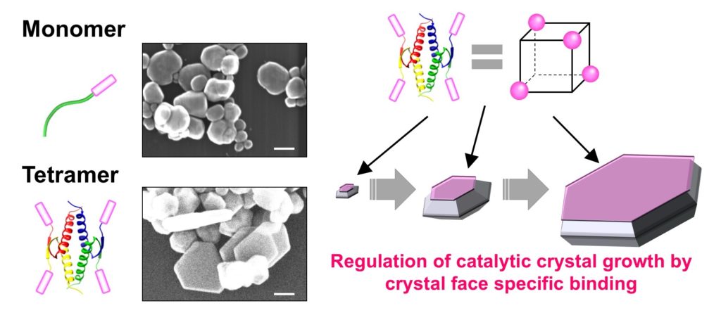
The image and caption helped me with better understanding the technique described in the press release,

The RNP-ssODN is designed to ensure the CRISPR-Cas9 molecule is encapsulated by the LNP. Once inside the cells, the ssODN dissociates and CRISPR-Cas9 can carry out its effect. (Haruno Onuma, Yusuke Sato, Hideyoshi Harashima. Journal of Controlled Release. February 10, 2023).

Here’s a link to and a citation for the paper,

Lipid nanoparticle-based ribonucleoprotein delivery for in vivo genome editing by Haruno Onuma, Yusuke Sato, Hideyoshi Harashima. Journal of Controlled Release Volume 355, March 2023, Pages 406-416 DOI: https://doi.org/10.1016/j.jconrel.2023.02.008

This paper is behind a paywall.

Put a ring on it: preventing clumps of gold nanoparticles

Caption: A comparison of how linear PEG (left) and cyclic PEG (right) attach to a gold nanoparticle Credit: Yubo Wang, Takuya Yamamoto

A January 20, 2021 news item on phys.org focuses on work designed to stop gold nanoparticles from clumping together (Note: A link has been removed),

Hokkaido University scientists have found a way to prevent gold nanoparticles from clumping, which could help towards their use as an anti-cancer therapy.

Attaching ring-shaped synthetic compounds to gold nanoparticles helps them retain their essential light-absorbing properties, Hokkaido University researchers report in the journal Nature Communications.

A January 20, 2021 Hokkaido University press release (also on EurekAlert but published Jan. 21, 2020), which originated the news item, elaborates on the work,

Metal nanoparticles have unique light-absorbing properties, making them interesting for a wide range of optical, electronic and biomedical applications. For example, if delivered to a tumour, they could react with applied light to kill cancerous tissue. A problem with this approach, though, is that they easily clump together in solution, losing their ability to absorb light. This clumping happens in response to a variety of factors, including temperature, salt concentration and acidity.

Scientists have been trying to find ways to ensure nanoparticles stay dispersed in their target environments. Covering them with polyethylene glycol, otherwise known as PEG, has been relatively successful at this in the case of gold nanoparticles. PEG is biocompatible and can prevent gold surfaces from clumping together in the laboratory and in living organisms, but improvements are still needed.

Applied chemist Takuya Yamamoto and colleagues at Hokkaido University, The University of Tokyo, and Tokyo Institute of Technology found that mixing gold nanoparticles with ring-shaped PEG, rather than the normally linear PEG, significantly improved dispersion. The ‘cyclic-PEG’ (c-PEG) attaches to the surfaces of the nanoparticles without forming chemical bonds with them, a process called physisorption. The coated nanoparticles remained dispersed when frozen, freeze-dried and heated.

The team tested the c-PEG-covered gold nanoparticles in mice and found that they cleared slowly from the blood and accumulated better in tumours compared to gold nanoparticles coated with linear PEG. However, accumulation was lower than desired levels, so the researchers recommend further investigations to fine-tune the nanoparticles for this purpose.

Associate Professor Takuya Yamamoto is part of the Laboratory of Chemistry of Molecular Assemblies at Hokkaido University, where he studies the properties and applications of various cyclic chemical compounds.

Here’s a link to and a citation for the paper,

Enhanced dispersion stability of gold nanoparticles by the physisorption of cyclic poly(ethylene glycol) by Yubo Wang, Jose Enrico Q. Quinsaat, Tomoko Ono, Masatoshi Maeki, Manabu Tokeshi, Takuya Isono, Kenji Tajima, Toshifumi Satoh, Shin-ichiro Sato, Yutaka Miura & Takuya Yamamoto. Nature Communications volume 11, Article number: 6089 (2020) DOI: https://doi.org/10.1038/s41467-020-19947-8 Published: 30 November 2020

This paper is open access.

Hydrogel (a soft, wet material) can memorize, retrieve, and forget information like a human brain

This is fascinating and it’s not a memristor. (You can find out more about memristors here on the Nanowerk website). Getting back to the research, scientists at Hokkaido University (Japan) are training squishy hydrogel to remember according to a July 28, 2020 news item on phys.org (Note: Links have been removed),

Hokkaido University researchers have found a soft and wet material that can memorize, retrieve, and forget information, much like the human brain. They report their findings in the journal Proceedings of the National Academy of Sciences (PNAS).

The human brain learns things, but tends to forget them when the information is no longer important. Recreating this dynamic memory process in manmade materials has been a challenge. Hokkaido University researchers now report a hydrogel that mimics the dynamic memory function of the brain: encoding information that fades with time depending on the memory intensity.

Hydrogels are flexible materials composed of a large percentage of water—in this case about 45%—along with other chemicals that provide a scaffold-like structure to contain the water. Professor Jian Ping Gong, Assistant Professor Kunpeng Cui and their students and colleagues in Hokkaido University’s Institute for Chemical Reaction Design and Discovery (WPI-ICReDD) are seeking to develop hydrogels that can serve biological functions.

“Hydrogels are excellent candidates to mimic biological functions because they are soft and wet like human tissues,” says Gong. “We are excited to demonstrate how hydrogels can mimic some of the memory functions of brain tissue.”

Caption: The hydrogel’s memorizing-forgetting behavior is achieved based on fast water uptake (swelling) at high temperature and slow water release (shrinking) at low temperature, which is enabled by dynamic bonds in the gel. The swelling part turns from transparent to opaque when cooled, enabling memory retrieval. (Chengtao Yu et al., PNAS, July 27, 2020) Credit: Chengtao Yu et al., PNAS, July 27, 2020

A July 27, 2020 Hokkaido University press release (also on EurekAlert but published July 28, 2020), which originated the news item, investigates just how the scientists trained the hydrogel,

In this study, the researchers placed a thin hydrogel between two plastic plates; the top plate had a shape or letters cut out, leaving only that area of the hydrogel exposed. For example, patterns included an airplane and the word “GEL.” They initially placed the gel in a cold water bath to establish equilibrium. Then they moved the gel to a hot bath. The gel absorbed water into its structure causing a swell, but only in the exposed area. This imprinted the pattern, which is like a piece of information, onto the gel. When the gel was moved back to the cold water bath, the exposed area turned opaque, making the stored information visible, due to what they call “structure frustration.” At the cold temperature, the hydrogel gradually shrank, releasing the water it had absorbed. The pattern slowly faded. The longer the gel was left in the hot water, the darker or more intense the imprint would be, and therefore the longer it took to fade or “forget” the information. The team also showed hotter temperatures intensified the memories.

“This is similar to humans,” says Cui. “The longer you spend learning something or the stronger the emotional stimuli, the longer it takes to forget it.”

The team showed that the memory established in the hydrogel is stable against temperature fluctuation and large physical stretching. More interestingly, the forgetting processes can be programmed by tuning the thermal learning time or temperature. For example, when they applied different learning times to each letter of “GEL,” the letters disappeared sequentially.

The team used a hydrogel containing materials called polyampholytes or PA gels. The memorizing-forgetting behavior is achieved based on fast water uptake and slow water release, which is enabled by dynamic bonds in the hydrogels. “This approach should work for a variety of hydrogels with physical bonds,” says Gong.

“The hydrogel’s brain-like memory system could be explored for some applications, such as disappearing messages for security,” Cui added.

Here’s a link to and a citation for the paper,

Hydrogels as dynamic memory with forgetting ability by Chengtao Yu, Honglei Guo, Kunpeng Cui, Xueyu Li, Ya Nan Ye, Takayuki Kurokawa, and Jian Ping Gong. PNAS August 11, 2020 117 (32) 18962-18968 DOI: https://doi.org/10.1073/pnas.2006842117 First published July 27, 2020

This paper is behind a paywall.

Controlling the nanostructure of inorganic materials with tumor suppressor proteins

A May 3, 2017 news item on Nanowerk announces research from Japan on using tumor suppressor proteins to control nanostructures,

A new method combining tumor suppressor protein p53 and biomineralization peptide BMPep successfully created hexagonal silver nanoplates, suggesting an efficient strategy for controlling the nanostructure of inorganic materials.

Precise control of nanostructures is a key factor to form functional nanomaterials. Biomimetic approaches are considered effective for fabricating nanomaterials because biomolecules are able to bind with specific targets, self-assemble, and build complex structures. Oligomerization, or the assembly of biomolecules, is a crucial aspect of natural materials that form higher-ordered structures.

A May 3,2017 Hokkaido University research press release, which originated the news item, delves into the details,

Some peptides are known to bind with a specific inorganic substance, such as silver, and enhance its crystal formation. This phenomenon, called peptide-mediated biomineralization, could be used as a biomimetic approach to create functional inorganic structures. Controlling the spatial orientation of the peptides could yield complex inorganic structures, but this has long been a great challenge.

A team of researchers led by Hokkaido University Professor Kazuyasu Sakaguchi has succeeded in controlling the oligomerization of the silver biomineralization peptide (BMPep) which led to the creation of hexagonal silver nanoplates.

The team utilized the well-known tumor suppressor protein p53 which has been known to form tetramers through its tetramerization domain (p53Tet). “The unique symmetry of the p53 tetramer is an attractive scaffold to be used in controlling the overall oligomerization state of the silver BMPep such as its spatial orientation, geometry, and valency,” says Sakaguchi.

In the experiments, the team successfully created silver BMPep fused with p53Tet. This resulted in the formation of BMPep tetramers which yielded hexagonal silver nanoplates. They also found that the BMPep tetramers have enhanced specificity to the structured silver surface, apparently regulating the direction of crystal growth to form hexagonal nanoplates. Furthermore, the tetrameric peptide acted as a catalyst, controlling the silver’s crystal growth without consuming the peptide.

“Our novel method can be applied to other biomineralization peptides and oligomerization proteins, thus providing an efficient and versatile strategy for controlling nanostructures of various inorganic materials. The production of tailor-made nanomaterials is now more feasible,” Sakaguchi commented.

monomeric and tetrameric biomineralization peptides

(Left panels) Schematic illustrations of monomeric and tetrameric biomineralization peptides fused with p53Tet and electron microscopy images of silver nanostructures formed by the biomineralization peptides. Scale bar = 100 nm. (Right) The proposed model in which tetrameric biomineralization peptides regulate the direction of crystal growth and therefore its nanostructure.

Here’s a link to and a citation for the paper,

Oligomerization enhances the binding affinity of a silver biomineralization peptide and catalyzes nanostructure formation by Tatsuya Sakaguchi, Jose Isagani B. Janairo, Mathieu Lussier-Price, Junya Wada, James G. Omichinski, & Kazuyasu Sakaguchi. Scientific Reports 7, Article number: 1400 (2017)  doi:10.1038/s41598-017-01442-8 Published online: 03 May 2017

This paper is open access.

Getting one step closer to molecular robots

A July 5, 2016 news item on ScienceDaily announced research from Hokkaido University (Japan),

Scientists at Japan’s Hokkaido University have developed light-powered molecular motors that repetitively bend and unbend, bringing us closer to molecular robots.

A July 6, 2016 Hokkaido University press release (also on EurekAlert), which originated the news item, expands on the theme,

Researchers are working on mimicking cellular systems to develop molecular motors that can move or even deliver drugs to target tissues. Engineering such motors may ultimately lead to molecular robots that can execute more complex tasks. To this end, researchers must find ways to convert motion at the molecular level to motion at the macroscopic level. They also must find ways to cause chemical reactions to repeat autonomously and continuously.
Yoshiyuki Kageyama, Sadamu Takeda and colleagues at Hokkaido University’s Department of Chemistry have successfully created a chemical compound, or a crystalline assembly, which autonomously repeated flipping under blue light.

The team made crystals composed of an organic compound, called azobenzene, commonly used in dye manufacturing, and oleic acid, commonly found in cooking oil. Azobenzene molecules take two structurally different forms: cis and trans. They repetitively convert from one form to the other under blue right. The scientists tested if this would influence the structure of the azobenzene-oleic acid crystal, which contained unequal amounts of cis– and trans-azobenzene.

By applying blue light to the crystals in solution, the team observed, under a microscope, an oscillatory bending-unbending motion of the thin crystals, suggesting the existence of two stable structures, bent or unbent, depending on the cis/trans ratio. The frequency of the motion increased when the light intensity was increased. Some crystal complexes even exhibited ‘swimming-like’ motions in the water. Previously reported light-responsive materials have been limited in their ability to deform. The properties of the compounds in the Hokkaido University-developed crystals, however, allowed for a two-step switching mechanism, resulting in regular repetitive oscillations.

Schematic illustration of each step of the self-oscillatory motion. (Ikegami T. et. al., Angewandte Chemie International Edition, May 19, 2016)

Schematic illustration of each step of the self-oscillatory motion. (Ikegami T. et. al., Angewandte Chemie International Edition, May 19, 2016)

“The ability to self-organize rhythmic motions, such as the repetitive flipping motion we observed, is one of the fundamental characteristics of living organisms”, says Kageyama. “This mechanism can be used in the future to develop bio-inspired molecular motors and robots that will find applications in wide areas, including medicine”.

You can observe the flipping here in this video provided by Hokkaido University,

Here’s a link to and a citation for the paper,

Dissipative and Autonomous Square-Wave Self-Oscillation of a Macroscopic Hybrid Self-Assembly under Continuous Light Irradiation by Tomonori Ikegami, Dr. Yoshiyuki Kageyama, Kazuma Obara, and Prof. Sadamu Takeda. Angewandte Chemie International Edition Volume 55, Issue 29, pages 8239–8243, July 11, 2016 DOI: 10.1002/anie.201600218 Version of Record online: 19 MAY 2016

© 2016 WILEY-VCH Verlag GmbH & Co. KGaA, Weinheim

This paper is behind a paywall.

Gold nanoparticles and two different collective oscillations

An April 27, 2016 news item on phys.org describes research into gold nanoparticles and Surface Plasmon Resonance at Hokkaido University and the University of Tsukuba (Japan),

The research group of Professor Hiroaki Misawa of Research Institute for Electronic Science, Hokkaido University and Assistant Professor Atsushi Kubo of the Faculty of Pure and Applied Sciences, University of Tsukuba, have successfully observed the dephasing time of the two different types of collective motions of electrons generated on the surface of a gold nanoparticle for the first time in the world, by combining a laser that emits ultrashort light pulses with a photoemission electron microscope.

An April 26, 2016 Hokkaido University press release, which originated the news item, explains further,

When gold is reduced to the size in nanometer scale, its color is red instead of gold. When gold nanoparticles are exposed to light, the collective oscillations of electrons existing on the localized surface of the gold causes red light to be strongly absorbed and dispersed.

This phenomenon is called Surface Plasmon Resonance. The red color of stained glass is also a result of this phenomenon. Recently, gold nanoparticles have been widely used in various fields, such as application in pregnancy tests.

This collective oscillations of electrons on the surface of gold nanoparticles caused by light was considered to be a phenomenon that sustained only for an extremely short time, and difficult to measure due to this shortness.

Our research group developed a methodology to measure the dephasing time of the collective oscillations of electrons occurring on the surface of gold nanoparticles by combining a laser that emits ultrashort light pulses of a few femtoseconds (1 femtosecond: 1´10-15 seconds), and a photoemission electron microscope in high spatial resolution.

When measured by this technique, the different dephasing times of the two different collective oscillations, namely dipole and quadrupole surface plasmon modes, could be resolved and identified as 5 femtoseconds and 9 femtoseconds, respectively.

Research using gold nanoparticles as optical antennae to harvest light for photovoltaic cell and an artificial photosynthesis system that can split water to obtain hydrogen is progressing. The successful measurement of the dephasing time of the collective oscillations of electrons is considered to be a useful guideline in developing these systems.

Here’s a link to and a citation for the paper,

Dissecting the Few-Femtosecond Dephasing Time of Dipole and Quadrupole Modes in Gold Nanoparticles Using Polarized Photoemission Electron Microscopy by Quan Sun†, Han Yu, Kosei Ueno, Atsushi Kubo, Yasutaka Matsuo, and Hiroaki Misawa. ACS Nano, 2016, 10 (3), pp 3835–3842 DOI: 10.1021/acsnano.6b00715Publication Date (Web): February 15, 2016

Copyright © 2016 American Chemical Society

This paper appears to be open access.

3D imaging biological cells with picosecond ultrasonics (acoustic imaging)

An April 22, 2015 news item on Nanowerk describes an acoustic imaging technique that’s been newly applied to biological cells,

Much like magnetic resonance imaging (MRI) is able to scan the interior of the human body, the emerging technique of “picosecond ultrasonics,” a type of acoustic imaging, can be used to make virtual slices of biological tissues without destroying them.

Now a team of researchers in Japan and Thailand has shown that picosecond ultrasonics can achieve micron resolution of single cells, imaging their interiors in slices separated by 150 nanometers — in stark contrast to the typical 0.5-millimeter spatial resolution of a standard medical MRI scan. This work is a proof-of-principle that may open the door to new ways of studying the physical properties of living cells by imaging them in vivo.

An April 20, 2015 American Institute of Physics news release, which originated the news item, provides a description of picosecond ultrasonics and more details about the research,

Picosecond ultrasonics has been used for decades as a method to explore the mechanical and thermal properties of materials like metals and semiconductors at submicron scales, and in recent years it has been applied to biological systems as well. The technique is suited for biology because it’s sensitive to sound velocity, density, acoustic impedance and the bulk modulus of cells.

This week, in a story appearing on the cover of the journal Applied Physics Letters, from AIP Publishing, researchers from Walailak University in Thailand and Hokkaido University in Japan describe the first known demonstration of 3-D cell imaging using picosecond ultrasonics.

Their work centers on imaging two types of mammalian biological tissue — a bovine aortic endothelial cell, a type of cell that lines a cow’s main artery blood vessel, and a mouse “adipose” fat cell. Endothelial cells were chosen because they play a key role in the physiology of blood cells and are useful in the study of biomechanics. Fat cells, on the other hand, were studied to provide an interesting comparison with varying cell geometries and contents.

How the Work was Done

The team accomplished the imaging by first placing a cell in solution on a titanium-coated sapphire substrate and then scanning a point source of high-frequency sound generated by using a beam of focused ultrashort laser pulses over the titanium film. This was followed by focusing another beam of laser pulses on the same point to pick up tiny changes in optical reflectance caused by the sound traveling through the cell tissue.

“By scanning both beams together, we’re able to build up an acoustic image of the cell that represents one slice of it,” explained co-author Professor Oliver B. Wright, who teaches in the Division of Applied Physics, Faculty of Engineering at Hokkaido University. “We can view a selected slice of the cell at a given depth by changing the timing between the two beams of laser pulses.”

The team’s work is particularly noteworthy because “in spite of much work imaging cells with more conventional acoustic microscopes, the time required for 3-D imaging probably remains too long to be practical,” Wright said. “Building up a 3-D acoustic image, in principle, allows you to see the 3-D relative positions of cell organelles without killing the cell. In our experiments in vitro, while we haven’t yet resolved the cell contents — possibly because cell nuclei weren’t contained within the slices we viewed — it should be possible in the future with various improvements to the technique.”

: Fluorescence micrographs of fat and endothelial cells superimposed on differential-interference and phase-contrast images, respectively.

Fluorescence micrographs of fat and endothelial cells superimposed on differential-interference and phase-contrast images, respectively. The nuclei are stained blue in the micrographs. The image on the right is a picosecond-ultrasonic image of a single endothelial cell with approximately 1-micron lateral and 150-nanometer depth resolutions. Deep blue corresponds to the lowest ultrasonic amplitude.
CREDIT: O. Wright/Hokkaido University

So far, the team has used infrared light to generate sound waves within the cell, “limiting the lateral spatial resolution to about one micron,” Wright explains. “By using an ultraviolet-pulsed laser, we could improve the lateral resolution by about a factor of three — and greatly improve the image quality. And, switching to a diamond substrate instead of sapphire would allow better heat conduction away from the probed area, which, in turn, would enable us to increase the laser power and image quality.”

So lowering the laser power or using substrates with higher thermal conductivity may soon open the door to in vivo imaging, which would be invaluable for investigating the mechanical properties of cell organelles within both vegetal and animal cells.

What’s next for the team? “The method we use to image the cells now actually involves a combination of optical and elastic parameters of the cell, which can’t be easily distinguished,” Wright said. “But we’ve thought of a way to separate them, which will allow us to measure the cell mechanical properties more accurately. So we’ll try this method in the near future, and we’d also like to try our method on single-celled organisms or even bacteria.”

Here’s a link to and a citation for the paper,

Three-dimensional imaging of biological cells with picosecond ultrasonics by Sorasak Danworaphong, Motonobu Tomoda, Yuki Matsumoto, Osamu Matsuda, Toshiro Ohashi, Hiromu Watanabe, Masafumi Nagayama, Kazutoshi Gohara, Paul H. Otsuka, and Oliver B. Wright. Appl. Phys. Lett. 106, 163701 (2015); http://dx.doi.org/10.1063/1.4918275

This paper is open access.

This research reminded me of a data sonification project that I featured in a Feb. 7, 2014 post which includes an embedded sound file of symphonic music based on data from NASA’s (US National Aeronautics and Space Administration) Voyager spacecraft.

Speeding up the process for converting carbon dioxide into hydrocarbon fuel

This is a personal thrill; it’s the first time in seven years that I’ve received a press release directly from an institution in Asia.

A March 10, 2015 MANA, the International Center for Materials Nanoarchitectonics at NIMS (National Institute for Materials Science) press release announces and describes hydrocarbon fuel research from Japan and China first published online in Nov. 2014 and later in print in January 2015,

A combination of semiconductor catalysts, optimum catalyst shape, gold-copper co-catalyst alloy nanoparticles and hydrous hydrazine reducing agent enables an increase of hydrocarbon generation from CO2 by a factor of ten.

“Solar-energy-driven conversion of CO2 into hydrocarbon fuels can simultaneously generate chemical fuels to meet energy demand and mitigate rising CO2 levels,” explain Jinhua Ye and her colleagues at the International Center for Materials Nanoarchitectonics in their latest report. Now the research team have identified the conditions and catalysts that will maximise the yield of hydrocarbons from CO2, generating ten times previously reported production rates.

Carbon dioxide can be converted into a hydrocarbon by means of ‘reduction reactions’ -a type of reaction that involves reducing the oxygen content of a molecule, increasing the hydrogen content or increasing the electrons. In photocatalytic reduction of CO2 light activates the catalyst for the reaction.

Ye and his team introduced four approaches that each contributed to an increased reaction rate. First, they combined two known semiconductor photocatalysts strontium titanate (STO) and titania [titanium dioxide] (TiO2) – which led to the separation of the charges generated by light and hence a more effective photocatalyst. Second, the high surface area of the nanotubes was made greater by holes in the tube surfaces, which enhances catalysis by increasing the contact between the gases and catalysts. Third, the tubes were decorated with gold-copper (Au3Cu) nanoparticle co-catalysts to further enhance the catalysis, and fourth, they used hydrous hydrazine (N2H4•H2O) as the source of hydrogen.

Although the high hydrogen content of hydrous hydrazine is widely recognised in the context of hydrogen storage there are no previous reports of its use for reduction reactions. The researchers demonstrated that the reducing properties of hydrous hydrazine were so great that oxidation of the co-catalytic nanoparticles – a problem when water or hydrogen are used – was avoided.

The researchers conclude their report, “This opens a feasible route to enhance the photocatalytic efficiency, which also aids the development of photocatalysts and co-catalysts.”

Affiliations

The researchers on this project are associated with the following institutions:

International Center for Materials Nanoarchitectonics (MANA), and the Environmental Remediation Materials Unit,  National Institute for Materials Science (NIMS) 1-1 Namiki, Tsukuba, Ibaraki 305-0044, Japan

Graduate School of Chemical Science and Engineering, Hokkaido University, Sapporo 060-0814, Japan

TU-NIMS Joint Research Center, School of Material Science and Engineering, Tianjin University 92 Weijin Road, Tianjin,  P.R. China

Here’s a link to and a citation for the paper,

Photocatalytic Reduction of Carbon Dioxide by Hydrous Hydrazine over Au–Cu Alloy Nanoparticles Supported on SrTiO3/TiO2 Coaxial Nanotube Arrays by Dr. Qing Kang, Dr. Tao Wang, Dr. Peng Li, Dr. Lequan Liu, Dr. Kun Chang, Mu Li, and Prof. Jinhua Ye. Angewandte Chemie International Edition Volume 54, Issue 3, pages 841–845, January 12, 2015 DOI: 10.1002/anie.201409183 Article first published online: 24 NOV 2014

© 2015 WILEY-VCH Verlag GmbH & Co. KGaA, Weinheim

This research is behind a paywall.

The memristor rises; commercialization and academic research in the US; carbon nanotubes could be made safer than we thought

In 2008, two memristor papers were published in Nature and Nature Nanotechnology, respectively. In the first (Nature, May 2008 [article still behind a paywall], a team at HP Labs claimed they had proved the existence of memristors (a fourth member of electrical engineering’s ‘Holy Trinity of the capacitor, resistor, and inductor’). In the second paper (Nature Nanotechnology, July 2008 [article still behind a paywall]) the team reported that they had achieved engineering control.

I mention this because (a) there’s some new excitement about memristors and (b) I love the story (you can read my summary of the 2008 story here on the Nanotech Mysteries wiki).

Unbeknownst to me in 2008, there was another team, located in Japan, whose work  on slime mould inspired research by a group at the University of California San Diego (UC San Diego)  which confirmed theorist Leon Chua’s (he first suggested memristors existed in 1971) intuition that biological organisms used memristive systems to learn. From an article (Synapse on a Chip) by Surf daddy Orca on the HPlus magazine site,

Experiments with slime molds in 2008 by Tetsu Saisuga at Hokkaido University in Sapporo sparked additional research at the University of California, San Diego by Max Di Ventra. Di Ventra was familiar with Chua’s work and built a memristive circuit that was able to learn and predict future signals. This ability turns out to be similar to the electrical activity involved in the ebb and flow of potassium and sodium ions across cellular membranes: synapses altering their response according to the frequency and strength of signals. New Scientist reports that Di Ventra’s work confirmed Chua’s suspicions that “synapses were memristors.” “The ion channel was the missing circuit element I was looking for,” says Chua, “and it already existed in nature.”

Fast forward to 2010 and a team at the University of Michigan led by Dr. Wei Lu showing how synapses behave like memristors (published in Nano Letters, DOI: 10.1021/nl904092h [article behind paywall]). (Fromthe  HPlus site article)

Scientific American describes a US military-funded project that is trying to use the memristor “to make neural computing a reality.” DARPA’s Systems of Neuromorphic Adaptive Plastic Scalable Electronics Program (SyNAPSE) is funded to create “electronic neuromorphic machine technology that is scalable to biological levels.”

I’m not sure if the research in Michigan and elsewhere is being funded by DARPA (the US Dept. of Defense’s Defense Advanced Research Project Agency) although it seems likely.

In the short term, scientists talk about energy savings (no need to reboot your computer when you turn it back on). In the longer term, they talk about hardware being able to learn. (Thanks to the Foresight Institute for the latest update on the memristor story and the pointer to HPlus.) Do visit the HPlus site as there are some videos of scientists talking about memristors and additional information (there’s yet another team working on research that is tangentially related).

Commercializing academic research in US

Thanks to Dave Bruggeman at the Pasco Phronesis blog who’s posted some information about a White House Request for Information (RFI) on commercializing academic research. This is of particular interest not just because of the discussion about innovation in Canada but also because the US National Nanotechnology Initiative’s report to PCAST (President’s Council of Advisors on Science and Technology, my comments about the webcast of the proceedings here). From the Pasco Phronesis posting about the NNI report,

While the report notes that the U.S. continues to have a strong nanotechnology sector and corresponding support from the government. However, as with most other economic and research sectors, the rest of the world is catching up, or spending enough to try and catch up to the United States.

According to the report, more attention needs to be paid to commercialization efforts (a concern not unique to nanotechnology).

I don’t know how long the White House’s RFI has been under development but it was made public at the end of March 2010 just weeks after the latest series of reports to PCAST. As for the RFI itself, from the Pasco Phronesis posting about it,

The RFI questions are organized around two basic concerns:

  • Seeking ideas for supporting the commercialization and diffusion of university research. This would include best practices, useful models, metrics (with evidence of their success), and suggested changes in federal policy and/or research funding. In addition, the RFI is interested in how commercialization ecosystems can be developed where none exist.
  • Collecting data on private proof of concept centers (POCCs). These entities seek to help get research over the so-called “Valley of Death” between demonstrable research idea and final commercial product. The RFI is looking for similar kinds of information as for commercialization in general: best practices, metrics, underlying conditions that facilitate such centers.

I find the news of this RFI a little surprising since I had the impression that commercialization of academic research in the US is far more advanced than it is here in Canada. Mind you, that impression is based on a conversation I had with a researcher a year ago who commented that his mentor at a US university rolled out more than 1 start up company every year. As I understand it researchers in Canada may start up one or two companies in their career but never a series of them.

Carbon nanotubes, is exposure ok?

There’s some new research which suggests that carbon nanotubes can be broken down by an enzyme. From the news item on Nanowerk,

A team of Swedish and American scientists has shown for the first time that carbon nanotubes can be broken down by an enzyme – myeloperoxidase (MPO) – found in white blood cells. Their discoveries are presented in Nature Nanotechnology (“Carbon nanotubes degraded by neutrophil myeloperoxidase induce less pulmonary inflammation”) and contradict what was previously believed, that carbon nanotubes are not broken down in the body or in nature. The scientists hope that this new understanding of how MPO converts carbon nanotubes into water and carbon dioxide can be of significance to medicine.

“Previous studies have shown that carbon nanotubes could be used for introducing drugs or other substances into human cells,” says Bengt Fadeel, associate professor at the Swedish medical university Karolinska Institutet. “The problem has been not knowing how to control the breakdown of the nanotubes, which can caused unwanted toxicity and tissue damage. Our study now shows how they can be broken down biologically into harmless components.”

I believe they tested single-walled carbon nanotubes (CNTs) only as the person who wrote the news release seems unaware that mutil-walled CNTs also exist. In any event, this could be very exciting if this research holds up under more testing.