Tag Archives: Hongyan Gao

Tissue-like bioelectronic mesh system capable of growing with cardiac tissues

Graphene, heralded for its biocompatibility, features in a March 22, 2024 news item on ScienceDaily about research about biosensing and a mesh system that can grow,

A team of engineers has recently built a tissue-like bioelectronic mesh system integrated with an array of atom-thin graphene sensors that can simultaneously measure both the electrical signal and the physical movement of cells in lab-grown human cardiac tissue. This tissue-like mesh can grow along with the cardiac cells, allowing researchers to observe how the heart’s mechanical and electrical functions change during the developmental process. The new device is a boon for those studying cardiac disease as well as those studying the potentially toxic side-effects of many common drug therapies.

Caption: A bioelectronic mesh, studded with graphene sensors (red), can measure the electrical signal and movement of cardiac tissue (purple and green) at the same time. Credit: Gao et al., 10.1038/s41467-024-46636-7

A March 21, 2024 University of Massachusetts at Amherst news release (also on EurekAlert), which originated the news item, delves further into the topics of heart disease and biosensor implants, Note: Links have been removed,

Cardiac disease is the leading cause of human morbidity and mortality across the world. The heart is also very sensitive to therapeutic drugs, and the pharmaceutical industry spends millions of dollars in testing to make sure that its products are safe. However, ways to effectively monitor living cardiac tissue are extremely limited.

In part, this is because it is very risky to implant sensors in a living heart, but also because the heart is a complex kind of muscle with more than one thing that needs monitoring. “Cardiac tissue is very special,” says Jun Yao, associate professor of electrical and computer engineering in UMass Amherst’s College of Engineering and the paper’s senior author. “It has a mechanical activity—the contractions and relaxations that pump blood through our body—coupled to an electrical signal that controls that activity.”

But today’s sensors can typically only measure one characteristic at a time, and a two-sensor device that could measure both charge and movement would be so bulky as to impede the cardiac tissue’s function. Until now, there was no single sensor capable of measuring the heart’s dual properties without interfering with its functioning.

The new device is built of two critical components, explains lead author Hongyan Gao, who is pursuing his Ph.D. in electrical engineering at UMass Amherst. The first is a three-dimensional cardiac microtissue (CMT), grown in a lab from human stem cells under the guidance of co-author Yubing Sun, associate professor of mechanical and industrial engineering at UMass Amherst. CMT has become the preferred model for in vitro testing because it is the closest analog yet to a full-size, living human heart. However, because CMT is grown in a test tube, it has to mature, a process that takes time and can be easily disrupted by a clumsy sensor.

The second critical component involves graphene—a pure-carbon substance only one atom thick. Graphene has a few surprising quirks to its nature that make it perfect for a cardiac sensor. Graphene is electrically conductive, and so it can sense the electrical charges shooting through cardiac tissue. It is also piezoresistive, which means that as it is stretched—say, by the beating of a heart—its electrical resistance increases. And because graphene is impossibly thin, it can register even the tiniest flutter of muscle contraction or relaxation and can do so without impeding the heart’s function, all through the maturation process. Co-author Jing Kong, professor of electrical engineering at MIT, and her group supplied this critical graphene material.

“Although there have already been many applications for graphene, it is wonderful to see that it can be used in this critical need, which takes advantage of graphene’s different characteristics,” says Kong.

Gao, Yao and their colleagues then embedded a series of graphene sensors in a soft, stretchable porous mesh scaffold they developed that has close structural and mechanical properties to human tissue and which can be applied non-invasively to cardiac tissue.

“No one has ever done this before,” says Gao. “Graphene can survive in a biological environment without degrading for a very long time and not lose its conductivity, so we can monitor the CMT across its entire maturation process.”

“This is crucial for a number of reasons,” adds Yao. “Our sensor can give real-time feedback to scientists and drug researchers, and it can do so in a cost-effective way. We take pride in using the insights of electrical engineering to help build tools that can be useful to a wide range of researchers.”

In the future, Gao says, he hopes to be able to adapt his sensor to grander scales, even to in vivo monitoring, which would provide the best-possible data to help solve cardiac disease.

This research was supported by the Army Research Office, the National Institutes of Health, the U.S. National Science Foundation, the Semiconductor Research Corporation, and the Link Foundation, as well as the Institute for Applied Life Sciences at UMass Amherst.

Here’s a link to and a citation for the paper,

Graphene-integrated mesh electronics with converged multifunctionality for tracking multimodal excitation-contraction dynamics in cardiac microtissues by Hongyan Gao, Zhien Wang, Feiyu Yang, Xiaoyu Wang, Siqi Wang, Quan Zhang, Xiaomeng Liu, Yubing Sun, Jing Kong & Jun Yao. Nature Communications volume 15, Article number: 2321 (2024) DOI: https://doi.org/10.1038/s41467-024-46636-7 Published: 14 March 2024

This paper is open access.

Neuromorphic computing with voltage usage comparable to human brains

Part of neuromorphic computing’s appeal is the promise of using less energy because, as it turns out, the human brain uses small amounts of energy very efficiently. A team of researchers at the University of Massachusetts at Amherst have developed function in the same range of voltages as the human brain. From an April 20, 2020 news item on ScienceDaily,

Only 10 years ago, scientists working on what they hoped would open a new frontier of neuromorphic computing could only dream of a device using miniature tools called memristors that would function/operate like real brain synapses.

But now a team at the University of Massachusetts Amherst has discovered, while on their way to better understanding protein nanowires, how to use these biological, electricity conducting filaments to make a neuromorphic memristor, or “memory transistor,” device. It runs extremely efficiently on very low power, as brains do, to carry signals between neurons. Details are in Nature Communications.

An April 20, 2020 University of Massachusetts at Amherst news release (also on EurekAlert), which originated the news items, dives into detail about how these researchers were able to achieve bio-voltages,

As first author Tianda Fu, a Ph.D. candidate in electrical and computer engineering, explains, one of the biggest hurdles to neuromorphic computing, and one that made it seem unreachable, is that most conventional computers operate at over 1 volt, while the brain sends signals called action potentials between neurons at around 80 millivolts – many times lower. Today, a decade after early experiments, memristor voltage has been achieved in the range similar to conventional computer, but getting below that seemed improbable, he adds.

Fu reports that using protein nanowires developed at UMass Amherst from the bacterium Geobacter by microbiologist and co-author Derek Lovely, he has now conducted experiments where memristors have reached neurological voltages. Those tests were carried out in the lab of electrical and computer engineering researcher and co-author Jun Yao.

Yao says, “This is the first time that a device can function at the same voltage level as the brain. People probably didn’t even dare to hope that we could create a device that is as power-efficient as the biological counterparts in a brain, but now we have realistic evidence of ultra-low power computing capabilities. It’s a concept breakthrough and we think it’s going to cause a lot of exploration in electronics that work in the biological voltage regime.”

Lovely points out that Geobacter’s electrically conductive protein nanowires offer many advantages over expensive silicon nanowires, which require toxic chemicals and high-energy processes to produce. Protein nanowires also are more stable in water or bodily fluids, an important feature for biomedical applications. For this work, the researchers shear nanowires off the bacteria so only the conductive protein is used, he adds.

Fu says that he and Yao had set out to put the purified nanowires through their paces, to see what they are capable of at different voltages, for example. They experimented with a pulsing on-off pattern of positive-negative charge sent through a tiny metal thread in a memristor, which creates an electrical switch.

They used a metal thread because protein nanowires facilitate metal reduction, changing metal ion reactivity and electron transfer properties. Lovely says this microbial ability is not surprising, because wild bacterial nanowires breathe and chemically reduce metals to get their energy the way we breathe oxygen.

As the on-off pulses create changes in the metal filaments, new branching and connections are created in the tiny device, which is 100 times smaller than the diameter of a human hair, Yao explains. It creates an effect similar to learning – new connections – in a real brain. He adds, “You can modulate the conductivity, or the plasticity of the nanowire-memristor synapse so it can emulate biological components for brain-inspired computing. Compared to a conventional computer, this device has a learning capability that is not software-based.”

Fu recalls, “In the first experiments we did, the nanowire performance was not satisfying, but it was enough for us to keep going.” Over two years, he saw improvement until one fateful day when his and Yao’s eyes were riveted by voltage measurements appearing on a computer screen.

“I remember the day we saw this great performance. We watched the computer as current voltage sweep was being measured. It kept doing down and down and we said to each other, ‘Wow, it’s working.’ It was very surprising and very encouraging.”

Fu, Yao, Lovely and colleagues plan to follow up this discovery with more research on mechanisms, and to “fully explore the chemistry, biology and electronics” of protein nanowires in memristors, Fu says, plus possible applications, which might include a device to monitor heart rate, for example. Yao adds, “This offers hope in the feasibility that one day this device can talk to actual neurons in biological systems.”

That last comment has me wondering about why you would want to have your device talk to actual neurons. For neuroprosthetics perhaps?

Here’s a link to and a citation for the paper,

Bioinspired bio-voltage memristors by Tianda Fu, Xiaomeng Liu, Hongyan Gao, Joy E. Ward, Xiaorong Liu, Bing Yin, Zhongrui Wang, Ye Zhuo, David J. F. Walker, J. Joshua Yang, Jianhan Chen, Derek R. Lovley & Jun Yao. Nature Communications volume 11, Article number: 1861 (2020) DOI: https://doi.org/10.1038/s41467-020-15759-y Published: 20 April 2020

This paper is open access.

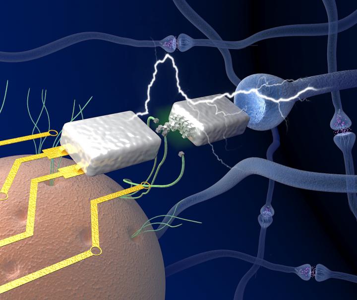
There is an illustration of the work

Caption: A graphic depiction of protein nanowires (green) harvested from microbe Geobacter (orange) facilitate the electronic memristor device (silver) to function with biological voltages, emulating the neuronal components (blue junctions) in a brain. Credit: UMass Amherst/Yao lab