Tag Archives: Howard Hughes Medical Institute

Congratulations to winners of 2020 Nobel Prize for Chemistry: Dr. Emmanuelle Charpentier & Dr. Jennifer A. Doudna (CRISPR-cas9)

It’s possible there’s a more dramatic development in the field of contemporary gene-editing but it’s indisputable that CRISPR (clustered regularly interspaced short palindromic repeats) -cas9 (CRISPR-associated 9 [protein]) ranks very highly indeed.

The technique, first discovered (or developed) in 2012, has brought recognition in the form of the 2020 Nobel Prize for Chemistry to CRISPR’s two discoverers, Emanuelle Charpentier and Jennifer Doudna.

An October 7, 2020 news item on phys.org announces the news,

The Nobel Prize in chemistry went to two researchers Wednesday [October 7, 2020] for a gene-editing tool that has revolutionized science by providing a way to alter DNA, the code of life—technology already being used to try to cure a host of diseases and raise better crops and livestock.

Emmanuelle Charpentier of France and Jennifer A. Doudna of the United States won for developing CRISPR-cas9, a very simple technique for cutting a gene at a specific spot, allowing scientists to operate on flaws that are the root cause of many diseases.

“There is enormous power in this genetic tool,” said Claes Gustafsson, chair of the Nobel Committee for Chemistry.

More than 100 clinical trials are underway to study using CRISPR to treat diseases, and “many are very promising,” according to Victor Dzau, president of the [US] National Academy of Medicine.

“My greatest hope is that it’s used for good, to uncover new mysteries in biology and to benefit humankind,” said Doudna, who is affiliated with the University of California, Berkeley, and is paid by the Howard Hughes Medical Institute, which also supports The Associated Press’ Health and Science Department.

The prize-winning work has opened the door to some thorny ethical issues: When editing is done after birth, the alterations are confined to that person. Scientists fear CRISPR will be misused to make “designer babies” by altering eggs, embryos or sperm—changes that can be passed on to future generations.

Unusually for phys.org, this October 7, 2020 news item is not a simple press/news release reproduced in its entirety but a good overview of the researchers’ accomplishments and a discussion of some of the issues associated with CRISPR along with the press release at the end.

I have covered some CRISPR issues here including intellectual property (see my March 15, 2017 posting titled, “CRISPR patent decision: Harvard’s and MIT’s Broad Institute victorious—for now‘) and designer babies (as exemplified by the situation with Dr. He Jiankui; see my July 28, 2020 post titled, “July 2020 update on Dr. He Jiankui (the CRISPR twins) situation” for more details about it).

An October 7, 2020 article by Michael Grothaus for Fast Company provides a business perspective (Note: A link has been removed),

Needless to say, research by the two scientists awarded the Nobel Prize in Chemistry today has the potential to change the course of humanity. And with that potential comes lots of VC money and companies vying for patents on techniques and therapies derived from Charpentier’s and Doudna’s research.

One such company is Doudna’s Editas Medicine [according to my search, the only company associated with Doudna is Mammoth Biosciences, which she co-founded], while others include Caribou Biosciences, Intellia Therapeutics, and Casebia Therapeutics. Given the world-changing applications—and the amount of revenue such CRISPR therapies could bring in—it’s no wonder that such rivalry is often heated (and in some cases has led to lawsuits over the technology and its patents).

As Doudna explained in her book, A Crack in Creation: Gene Editing and the Unthinkable Power to Control Evolution, cowritten by Samuel H. Sternberg …, “… —but we could also have woolly mammoths, winged lizards, and unicorns.” And as for that last part, she made clear, “No, I am not kidding.”

Everybody makes mistakes and the reference to Editas Medicine is the only error I spotted. You can find out more about Mammoth Biosciences here and while Dr. Doudna’s comment, “My greatest hope is that it’s used for good, to uncover new mysteries in biology and to benefit humankind,” is laudable it would seem she wishes to profit from the discovery. Mammoth Biosciences is a for-profit company as can be seen at the end of the Mammoth Biosciences’ October 7, 2020 congratulatory news release,

About Mammoth Biosciences

Mammoth Biosciences is harnessing the diversity of nature to power the next-generation of CRISPR products. Through the discovery and development of novel CRISPR systems, the company is enabling the full potential of its platform to read and write the code of life. By leveraging its internal research and development and exclusive licensing to patents related to Cas12, Cas13, Cas14 and Casɸ, Mammoth Biosciences can provide enhanced diagnostics and genome editing for life science research, healthcare, agriculture, biodefense and more. Based in San Francisco, Mammoth Biosciences is co-founded by CRISPR pioneer Jennifer Doudna and Trevor Martin, Janice Chen, and Lucas Harrington. The firm is backed by top institutional investors [emphasis mine] including Decheng, Mayfield, NFX, and 8VC, and leading individual investors including Brook Byers, Tim Cook, and Jeff Huber.

An October 7, 2029 Nobel Prize press release, which unleashed all this interest in Doudna and Charpentier, notes this,

Prize amount: 10 million Swedish kronor, to be shared equally between the Laureates.

In Canadian money that amount is $1,492,115.03 (as of Oct. 9, 2020 12:40 PDT when I checked a currency converter).

Ordinarily there’d be a mildly caustic comment from me about business opportunities and medical research but this is a time for congratulations to both Dr. Emanuelle Charpentier and Dr. Jennifer Doudna.
Are plants and brains alike?

The answer to the question about whether brains and plants are alike is the standard ‘yes and no’. That said, there are some startling similarities from a statistical perspective (from a July 6, 2017 Salk Institute news release (also received via email; Note: Links have been removed),

Plants and brains are more alike than you might think: Salk scientists discovered that the mathematical rules governing how plants grow are similar to how brain cells sprout connections. The new work, published in Current Biology on July 6, 2017, and based on data from 3D laser scanning of plants, suggests there may be universal rules of logic governing branching growth across many biological systems.

“Our project was motivated by the question of whether, despite all the diversity we see in plant forms, there is some form or structure they all share,” says Saket Navlakha, assistant professor in Salk’s Center for Integrative Biology and senior author of the paper. “We discovered that there is—and, surprisingly, the variation in how branches are distributed in space can be described mathematically by something called a Gaussian function, which is also known as a bell curve.”

Being immobile, plants have to find creative strategies for adjusting their architecture to address environmental challenges, like being shaded by a neighbor. The diversity in plant forms, from towering redwoods to creeping thyme, is a visible sign of these strategies, but Navlakha wondered if there was some unseen organizing principle at work. To find out, his team used high-precision 3D scanning technology to measure the architecture of young plants over time and quantify their growth in ways that could be analyzed mathematically.

“This collaboration arose from a conversation that Saket and I had shortly after his arrival at Salk,” says Professor and Director of the Plant Molecular and Cellular Biology Laboratory Joanne Chory, who, along with being the Howard H. and Maryam R. Newman Chair in Plant Biology, is also a Howard Hughes Medical Investigator and one of the paper’s coauthors. “We were able to fund our studies thanks to Salk’s innovation grant program and the Howard Hughes Medical Institute.”

The team began with three agriculturally valuable crops: sorghum, tomato and tobacco. The researchers grew the plants from seeds under conditions the plants might experience naturally (shade, ambient light, high light, high heat and drought). Every few days for a month, first author Adam Conn scanned each plant to digitally capture its growth. In all, Conn scanned almost 600 plants.

“We basically scanned the plants like you would scan a piece of paper,” says Conn, a Salk research assistant. “But in this case the technology is 3D and allows us to capture a complete form—the full architecture of how the plant grows and distributes branches in space.”

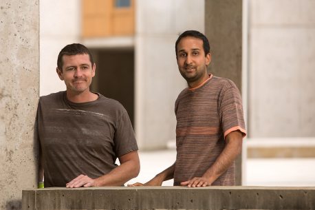
From left: Adam Conn and Saket Navlakha
From left: Adam Conn and Saket Navlakha Credit: Salk Institute

Each plant’s digital representation is called a point cloud, a set of 3D coordinates in space that can be analyzed computationally. With the new data, the team built a statistical description of theoretically possible plant shapes by studying the plant’s branch density function. The branch density function depicts the likelihood of finding a branch at any point in the space surrounding a plant.

This model revealed three properties of growth: separability, self-similarity and a Gaussian branch density function. Separability means that growth in one spatial direction is independent of growth in other directions. According to Navlakha, this property means that growth is very simple and modular, which may let plants be more resilient to changes in their environment. Self-similarity means that all the plants have the same underlying shape, even though some plants may be stretched a little more in one direction, or squeezed in another direction. In other words, plants don’t use different statistical rules to grow in shade than they do to grow in bright light. Lastly, the team found that, regardless of plant species or growth conditions, branch density data followed a Gaussian distribution that is truncated at the boundary of the plant. Basically, this says that branch growth is densest near the plant’s center and gets less dense farther out following a bell curve.

The high level of evolutionary efficiency suggested by these properties is surprising. Even though it would be inefficient for plants to evolve different growth rules for every type of environmental condition, the researchers did not expect to find that plants would be so efficient as to develop only a single functional form. The properties they identified in this work may help researchers evaluate new strategies for genetically engineering crops.

Previous work by one of the paper’s authors, Charles Stevens, a professor in Salk’s Molecular Neurobiology Laboratory, found the same three mathematical properties at work in brain neurons. “The similarity between neuronal arbors and plant shoots is quite striking, and it seems like there must be an underlying reason,” says Stevens. “Probably, they both need to cover a territory as completely as possible but in a very sparse way so they don’t interfere with each other.”

The next challenge for the team is to identify what might be some of the mechanisms at the molecular level driving these changes. Navlakha adds, “We could see whether these principles deviate in other agricultural species and maybe use that knowledge in selecting plants to improve crop yields.”

Should you not be able to access the news release, you can find the information in a July 6, 2017 news item on ScienceDaily.

For the paper, here’s a link and a citation,

A Statistical Description of Plant Shoot Architecture by Adam Conn, Ullas V. Pedmale4, Joanne Chory, Charles F. Stevens, Saket Navlakha. Current Biology DOI: http://dx.doi.org/10.1016/j.cub.2017.06.009 Publication stage: In Press Corrected Proof July 2017

This paper is behind a paywall.

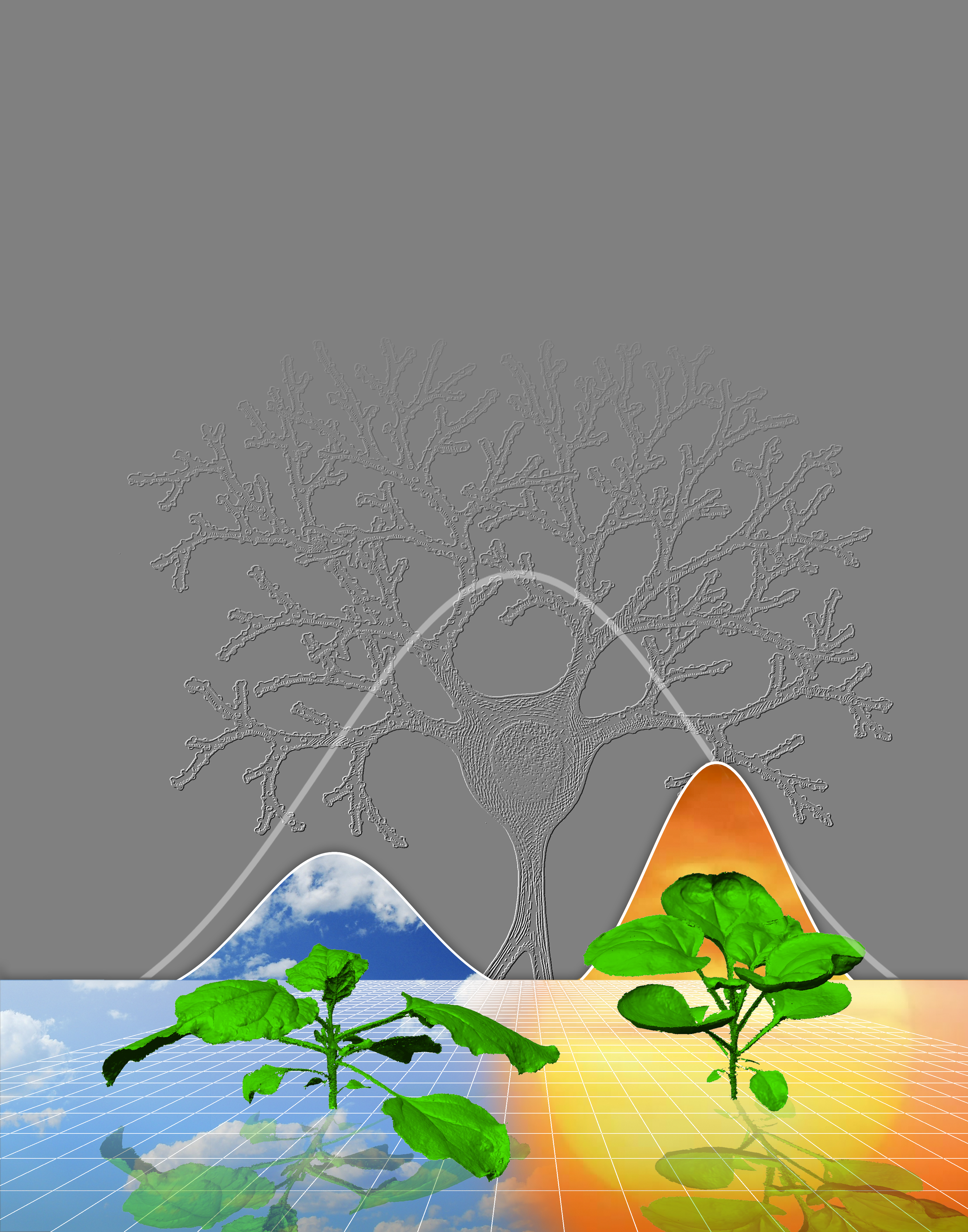
Here’s an image that illustrates the principles the researchers are attempting to establish,

This illustration represents how plants use the same rules to grow under widely different conditions (for example, cloudy versus sunny), and that the density of branches in space follows a Gaussian (“bell curve”) distribution, which is also true of neuronal branches in the brain. Credit: Salk Institute

Curiosity may not kill the cat but, in science, it might be an antidote to partisanship

I haven’t stumbled across anything from the Cultural Cognition Project at Yale Law School in years so before moving onto their latest news, here’s more about the project,

The Cultural Cognition Project is a group of scholars interested in studying how cultural values shape public risk perceptions and related policy beliefs. Cultural cognition refers to the tendency of individuals to conform their beliefs about disputed matters of fact (e.g., whether global warming is a serious threat; whether the death penalty deters murder; whether gun control makes society more safe or less) to values that define their cultural identities.Project members are using the methods of various disciplines — including social psychology, anthropology, communications, and political science — to chart the impact of this phenomenon and to identify the mechanisms through which it operates. The Project also has an explicit normative objective: to identify processes of democratic decisionmaking by which society can resolve culturally grounded differences in belief in a manner that is both congenial to persons of diverse cultural outlooks and consistent with sound public policymaking.

It’s nice to catch up with some of the project’s latest work, from a Jan. 26, 2017 Yale University news release (also on EurekAlert),

Disputes over science-related policy issues such as climate change or fracking often seem as intractable as other politically charged debates. But in science, at least, simple curiosity might help bridge that partisan divide, according to new research.

In a study slated for publication in the journal Advances in Political Psychology, a Yale-led research team found that people who are curious about science are less polarized in their views on contentious issues than less-curious peers.

In an experiment, they found out why: Science-curious individuals are more willing to engage with surprising information that runs counter to their political predispositions.

“It’s a well-established finding that most people prefer to read or otherwise be exposed to information that fits rather than challenges their political preconceptions,” said research team leader Dan Kahan, Elizabeth K. Dollard Professor of Law and professor of psychology at Yale Law School. “This is called the echo-chamber effect.”

But science-curious individuals are more likely to venture out of that chamber, he said.

“When they are offered the choice to read news articles that support their views or challenge them on the basis of new evidence, science-curious individuals opt for the challenging information,” Kahan said. “For them, surprising pieces of evidence are bright shiny objects — they can’t help but grab at them.”

Kahan and other social scientists previously have shown that information based on scientific evidence can actually intensify — rather than moderate — political polarization on contentious topics such as gun control, climate change, fracking, or the safety of certain vaccines. The new study, which assessed science knowledge among subjects, reiterates the gaping divide separating how conservatives and liberals view science.

Republicans and Democrats with limited knowledge of science were equally likely to agree or disagree with the statement that “there is solid evidence that global warming is caused by human activity. However, the most science-literate conservatives were much more likely to disagree with the statement than less-knowledgeable peers. The most knowledgeable liberals almost universally agreed with the statement.

“Whatever measure of critical reasoning we used, we always observed this depressing pattern: The members of the public most able to make sense of scientific evidence are in fact the most polarized,” Kahan said.

But knowledge of science, and curiosity about science, are not the same thing, the study shows.

The team became interested in curiosity because of its ongoing collaborative research project to improve public engagement with science documentaries involving the Cultural Cognition Project at Yale Law School, the Annenberg Public Policy Center of the University of Pennsylvania, and Tangled Bank Studios at the Howard Hughes Medical Institute.

They noticed that the curious — those who sought out science stories for personal pleasure — not only were more interested in viewing science films on a variety of topics but also did not display political polarization associated with contentious science issues.

The new study found, for instance, that a much higher percentage of curious liberals and conservatives chose to read stories that ran counter to their political beliefs than did their non-curious peers.

“As their science curiosity goes up, the polarizing effects of higher science comprehension dissipate, and people move the same direction on contentious policies like climate change and fracking,” Kahan said.

It is unclear whether curiosity applied to other controversial issues can minimize the partisan rancor that infects other areas of society. But Kahan believes that the curious from both sides of the political and cultural divide should make good ambassadors to the more doctrinaire members of their own groups.

“Politically curious people are a resource who can promote enlightened self-government by sharing scientific information they are naturally inclined to learn and share,” he said.

Here’s my standard link to and citation for the paper,

Science Curiosity and Political Information Processing by Dan M. Kahan, Asheley R Landrum, Katie Carpenter, Laura Helft, and Kathleen Hall Jamieson. Political Psychology Volume 38, Issue Supplement S1 February 2017 Pages 179–199 DOI: 10.1111/pops.12396View First published: 26 January 2017

This paper is open and it can also be accessed here.

I last mentioned Kahan and The Cultural Cognition Project in an April 10, 2014 posting (scroll down about 45% of the way) about responsible science.

Using light to make gold crystal nanoparticles

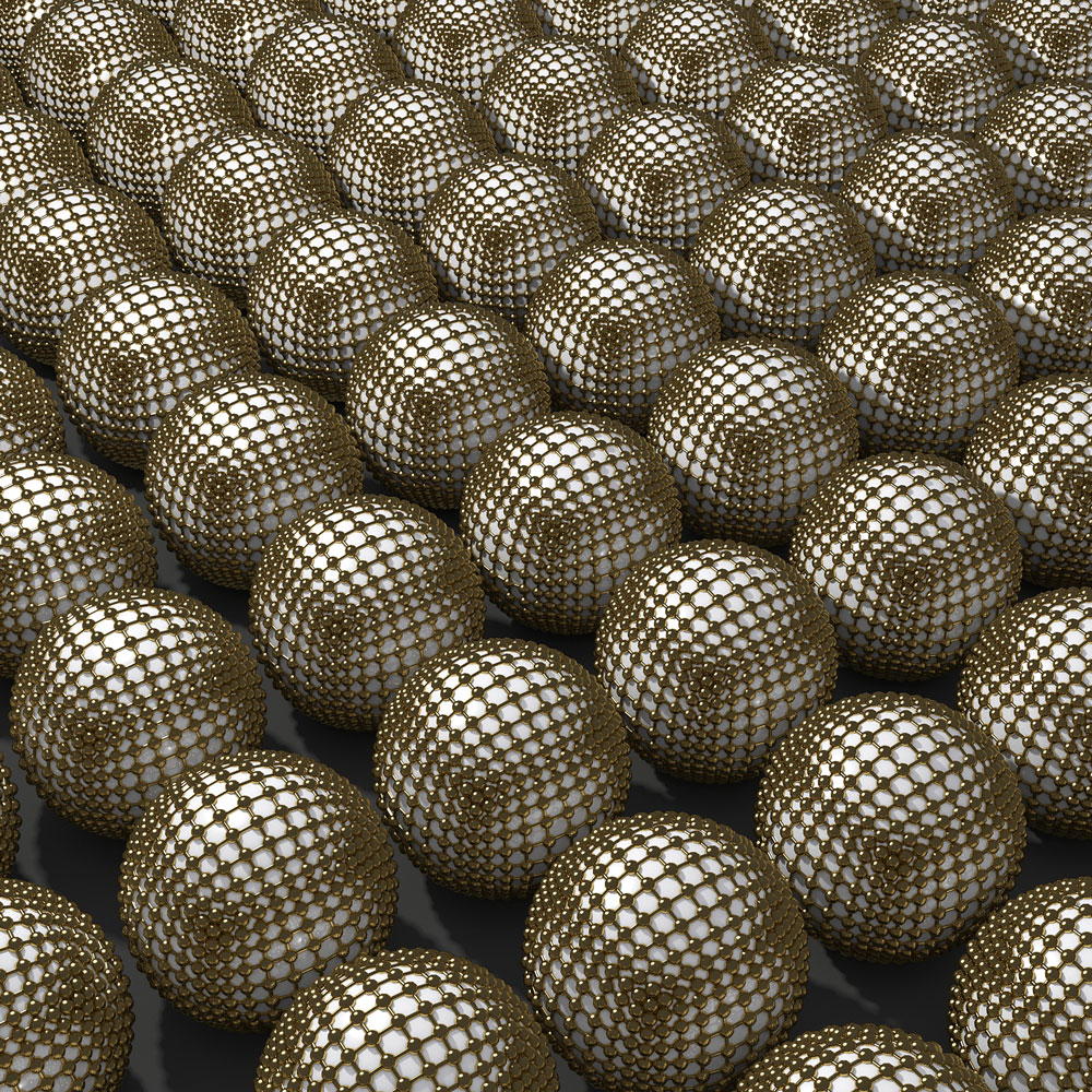
Gold crystal nanoparticles? Courtesy: University of Florida

Gold crystal nanoparticles? Courtesy: University of Florida

A team from the University of Florida has used gold instead of silver in a process known as plasmon-driven synthesis. From a July 8, 2016 news item on phys.org,

A team of University of Florida researchers has figured out how gold can be used in crystals grown by light to create nanoparticles, a discovery that has major implications for industry and cancer treatment and could improve the function of pharmaceuticals, medical equipment and solar panels.

A July 6, 2016 University of Florida news release, which originated the news item, provides more detail,

Nanoparticles can be “grown” in crystal formations with special use of light, in a process called plasmon-driven synthesis. However, scientists have had limited control unless they used silver, but silver limits the uses for medical technology. The team is the first to successfully use gold, which works well within the human body, with this process.

“How does light actually play a role in the synthesis? [This knowledge] was not well developed,” said David Wei, an associate professor of chemistry who led the research team. “Gold was the model system to demonstrate this.”

Gold is highly desired for nanotechnology because it is malleable, does not react with oxygen and conducts heat well. Those properties make gold an ideal material for nanoparticles, especially those that will be placed in the body.

When polyvinylpyrrolidone, or PVP, a substance commonly found in pharmaceutical tablets, is used in the plasmon-driven synthesis, it enables scientists to better control the growth of crystals. In Wei’s research, PVP surprised the team by showing its potential to relay light-generated “hot” electrons to a gold surface to grow the crystals.

The research describes the first plasmonic synthesis strategy that can make high-yield gold nanoprisms. Even more exciting, the team has demonstrated that visible-range and low-power light can be used in the synthesis. Combined with nanoparticles being used in solar photovoltaic devices, this method can even harness solar energy for chemical synthesis, to make nanomaterials or for general applications in chemistry.

Wei has spent the last decade working in nanotechnology. He is intrigued by its applications in photochemistry and biomedicine, especially in targeted drug delivery and photothermal therapeutics, which is crucial to cancer treatment. His team includes collaborators from Pacific Northwest National Laboratory, where he has worked as a visiting scholar, and Brookhaven National Laboratory. In addition, the project has provided an educational opportunity for chemistry students: one high school student (through UF’s Student Science Training Program), two University scholars who also [sic] funded by the Howard Hughes Medical Institute, five graduate students and two postdocs.

Here’s a link to and a citation for the paper,

Polyvinylpyrrolidone-induced anisotropic growth of gold nanoprisms in plasmon-driven synthesis by Yueming Zhai, Joseph S. DuChene, Yi-Chung Wang, Jingjing Qiu, Aaron C. Johnston-Peck, Bo You, Wenxiao Guo, Benedetto DiCiaccio, Kun Qian, Evan W. Zhao, Frances Ooi, Dehong Hu, Dong Su, Eric A. Stach, Zihua Zhu, & Wei David Wei. Nature Materials (2016) doi:10.1038/nmat4683 Published online 04 July 2016

This paper is behind a paywall.

Cell-to-cell communication via nanotubes

It turns out that the cells communicating with each other are located in fruit flies. So, it’s perhaps not quite as exciting as one might have imagined, nonetheless, a July 1, 2015 news item on ScienceDaily provides some intriguing insights into cell communication,

When it comes to communicating with each other, some cells may be more “old school” than was previously thought.

Certain types of stem cells use microscopic, threadlike nanotubes to communicate with neighboring cells, like a landline phone connection, rather than sending a broadcast signal, researchers at University of Michigan Life Sciences Institute and University of Texas Southwestern Medical Center have discovered.

The findings, which are scheduled for online publication July 1 in Nature, offer new insights on how stem cells retain their identities when they divide to split off a new, specialized cell.

The fruit-fly research also suggests that short-range, cell-to-cell communication may rely on this type of direct connection more than was previously understood, said co-senior author Yukiko Yamashita, a U-M developmental biologist whose lab is located at the Life Sciences Institute.

A July 1, 2015 University of Michigan news release (also on EurekAlert), which originated the news item, expands on the theme,

“There are trillions of cells in the human body, but nowhere near that number of signaling pathways,” she said. “There’s a lot we don’t know about how the right cells get just the right messages to the right recipients at the right time.”

The nanotubes had actually been hiding in plain sight.

The investigation began when a postdoctoral researcher in Yamashita’s lab, Mayu Inaba, approached her mentor with questions about tiny threads of connection she noticed in an image of fruit fly reproductive stem cells, which are also known as germ line cells. The projections linked individual stem cells back to a central hub in the stem cell “niche.” Niches create a supportive environment for stem cells and help direct their activity.

Yamashita, a Howard Hughes Medical Institute investigator, MacArthur Fellow and an associate professor at the U-M Medical School, looked through her old image files and discovered that the connections appeared in numerous images.

“I had seen them, but I wasn’t seeing them,” Yamashita said. “They were like a little piece of dust on an otherwise normal picture. After we presented our findings at meetings, other scientists who work with the same cells would say, ‘We see them now, too.'”

It’s not surprising that the minute structures went overlooked for so long. Each one is about 3 micrometers long; by comparison, a piece of paper is 100 micrometers thick.

While the study looked specifically at reproductive cells in male Drosophila fruit flies, there have been indications of similar structures in other contexts, including mammalian cells, Yamashita said.

Fruit flies are an important model for this type of investigation, she added. If one was to start instead with human cells, one might find something, but the system’s greater complexity would make it far more difficult to tease apart the underlying mechanisms.

The findings shed new light on a key attribute of stem cells: their ability to make new specialized cells while still retaining their identity as stem cells.

Germ line stem cells typically divide asymmetrically. In the male fruit fly, when a stem cell divides, one part stays attached to the hub and remains a stem cell. The other part moves away from the hub and begins differentiation into a fly sperm cell.

Until the discovery of the nanotubes, scientists had been puzzled as to how cellular signals guiding identity could act on one of the cells but not the other, said collaborator Michael Buszczak, an associate professor of molecular biology at UT Southwestern, who shares corresponding authorship of the paper and currently co-mentors Inaba with Yamashita.

The researchers conducted experiments that showed disruption of nanotube formation compromised the ability of the germ line stem cells to renew themselves.

I gather the fruit fly research offers the basis for more extensive investigations into other species and their cell-to-cell communication.

Here’s a link to and a citation for the paper,

Nanotubes mediate niche–stem-cell signalling in the Drosophila testis by Mayu Inaba, Michael Buszczak, & Yukiko M. Yamashita. Nature (2015) doi:10.1038/nature14602 Published online 01 July 2015

This paper is behind a paywall.

Animal-based (some of it ‘fishy’) sunscreen from Oregon State University

In the Northern Hemisphere countries it’s time to consider one’s sunscreen options.While this Oregon State University into animal-based sunscreens is intriguing,  market-ready options likely won’t be available for quite some time. (There is a second piece of related research, more ‘fishy’ in nature [pun], featured later in this post.) From a May 12, 2015 Oregon State University news release,

Researchers have discovered why many animal species can spend their whole lives outdoors with no apparent concern about high levels of solar exposure: they make their own sunscreen.

The findings, published today in the journal eLife by scientists from Oregon State University, found that many fish, amphibians, reptiles, and birds can naturally produce a compound called gadusol, which among other biologic activities provides protection from the ultraviolet, or sun-burning component of sunlight.

The researchers also believe that this ability may have been obtained through some prehistoric, natural genetic engineering.

Here’s an amusing image to illustrate the researchers’ point,

Gadusol is the gene found in some animals which gives natural sun protection. Courtesy: Oregon State University

Gadusol is the gene found in some animals which gives natural sun protection.
Courtesy: Oregon State University

The news release goes on to describe gadusol and its believed evolutionary pathway,

The gene that provides the capability to produce gadusol is remarkably similar to one found in algae, which may have transferred it to vertebrate animals – and because it’s so valuable, it’s been retained and passed along for hundreds of millions of years of animal evolution.

“Humans and mammals don’t have the ability to make this compound, but we’ve found that many other animal species do,” said Taifo Mahmud, a professor in the OSU College of Pharmacy, and lead author on the research.

The genetic pathway that allows gadusol production is found in animals ranging from rainbow trout to the American alligator, green sea turtle and a farmyard chicken.

“The ability to make gadusol, which was first discovered in fish eggs, clearly has some evolutionary value to be found in so many species,” Mahmud said. “We know it provides UV-B protection, it makes a pretty good sunscreen. But there may also be roles it plays as an antioxidant, in stress response, embryonic development and other functions.”

In their study, the OSU researchers also found a way to naturally produce gadusol in high volumes using yeast. With continued research, it may be possible to develop gadusol as an ingredient for different types of sunscreen products, cosmetics or pharmaceutical products for humans.

A conceptual possibility, Mahmud said, is that ingestion of gadusol could provide humans a systemic sunscreen, as opposed to a cream or compound that has to be rubbed onto the skin.

The existence of gadusol had been known of in some bacteria, algae and other life forms, but it was believed that vertebrate animals could only obtain it from their diet. The ability to directly synthesize what is essentially a sunscreen may play an important role in animal evolution, and more work is needed to understand the importance of this compound in animal physiology and ecology, the researchers said.

Here’s a link to and a citation for the paper,

De novo synthesis of a sunscreen compound in vertebrates by Andrew R Osborn, Khaled H Almabruk, Garrett Holzwarth, Shumpei Asamizu, Jane LaDu, Kelsey M Kean, P Andrew Karplus, Robert L Tanguay, Alan T Bakalinsky, and Taifo Mahmud. eLife 2015;4:e05919 DOI: http://dx.doi.org/10.7554/eLife.05919 Published May 12, 2015

This is an open access paper.

The second piece of related research, also published yesterday on May 12, 2015, comes from a pair of scientists at Harvard University. From a May 12, 2015  eLife news release on EurekAlert,

Scientists from Oregon State University [two authors are listed for the ‘zebrafish’ paper and both are from Harvard University] have discovered that fish can produce their own sunscreen. They have copied the method used by fish for potential use in humans.

In the study published in the journal eLife, scientists found that zebrafish are able to produce a chemical called gadusol that protects against UV radiation. They successfully reproduced the method that zebrafish use by expressing the relevant genes in yeast. The findings open the door to large-scale production of gadusol for sunscreen and as an antioxidant in pharmaceuticals.

Gadusol was originally identified in cod roe and has since been discovered in the eyes of the mantis shrimp, sea urchin eggs, sponges, and in the dormant eggs and newly hatched larvae of brine shrimps. It was previously thought that fish can only acquire the chemical through their diet or through a symbiotic relationship with bacteria.

Marine organisms in the upper ocean and on reefs are subject to intense and often unrelenting sunlight. Gadusol and related compounds are of great scientific interest for their ability to protect against DNA damage from UV rays. There is evidence that amphibians, reptiles, and birds can also produce gadusol, while the genetic machinery is lacking in humans and other mammals.

The team were investigating compounds similar to gadusol that are used to treat diabetes and fungal infections. It was believed that the biosynthetic enzyme common to all of them, EEVS, was only present in bacteria. The scientists were surprised to discover that fish and other vertebrates contain similar genes to those that code for EEVS.

Curious about their function in animals, they expressed the zebrafish gene in E. coli and analysis suggested that fish combine EEVS with another protein, whose production may be induced by light, to produce gadusol. To check that this combination is really sufficient, the scientists transferred the genes to yeast and set them to work to see what they would create. This confirmed the production of gadusol. Its successful production in yeast provides a viable route to commercialisation.

As well as providing UV protection, gadusol may also play a role in stress responses, in embryonic development, and as an antioxidant.

Here’s a link to and a citation for the second paper from this loosely affiliated team of Oregon State University and Harvard University researchers,

Biochemistry: Shedding light on sunscreen biosynthesis in zebrafish by Carolyn A Brotherton and Emily P Balskus. eLife 2015;4:e07961 DOI: http://dx.doi.org/10.7554/eLife.07961 Published May 12, 2015

This paper, too, is open access.

One final bit and this is about the journal, eLife, from their news release on EurekAlert,

About eLife

eLife is a unique collaboration between the funders and practitioners of research to improve the way important research is selected, presented, and shared. eLife publishes outstanding works across the life sciences and biomedicine — from basic biological research to applied, translational, and clinical studies. eLife is supported by the Howard Hughes Medical Institute, the Max Planck Society, and the Wellcome Trust. Learn more at elifesciences.org.

It seems this journal is a joint, US (Howard Hughes Medical Institute), German (Max Planck Society), UK (Wellcome Trust) effort.

Protein nanomachines at the University of Washington

Scouring pad or protein nanomachine?

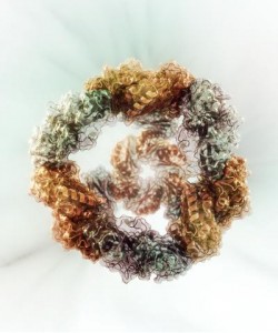
Caption: This is a computational model of a successfully designed two-component protein nanocage with tetrahedral symmetry. Credit: Dr. Vikram Mulligan

Caption: This is a computational model of a successfully designed two-component protein nanocage with tetrahedral symmetry.
Credit: Dr. Vikram Mulligan

This illustration of a protein nanocage reminded me of a type of scouring pad, which come to think of it, I haven’t seen in any stores for some years. Getting back on topic, this nanocage is a first step to building nanomachines according to a June 5, 2014 news item on Nanowerk,

A route for constructing protein nanomachines engineered for specific applications may be closer to reality.

Biological systems produce an incredible array of self-assembling, functional protein tools. Some examples of these nanoscale protein materials are scaffolds to anchor cellular activities, molecular motors to drive physiological events, and capsules for delivering viruses into host cells.

Scientists inspired by these sophisticated molecular machines want to build their own, with forms and functions customized to tackle modern-day challenges. The ability to design new protein nanostructures could have useful implications in targeted delivery of drugs, in vaccine development and in plasmonics, which is manipulating electromagnetic signals to guide light diffraction for information technologies, energy production or other uses.

A recently developed computational method may be an important step toward that goal. The project was led by the University of Washington’s [Washington state] Neil King, translational investigator; Jacob Bale, graduate student in Molecular and Cellular Biology; and William Sheffler in David Baker’s laboratory at the University of Washington Institute for Protein Design, in collaboration with colleagues at UCLA [University of California at Los Angeles] and Janelia Farm.

The work is based in the Rosetta macromolecular modeling package developed by Baker and his colleagues. The program was originally created to predict natural protein structures from amino acid sequences. Researchers in the Baker lab and around the world are increasingly using Rosetta to design new protein structures and sequences aimed at solving real-world problems.

A June 4 (?), 2014 University of Washington news release by Leila Gray (also on EurekAlert), which originated the news item, provides more detail about the models and what the scientists hope to accomplish,

“Proteins are amazing structures that can do remarkable things,” King said, “they can respond to changes in their environment. Exposure to a particular metabolite or a rise in temperature, for example, can trigger an alteration in a particular protein’s shape and function.” People often call proteins the building blocks of life.

“But unlike, say, a PVC pipe,” King said, “they are not simply construction material.” They are also construction (and demolition) workers — speeding up chemical reactions, breaking down food, carrying messages, interacting with each other, and performing countless other duties vital to life.

With the new software the scientists were able to create five novel, 24-subunit cage-like protein nanomaterials. Importantly, the actual structures, the researchers observed, were in very close agreement with their computer modeling.

Their method depends on encoding pairs of protein amino acid sequences with the information needed to direct molecular assembly through protein-protein interfaces. The interfaces not only provide the energetic forces that drive the assembly process, they also precisely orient the pairs of protein building blocks with the geometry required to yield the desired cage-like symmetric architectures.

Creating this cage-shaped protein, the scientists said, may be a first step towards building nano-scale containers. [emphasis mine] King said he looks forward to a time when cancer-drug molecules will be packaged inside of designed nanocages and delivered directly to tumor cells, sparing healthy cells.

“The problem today with cancer chemotherapy is that it hits every cell and makes the patient feel sick,” King said. Packaging the drugs inside customized nanovehicles with parking options restricted to cancer sites might circumvent the side effects.

The scientists note that combining just two types of symmetry elements, as in this study, can in theory give rise to a range of symmetrical shapes, such as cubic point groups, helices, layers, and crystals.

King explained that the immune system responds to repetitive, symmetric patterns, such as those on the surface of a virus or disease bacteria. Building nano-decoys may be a way train the immune system to attack certain types of pathogens.

“This concept may become the foundation for vaccines based on engineered nanomaterials,” King said. Further down the road, he and Bale anticipate that these design methods might also be useful for developing new clean energy technologies.

The scientists added in their report, “The precise control over interface geometry offered by our method enables the design of two-component protein nanomaterials with diverse nanoscale features, such as surfaces, pores, and internal volumes, with high accuracy.”

They went on to say that the combinations possible with two-component materials greatly expand the number and variety of potential nanomaterials that could be designed.

It may be possible to produce nanomaterials in a variety of sizes, shapes and arrangements, and also move on to construct increasingly more complex materials from more than two components.

The researchers emphasized that the long-term goal of such structures is not to be static. The hope is that they will mimic or go beyond the dynamic performance of naturally occurring protein assemblies, and that eventually novel molecular protein machines could be manufactured with programmable functions. [emphasis mine]

The researchers pointed out that although designing proteins and protein-based nanomaterials is very challenging due to the relative complexity of protein structures and interactions, there are now more than a handful of laboratories around the world making major strides in this field. Each of the leading contributors have key strengths, they said. The strengths of the UW team is in the accuracy of the match of the designed proteins to the computational models and the predictability of the results.

It seems like it’s going to be several years before we have protein nanomachines. Here’s a link to and a citation for the research paper,

Accurate design of co-assembling multi-component protein nanomaterials by Neil P. King, Jacob B. Bale, William Sheffler, Dan E. McNamara, Shane Gonen, Tamir Gonen, Todd O. Yeates, & David Baker. Nature 510, 103–108 (05 June 2014) doi:10.1038/nature13404 Published online 25 May 2014

This paper is behind a paywall but there is a free preview via ReadCube Access.

For anyone curious about the Rosetta macromolecular modeling package used in this work, you can find out more here at the Rosetta Commons website.  As for Janelia Farm, it is a research center in Virginia and is part of the Howard Hughes Medical Institute.