Tag Archives: hydrogels

Ionic skin for ‘smart’ skin

An April 28, 2022 University of British Columbia (UBC) news release (also on EurekAlert) announces a step forward in the attempt to create ‘smart’ skin, Note: Links have been removed,

In the quest to build smart skin that mimics the sensing capabilities of natural skin, ionic skins have shown significant advantages. They’re made of flexible, biocompatible hydrogels that use ions to carry an electrical charge. In contrast to smart skins made of plastics and metals, the hydrogels have the softness of natural skin. This offers a more natural feel to the prosthetic arm or robot hand they are mounted on, and makes them comfortable to wear.

These hydrogels can generate voltages when touched, but scientists did not clearly understand how — until a team of researchers at UBC devised a unique experiment, published today in Science.

“How hydrogel sensors work is they produce voltages and currents in reaction to stimuli, such as pressure or touch – what we are calling a piezoionic effect. But we didn’t know exactly how these voltages are produced,” said the study’s lead author Yuta Dobashi, who started the work as part of his master’s in biomedical engineering at UBC.

Working under the supervision of UBC researcher Dr. John Madden, Dobashi devised hydrogel sensors containing salts with positive and negative ions of different sizes. He and collaborators in UBC’s physics and chemistry departments applied magnetic fields to track precisely how the ions moved when pressure was applied to the sensor.

“When pressure is applied to the gel, that pressure spreads out the ions in the liquid at different speeds, creating an electrical signal. Positive ions, which tend to be smaller, move faster than larger, negative ions. This results in an uneven ion distribution which creates an electric field, which is what makes a piezoionic sensor work.”

The researchers say this new knowledge confirms that hydrogels work in a similar way to how humans detect pressure, which is also through moving ions in response to pressure, inspiring potential new applications for ionic skins.

“The obvious application is creating sensors that interact directly with cells and the nervous system, since the voltages, currents and response times are like those across cell membranes,” says Dr. Madden, an electrical and computer engineering professor in UBC’s faculty of applied science. “When we connect our sensor to a nerve, it produces a signal in the nerve. The nerve, in turn, activates muscle contraction.”

“You can imagine a prosthetic arm covered in an ionic skin. The skin senses an object through touch or pressure, conveys that information through the nerves to the brain, and the brain then activates the motors required to lift or hold the object. With further development of the sensor skin and interfaces with nerves, this bionic interface is conceivable.”

Another application is a soft hydrogel sensor worn on the skin that can monitor a patient’s vital signs while being totally unobtrusive and generating its own power.

Dobashi, who’s currently completing his PhD work at the University of Toronto, is keen to continue working on ionic technologies after he graduates.

“We can imagine a future where jelly-like ‘iontronics’ are used for body implants. Artificial joints can be implanted, without fear of rejection inside the human body. Ionic devices can be used as part of artificial knee cartilage, adding a smart sensing element.  A piezoionic gel implant might release drugs based on how much pressure it senses, for example.”

Dr. Madden added that the market for smart skins is estimated at $4.5 billion in 2019 and it continues to grow. “Smart skins can be integrated into clothing or placed directly on the skin, and ionic skins are one of the technologies that can further that growth.”

The research includes contributions from UBC chemistry PhD graduate Yael Petel and Carl Michal, UBC professor of physics, who used the interaction between strong magnetic fields and the nuclear spins of ions to track ion movements within the hydrogels. Cédric Plesse, Giao Nguyen and Frédéric Vidal at CY Cergy Paris University in France helped develop a new theory on how the charge and voltage are generated in the hydrogels.

Interview language(s): English (Dobashi, Madden), French (Plesse, Madden), Japanese (Dobashi)

Here’s a link to and a citation for the paper,

Piezoionic mechanoreceptors: Force-induced current generation in hydrogels by
Yuta Dobashi, Dickson Yao, Yael Petel, Tan Ngoc Nguyen, Mirza Saquib Sarwar, Yacine Thabet, Cliff L. W. Ng, Ettore Scabeni Glitz, Giao Tran Minh Nguyen, Cédric Plesse, Frédéric Vidal, Carl A. Michal and John D. W. Madden. Science • 28 Apr 2022 • Vol 376, Issue 6592 • pp. 502-507 • DOI: 10.1126/science.aaw1974

This paper is behind a paywall.

Removing vandals’ graffiti from street art with nanotechnology-enabled method and Happy Italian Research in the World Day and more …

Happy Italian Research in the World Day! Each year since 2018 this has been celebrated on the day that Leonardo da Vinci was born over 500 years ago on April 15. It’s also the start of World Creativity and Innovation Week (WCIW), April 15 – 21, 2021 with over 80 countries (Italy, The Gambia, Mauritius, Belarus, Iceland, US, Syria, Vietnam, Indonesia, Denmark, etc.) celebrating. By the way, April 21, 2021 is the United Nations’ World Creativity and Innovation Day. Now, onto some of the latest research, coming from Italy, on art conservation.

There’s graffiti and there’s graffiti as Michele Baglioni points out in an April 13, 2021 American Chemical Society (ACS) press conference (Rescuing street art from vandals’ graffiti) held during the ACS Spring 2021 Meeting being held online April 5-30, 2021.

An April 13, 2021 news item on ScienceDaily announced the research,

From Los Angeles and the Lower East Side of New York City to Paris and Penang, street art by famous and not-so-famous artists adorns highways, roads and alleys. In addition to creating social statements, works of beauty and tourist attractions, street art sometimes attracts vandals who add their unwanted graffiti, which is hard to remove without destroying the underlying painting. Now, researchers report novel, environmentally friendly techniques that quickly and safely remove over-paintings on street art.

A new eco-friendly method can remove the graffiti that this person is about to spray on the street art behind them. Credit: FOTOKITA/Shutterstock.com

An April 13, 2021 ACS news release (also on EurekAlert), which originated the news item, provides details about this latest work and how it fits into the field of art conservation,

“For decades, we have focused on cleaning or restoring classical artworks that used paints designed to last centuries,” says Piero Baglioni, Ph.D., the project’s principal investigator. “In contrast, modern art and street art, as well as the coatings and graffiti applied on top, use materials that were never intended to stand the test of time.”

Research fellow Michele Baglioni, Ph.D., (no relation to Piero Baglioni) and coworkers built on their colleagues’ work and designed a nanostructured fluid, based on nontoxic solvents and surfactants, loaded in highly retentive hydrogels that very slowly release cleaning agents to just the top layer — a few microns in depth. The undesired top layer is removed in seconds to minutes, with no damage or alteration to the original painting.

Street art and overlying graffiti usually contain one or more of three classes of paint binders — acrylic, vinyl or alkyd polymers. Because these paints are similar in composition, removing the top layer frequently damages the underlying layer. Until now, the only way to remove unwanted graffiti was by using chemical cleaners or mechanical action such as scraping or sand blasting. These traditional methods are hard to control and often damaged the original art.

“We have to know exactly what is going on at the surface of the paintings if we want to design cleaners,” explains Michele Baglioni, who is at the University of Florence (Italy). “In some respects, the chemistry is simple — we are using known surfactants, solvents and polymers. The challenge is combining them in the right way to get all the properties we need.”

Michele Baglioni and coworkers used Fourier transform infrared spectroscopy to characterize the binders, fillers and pigments in the three classes of paints. After screening for suitable low-toxicity, “green” solvents and biodegradable surfactants, he used small angle X-ray scattering analyses to study the behavior of four alkyl carbonate solvents and a biodegradable nonionic surfactant in water.

The final step was formulating the nanostructured cleaning combination. The system that worked well also included 2-butanol and a readily biodegradable alkyl glycoside hydrotrope as co-solvents/co-surfactants. Hydrotropes are water-soluble, surface-active compounds used at low levels that allow more concentrated formulations of surfactants to be developed. The system was then loaded into highly retentive hydrogels and tested for its ability to remove overpaintings on laboratory mockups using selected paints in all possible combinations.

After dozens of tests, which helped determine how long the gel should be applied and removed without damaging the underlying painting, he tested the gels on a real piece of street art in Florence, successfully removing graffiti without affecting the original work.

“This is the first systematic study on the selective and controlled removal of modern paints from paints with similar chemical composition,” Michele Baglioni says. The hydrogels can also be used for the removal of top coatings on modern art that were originally intended to preserve the paintings but have turned out to be damaging. The hydrogels will become available commercially from CSGI Solutions for Conservation of Cultural Heritage, a company founded by Piero Baglioni and others. CSGI, the Center for Colloid and Surface Science, is a university consortium mainly funded through programs of the European Union.

And, there was this after the end of the news release,

The researchers acknowledge support and funding from the European Union NANORESTART (Nanomaterials for the Restoration of Works of Art) Program [or NanoRestArt] and CSGI.

The NanoRestArt project has been mentioned here a number of times,

The project ended in November 2018 but the NanoRestArt website can still be accessed.

Congratulations to Molly Shoichet (her hydrogels are used in regenerative medicine and more) for winning the $1 million Gerhard Herzberg Canada Gold Medal

I imagine that most anyone who’s been in contact with Ms. Shoichet is experiencing a thrill on hearing this morning’s (November 10, 2020) news about winning Canada’s highest honour for science and engineering research. (Confession: she, very kindly, once gave me a brief interview for a posting on this blog, more about that later).

Why Molly Shoichet won the Gerhard Herzberg Canada Gold Medal

Emily Chung’s Nov. 10, 2020 news item on the Canadian Broadcasting Corporation (CBC) website announces the exciting news (Note: Links have been removed),

A Toronto chemical engineering professor has won the $1 million Gerhard Herzberg Canada Gold Medal, the country’s top science prize, for her work designing gels that mimic human tissues.

The Natural Sciences and Engineering Research Council of Canada (NSERC) announced Tuesday [Nov. 10, 2020] that Molly Shoichet, professor of chemical engineering and applied chemistry and Canada Research Chair in Tissue Engineering at the University of Toronto is this year’s recipient of the award, which recognizes “sustained excellence” and “overall influence” of research conducted in Canada in the natural sciences or engineering.

Shoichet’s hydrogels are used for drug development and  delivery and regenerative medicine to heal injuries and treat diseases.

NSERC said Shoichet’s work has led to the development of several “game-changing” applications of such materials. They “delivered a crucial breakthrough” by allowing cells to be grown in three dimensions as they do in the body, rather than the two dimensions they typically do in a petri dish.

Hydrogels are polymer materials — materials such as plastics, made of repeating units — that become swollen with water.

“If you’ve ever eaten Jell-o, that’s a hydrogel,” Shoichet said. Slime and the absorbent material inside disposable diapers are also hydrogels.

Shoichet was born in Toronto, and studied science and engineering at the Massachusetts Institute of Technology and the University of Massachusetts Amherst. After graduating, she worked in the biotech industry alongside “brilliant biologists,” she said. She noticed that the biologists’ research was limited by what types of materials were available.

As an engineer, she realized she could help by custom designing materials for biologists. She could make materials specifically suit their needs, to answer their specific questions by designing hydrogels to mimic particular tissues.

Her collaborations with biologists have also generated three spinoff companies, including AmacaThera, which was recently approved to run human trials of a long-acting anesthetic delivered with an injectable hydrogel to deal with post-surgical pain.

Shoichet noted that drugs given to deal with that kind of pain lead to a quarter of opioid addictions, which have been a deadly problem in Canada and around the world.

“What we’re really excited about is not only meeting that critical need of providing people with greater pain relief for a sustained period of time, but also possibly putting a dent in the operation,” she said. 

Liz Do’s Nov. 10, 2020 University of Toronto news release provides more details (Note: Links have been removed),

The  Herzberg Gold Medal is awarded by the Natural Sciences and Engineering Research Council (NSERC) in recognition of research contributions characterized by both excellence and influence.

“I was completely overwhelmed when I was told the good news,” says Shoichet. “There are so many exceptional people who’ve won this award and I admire them. To think of my peers putting me in that same category is really incredible.”

A pioneer in regenerative medicine, tissue engineering and drug delivery, Shoichet and her team are internationally known for their discovery and innovative use of 3D hydrogels.

“One of the challenges facing drug screening is that many of the drugs discovered work well in the lab, but not in people, and a possible explanation for this discrepancy is that these drugs are discovered in environments that do not reflect that of the body,” explains Shoichet.

Shoichet’s team has invented a series of biomaterials that provide a soft, three-dimensional environment in which to grow cells. These hydrogels — water-swollen materials — better mimic human tissue than hard two-dimensional plastic dishes that are typically used. “Now we can do more predictive drug screening,” says Shoichet.

Her lab is using these biomaterials to discover drugs for breast and brain cancer and a rare lung disease. Shoichet’s lab has been equally innovative in regenerative medicine strategies to promote repair of the brain after stroke and overcome blindness.

“Everything that we do is motivated by answering a question in biology, using our engineering and chemistry tools to answer those questions,” says Shoichet.

“The hope is that our contributions will ultimately make a positive impact in the cancer community and in treating diseases for which we can only slow the progression rather than stop and reverse, such as with blindness.”

Shoichet is also an advocate for and advisor on the fields of science and engineering. She has advised both federal and provincial governments through her service on Canada’s Science, Technology and Innovation Council and the Ontario Research Innovation Council. From 2014 to 2018, she was the Senior Advisor to the President on Science & Engineering Engagement at the University of Toronto. She is the co-founder of Research2Reality [emphasis mine], which uses social media to promote innovative research across the country. She also served as Ontario’s first Chief Scientist [emphasis mine], with a mandate to advance science and innovation in the province.

Shoichet is the only person to be elected a fellow of all three of Canada’s National Academies and is a foreign member of the U.S. National Academy of Engineering, and fellow of the Royal Society (UK) — the oldest and most prestigious academic society.

Doug Ford (premier of Ontario) and Molly Shoichet

She did serve as Ontario’s first Chief Scientist—for about six months (Nov. 2017 – July 2018). Molly Shoichet was fired when a new provincial government was elected in the summer of 2018. Here’s more about the incident from a July 4, 2018 article by Ryan Maloney for huffingtonpost.ca (Note: Links have been removed),

New Ontario Premier Doug Ford has fired the province’s first chief scientist.

Dr. Molly Shoichet, a renowned biomedical engineer who teaches at the University of Toronto, was appointed in November [2017] to advise the government and ensure science and research were at the forefront of decision-making.

Shoichet told HuffPost Canada in an email that the she does not believe the decision was about her, and “I don’t even know whether it was about this role.” She said she is disappointed but honoured to have served Ontarians, even for a short time.

Ford’s spokesman, Simon Jefferies told The Canadian Press Wednesday that the government is starting the process of “finding a suitable and qualified replacement.” [emphasis mine]

The move comes just days after Ford’s Progressive Conservatives officially took power in Canada’s largest province with a majority government.

Almost a year later, there was no replacement in sight according to a June 24, 2019 opinion piece by Kimberly Girling (then the Research and Policy Director of the Evidence for Democracy not-for-profit) for the star.com,

Premier Doug Ford, I’m concerned for your government.

I know you feel it too. Last week, one year into your mandate and faced with sharply declining polls after your first provincial budget, you conducted a major cabinet shuffle. This shuffle is clearly an attempt to “put the right people in the right place at the right time” and improve the outcomes of your cabinet. But I’m still concerned.

Since your election, your caucus has made many bold decisions. Unfortunately, it seems many are Ontarians unhappy with most of these decisions, and I’m not sure the current shuffle is enough to fix this.

[] I think you’re missing someone.

What about a Chief Scientist?

It isn’t a radical idea. Actually, you used to have one. Ontario’s first Chief Scientist, Dr. Molly Shoichet, was appointed to advise the government on science policy and champion science and innovation for Ontario. However, when your government was elected, you fired Dr. Shoichet within the first week.

It’s been a year, and so far we haven’t seen any attempts to fill this vacant position. [emphasis mine]

I wonder if Doug Ford and his crew regret the decision to fire Shoichet especially now that the province is suffering from a new peak in rising COVID-19 case numbers. These days government could do with a little bit of good news.

The only way we might ever know is if Doug Ford writes a memoir (in about 20 or 30 years from now).

Molly Shoichet, Research2Reality, and FrogHeart

A May 11, 2015 posting announced the launch of Research2Reality and it’s in this posting that I have a few comments from Molly Shoichet about co-founding a national science communication project. Given how busy she was at the time, I was amazed she took a few minutes to speak to me and took more time to make it possible for me to interview Raymond Laflamme (then director of the Institute for Quantum Computing at the University of Waterloo [Ontario]) and a prominent physicist.

Here are the comments Molly Shoichet offered (from the May 11, 2015 posting),

“I’m very excited about this and really hope that other people will be too,” says Shoichet. The audience for the Research2Reality endeavour is for people who like to know more and have questions when they see news items about science discoveries that can’t be answered by investigating mainstream media programmes or trying to read complex research papers.

This is a big undertaking. ” Mike [Mike MacMillan, co-founder] and I thought about this for about two years.” Building on the support they received from the University of Toronto, “We reached out to the vice-presidents of research at the top fifteen universities in the country.” In the end, six universities accepted the invitation to invest in this project,

Five years later, it’s still going.

Finally: Congratulations Molly Shoichet!
Dessert or computer screen?

Scientists at Japan’s University of Osaka have a technique for creating higher resolution computer and smart phone screens from the main ingredient for a dessert, nata de coco. From the nata de coco Wikipedia entry (Note: Links have been removed),

Nata de coco (also marketed as “coconut gel”) is a chewy, translucent, jelly-like food produced by the fermentation of coconut water,[1] which gels through the production of microbial cellulose by ‘Komagataeibacter xylinus’. Originating in the Philippines, nata de coco is most commonly sweetened as a candy or dessert, and can accompany a variety of foods, including pickles, drinks, ice cream, puddings, and fruit cocktails.[2]

An April 18, 2018 news item on Nanowerk announces the research (Note: A link has been removed),

A team at the Institute of Scientific and Industrial Research at Osaka University has determined the optical parameters of cellulose molecules with unprecedented precision. They found that cellulose’s intrinsic birefringence, which describes how a material reacts differently to light of various orientations, is powerful enough to be used in optical displays, such as flexible screens or electronic paper (ACS Macro Letters, “Estimation of the Intrinsic Birefringence of Cellulose Using Bacterial Cellulose Nanofiber Films”

An April 18, 2019 Osaka University press release on AlphaGalileo, which originated the news release, provides some historical context for the use of cellulose along with additional detail about the research,

Cellulose is an ancient material that may be poised for a major comeback. It has been utilized for millennia as the primary component of paper books, cotton clothing, and nata de coco, a tropical dessert made from coconut water. While books made of dead trees and plain old shirts might seem passé in world increasingly filled with tablets and smartphones, researchers at Osaka University have shown that cellulose might have just what it takes to make our modern electronic screens cheaper and provide sharper, more vibrant images.

Cellulose, a naturally occurring polymer, consists of many long molecular chains. Because of its rigidity and strength, cellulose helps maintain the structural integrity of the cell walls in plants. It makes up about 99% of the nanofibers that comprise nata de coco, and helps create its unique and tasty texture.

The team at Osaka University achieved better results using unidirectionally-aligned cellulose nanofiber films created by stretching hydrogels from nata de coco at various rates. Nata de coco nanofibers allow the cellulose chains to be straight on the molecular level, and this is helpful for the precise determination of the intrinsic birefringence–that is, the maximum birefringence of fully extended polymer chains. The researchers were also able to measure the birefringence more accurately through improvements in method. “Using high quality samples and methods, we were able to reliably determine the inherent birefringence of cellulose, for which very different values had been previously estimated,” says senior author Masaya Nogi.

The main application the researchers envision is as light compensation films for liquid crystal displays (LCDs), since they operate by controlling the brightness of pixels with filters that allow only one orientation of light to pass through. Potentially, any smartphone, computer, or television that has an LCD screen could see improved contrast, along with reduced color unevenness and light leakage with the addition of cellulose nanofiber films.

“Cellulose nanofibers are promising light compensation materials for optoelectronics, such as flexible displays and electronic paper, since they simultaneously have good transparency, flexibility, dimensional stability, and thermal conductivity,” says lead author Kojiro Uetani. “So look for this ancient material in your future high-tech devices.”

Here’s a link to and a citation for the paper,

Estimation of the Intrinsic Birefringence of Cellulose Using Bacterial Cellulose Nanofiber Films by Kojiro Uetani, Hirotaka Koga, and Masaya Nogi. ACS Macro Lett., 2019, 8 (3), pp 250–254 DOI: 10.1021/acsmacrolett.9b00024 Publication Date (Web): February 22, 2019 Copyright © 2019 American Chemical Society

This paper is behind a paywall.

Nanocellulosic 3D-printed ears

It’s been a while since I’ve had a story abut cellulose nanocrystals (CNC) and this one comes from Switzerland’s Empa (Swiss Federal Laboratories for Materials Science and Technology) in a January 15, 2019 news item on Nanowerk (Note: A link has been removed),

Cellulose obtained from wood has amazing material properties. Empa researchers are now equipping the biodegradable material with additional functionalities to produce implants for cartilage diseases using 3D printing (ACS Nano, “Dynamics of Cellulose Nanocrystal Alignment during 3D Printing”).

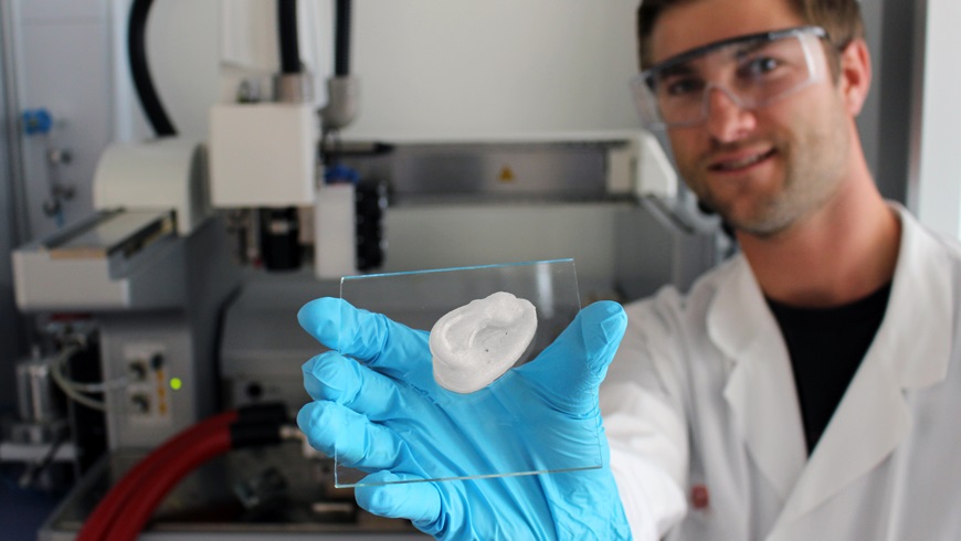
It all starts with an ear. Empa researcher Michael Hausmann removes the object shaped like a human ear from the 3D printer and explains: “In viscous state cellulose nanocrystals can easily be shaped together with nother biopolymers into complex 3-dimensional structures using a 3D printer, such as the Bioplotter.”

Once cross-linked, the structures remain stable despite their soft mechanical properties. Hausmann is currently investigating the characteristics of the nanocellulose composite hydrogels in order to further optimize their stability as well as the printing process. The researcher already used X-ray analysis to determine how cellulose is distributed and organized within the printed structures.

At this point in time the printed ear is entirely and solely made of cellulose nanocrystals and a biopolymer. However, the objective is to incorporate both human cells and therapeutics into the base structure in order to produce biomedical implants.

Here’s one of the researchers (Michael Hausmann) showing off their ‘ear’,

A 3D-printed ear: Empa researcher Michael Hausmann uses nanocellulose as the basis for novel implants (Image: Empa)

Doesn’t look like much does, eh? It’s scaffolding or, you could say, a kind of skeleton and a January 15, 2019 Empa press release, which originated the news item, describes it and explains how it will house new cells,

A new project is currently underway, looking into how chondrocytes (cartilage cells) can be integrated into the scaffold to yield artificial cartilage tissue. As soon as the colonization of the hydrogel with cells is established, nanocellulose based composites in the shape of an ear could serve as an implant for children with an inherited auricular malformation as for instance, in microtia, where the external ears are only incompletely developed. A reconstruction of the auricle can esthetically and medically correct the malformation; otherwise the hearing ability can be severely impaired. In the further course of the project, cellulose nanocrystals containing hydrogels will also be used for the replacement of articular cartilage (e.g. knee) in cases of joint wear due to, for example, chronic arthritis.

Once the artificial tissue has been implanted in the body, the biodegradable polymer material is expected to degrade over time. The cellulose itself is not degradable in the body, but biocompatible. However, it is not only its biocompatibility that makes nanocellulose the perfect material for implant scaffolds. “It is also the mechanical performance of cellulose nanocrystals that make them such promising candidates because the tiny but highly stable fibers can extremely well reinforce the produced implant,” said Hausmann.

Moreover, nanocellulose allows the incorporation of various functions by chemical modifications into the viscous hydrogel. Thus, the structure, the mechanical properties and the interactions of the nanocellulose with its environment can be specifically tailored to the desired end product. “For instance, we can incorporate active substances that promote the growth of chondrocytes or that sooth joint inflammation into the hydrogel,” says the Empa researcher.

And last but not least, as raw material cellulose is the most abundant natural polymer on earth. Therefore, the use of cellulose nanocrystals not only benefits from the mere elegance of the novel process but also from the availability of the raw material.

The white nanocellulose ear lies glossy on the glass carrier. Just out of the Bioplotter, it is already robust and dimensionally stable. Hausmann can give the go-ahead for the next steps. 

Here’s a link to and a citation for the paper,

Dynamics of Cellulose Nanocrystal Alignment during 3D Printing by Michael K. Hausmann, Patrick A. Rühs, Gilberto Siqueira, Jörg Läuger, Rafael Libanori, Tanja Zimmermann, and André R. Studart. ACS Nano, 2018, 12 (7), pp 6926–6937 DOI: 10.1021/acsnano.8b02366 Publication Date (Web): July 5, 2018

Copyright © 2018 American Chemical Society

This paper is behind a paywall.

A 3D printed ‘living’ tattoo

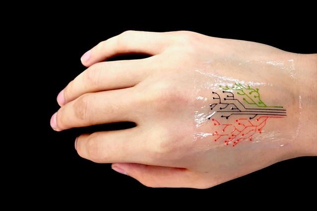
MIT engineers have devised a 3-D printing technique that uses a new kind of ink made from genetically programmed living cells. Courtesy of the researchers [and MIT]

If that image isn’t enough, there’s also a video abstract (I don’t think I’ve seen one of these before) for the paper,

For those who’d still like to read the text, here’s more from a December 5, 2017 MIT (Massachusetts Institute of Technology) news release (also on EurekAlert),

MIT engineers have devised a 3-D printing technique that uses a new kind of ink made from genetically programmed living cells.

The cells are engineered to light up in response to a variety of stimuli. When mixed with a slurry of hydrogel and nutrients, the cells can be printed, layer by layer, to form three-dimensional, interactive structures and devices.

The team has then demonstrated its technique by printing a “living tattoo” — a thin, transparent patch patterned with live bacteria cells in the shape of a tree. Each branch of the tree is lined with cells sensitive to a different chemical or molecular compound. When the patch is adhered to skin that has been exposed to the same compounds, corresponding regions of the tree light up in response.

The researchers, led by Xuanhe Zhao, the Noyce Career Development Professor in MIT’s Department of Mechanical Engineering, and Timothy Lu, associate professor of biological engineering and of electrical engineering and computer science, say that their technique can be used to fabricate “active” materials for wearable sensors and interactive displays. Such materials can be patterned with live cells engineered to sense environmental chemicals and pollutants as well as changes in pH and temperature.

What’s more, the team developed a model to predict the interactions between cells within a given 3-D-printed structure, under a variety of conditions. The team says researchers can use the model as a guide in designing responsive living materials.

Zhao, Lu, and their colleagues have published their results today [December 5, 2017] in the journal Advanced Materials. The paper’s co-authors are graduate students Xinyue Liu, Hyunwoo Yuk, Shaoting Lin, German Alberto Parada, Tzu-Chieh Tang, Eléonore Tham, and postdoc Cesar de la Fuente-Nunez.

A hardy alternative

In recent years, scientists have explored a variety of responsive materials as the basis for 3D-printed inks. For instance, scientists have used inks made from temperature-sensitive polymers to print heat-responsive shape-shifting objects. Others have printed photoactivated structures from polymers that shrink and stretch in response to light.

Zhao’s team, working with bioengineers in Lu’s lab, realized that live cells might also serve as responsive materials for 3D-printed inks, particularly as they can be genetically engineered to respond to a variety of stimuli. The researchers are not the first to consider 3-D printing genetically engineered cells; others have attempted to do so using live mammalian cells, but with little success.

“It turns out those cells were dying during the printing process, because mammalian cells are basically lipid bilayer balloons,” Yuk says. “They are too weak, and they easily rupture.”

Instead, the team identified a hardier cell type in bacteria. Bacterial cells have tough cell walls that are able to survive relatively harsh conditions, such as the forces applied to ink as it is pushed through a printer’s nozzle. Furthermore, bacteria, unlike mammalian cells, are compatible with most hydrogels — gel-like materials that are made from a mix of mostly water and a bit of polymer. The group found that hydrogels can provide an aqueous environment that can support living bacteria.

The researchers carried out a screening test to identify the type of hydrogel that would best host bacterial cells. After an extensive search, a hydrogel with pluronic acid was found to be the most compatible material. The hydrogel also exhibited an ideal consistency for 3-D printing.

“This hydrogel has ideal flow characteristics for printing through a nozzle,” Zhao says. “It’s like squeezing out toothpaste. You need [the ink] to flow out of a nozzle like toothpaste, and it can maintain its shape after it’s printed.”

From tattoos to living computers

Lu provided the team with bacterial cells engineered to light up in response to a variety of chemical stimuli. The researchers then came up with a recipe for their 3-D ink, using a combination of bacteria, hydrogel, and nutrients to sustain the cells and maintain their functionality.

“We found this new ink formula works very well and can print at a high resolution of about 30 micrometers per feature,” Zhao says. “That means each line we print contains only a few cells. We can also print relatively large-scale structures, measuring several centimeters.”

They printed the ink using a custom 3-D printer that they built using standard elements combined with fixtures they machined themselves. To demonstrate the technique, the team printed a pattern of hydrogel with cells in the shape of a tree on an elastomer layer. After printing, they solidified, or cured, the patch by exposing it to ultraviolet radiation. They then adhere the transparent elastomer layer with the living patterns on it, to skin.

To test the patch, the researchers smeared several chemical compounds onto the back of a test subject’s hand, then pressed the hydrogel patch over the exposed skin. Over several hours, branches of the patch’s tree lit up when bacteria sensed their corresponding chemical stimuli.

The researchers also engineered bacteria to communicate with each other; for instance they programmed some cells to light up only when they receive a certain signal from another cell. To test this type of communication in a 3-D structure, they printed a thin sheet of hydrogel filaments with “input,” or signal-producing bacteria and chemicals, overlaid with another layer of filaments of an “output,” or signal-receiving bacteria. They found the output filaments lit up only when they overlapped and received input signals from corresponding bacteria .

Yuk says in the future, researchers may use the team’s technique to print “living computers” — structures with multiple types of cells that communicate with each other, passing signals back and forth, much like transistors on a microchip.

“This is very future work, but we expect to be able to print living computational platforms that could be wearable,” Yuk says.

For more near-term applications, the researchers are aiming to fabricate customized sensors, in the form of flexible patches and stickers that could be engineered to detect a variety of chemical and molecular compounds. They also envision their technique may be used to manufacture drug capsules and surgical implants, containing cells engineered produce compounds such as glucose, to be released therapeutically over time.

“We can use bacterial cells like workers in a 3-D factory,” Liu says. “They can be engineered to produce drugs within a 3-D scaffold, and applications should not be confined to epidermal devices. As long as the fabrication method and approach are viable, applications such as implants and ingestibles should be possible.”

Here’s a link to and a citation for the paper,

3D Printing of Living Responsive Materials and Devices by Xinyue Liu, Hyunwoo Yuk, Shaoting Lin, German Alberto Parada, Tzu-Chieh Tang, Eléonore Tham, Cesar de la Fuente-Nunez, Timothy K. Lu, and Xuanhe Zhao. Advanced Materials DOI: 10.1002/adma.201704821 Version of Record online: 5 DEC 2017

© 2017 WILEY-VCH Verlag GmbH & Co. KGaA, Weinheim

This paper is behind a paywall.

A biocompatible (implantable) micromachine (microrobot)

I appreciate the detail and information in this well written Jan. 4, 2017 Columbia University news release (h/t Jan. 4, 2016 Nanowerk; Note: Links have been removed),

A team of researchers led by Biomedical Engineering Professor Sam Sia has developed a way to manufacture microscale-sized machines from biomaterials that can safely be implanted in the body. Working with hydrogels, which are biocompatible materials that engineers have been studying for decades, Sia has invented a new technique that stacks the soft material in layers to make devices that have three-dimensional, freely moving parts. The study, published online January 4, 2017, in Science Robotics, demonstrates a fast manufacturing method Sia calls “implantable microelectromechanical systems” (iMEMS).

By exploiting the unique mechanical properties of hydrogels, the researchers developed a “locking mechanism” for precise actuation and movement of freely moving parts, which can provide functions such as valves, manifolds, rotors, pumps, and drug delivery. They were able to tune the biomaterials within a wide range of mechanical and diffusive properties and to control them after implantation without a sustained power supply such as a toxic battery. They then tested the “payload” delivery in a bone cancer model and found that the triggering of release of doxorubicin from the device over 10 days showed high treatment efficacy and low toxicity, at 1/10 of the standard systemic chemotherapy dose.

“Overall, our iMEMS platform enables development of biocompatible implantable microdevices with a wide range of intricate moving components that can be wirelessly controlled on demand and solves issues of device powering and biocompatibility,” says Sia, also a member of the Data Science Institute. “We’re really excited about this because we’ve been able to connect the world of biomaterials with that of complex, elaborate medical devices. Our platform has a large number of potential applications, including the drug delivery system demonstrated in our paper which is linked to providing tailored drug doses for precision medicine.”

I particularly like this bit about hydrogels being a challenge to work with and the difficulties of integrating both rigid and soft materials,

Most current implantable microdevices have static components rather than moving parts and, because they require batteries or other toxic electronics, have limited biocompatibility. Sia’s team spent more than eight years working on how to solve this problem. “Hydrogels are difficult to work with, as they are soft and not compatible with traditional machining techniques,” says Sau Yin Chin, lead author of the study who worked with Sia. “We have tuned the mechanical properties and carefully matched the stiffness of structures that come in contact with each other within the device. Gears that interlock have to be stiff in order to allow for force transmission and to withstand repeated actuation. Conversely, structures that form locking mechanisms have to be soft and flexible to allow for the gears to slip by them during actuation, while at the same time they have to be stiff enough to hold the gears in place when the device is not actuated. We also studied the diffusive properties of the hydrogels to ensure that the loaded drugs do not easily diffuse through the hydrogel layers.”

The team used light to polymerize sheets of gel and incorporated a stepper mechanization to control the z-axis and pattern the sheets layer by layer, giving them three-dimensionality. Controlling the z-axis enabled the researchers to create composite structures within one layer of the hydrogel while managing the thickness of each layer throughout the fabrication process. They were able to stack multiple layers that are precisely aligned and, because they could polymerize a layer at a time, one right after the other, the complex structure was built in under 30 minutes.

Sia’s iMEMS technique addresses several fundamental considerations in building biocompatible microdevices, micromachines, and microrobots: how to power small robotic devices without using toxic batteries, how to make small biocompatible moveable components that are not silicon which has limited biocompatibility, and how to communicate wirelessly once implanted (radio frequency microelectronics require power, are relatively large, and are not biocompatible). The researchers were able to trigger the iMEMS device to release additional payloads over days to weeks after implantation. They were also able to achieve precise actuation by using magnetic forces to induce gear movements that, in turn, bend structural beams made of hydrogels with highly tunable properties. (Magnetic iron particles are commonly used and FDA-approved for human use as contrast agents.)

In collaboration with Francis Lee, an orthopedic surgeon at Columbia University Medical Center at the time of the study, the team tested the drug delivery system on mice with bone cancer. The iMEMS system delivered chemotherapy adjacent to the cancer, and limited tumor growth while showing less toxicity than chemotherapy administered throughout the body.

“These microscale components can be used for microelectromechanical systems, for larger devices ranging from drug delivery to catheters to cardiac pacemakers, and soft robotics,” notes Sia. “People are already making replacement tissues and now we can make small implantable devices, sensors, or robots that we can talk to wirelessly. Our iMEMS system could bring the field a step closer in developing soft miniaturized robots that can safely interact with humans and other living systems.”

Here’s a link to and a citation for the paper,

Additive manufacturing of hydrogel-based materials for next-generation implantable medical devices by Sau Yin Chin, Yukkee Cheung Poh, Anne-Céline Kohler, Jocelyn T. Compton, Lauren L. Hsu, Kathryn M. Lau, Sohyun Kim, Benjamin W. Lee, Francis Y. Lee, and Samuel K. Sia. Science Robotics  04 Jan 2017: Vol. 2, Issue 2, DOI: 10.1126/scirobotics.aah6451

This paper appears to be open access.

The researchers have provided a video demonstrating their work (you may want to read the caption below before watching),

Magnetic actuation of the Geneva drive device. A magnet is placed about 1cm below and without contact with the device. The rotating magnet results in the rotational movement of the smaller driving gear. With each full rotation of this driving gear, the larger driven gear is engaged and rotates by 60º, exposing the next reservoir to the aperture on the top layer of the device.

—Video courtesy of Sau Yin Chin/Columbia Engineering

You can hear some background conversation but it doesn’t seem to have been included for informational purposes.

Tiny, electrically conductive 3D-printed chair made from cellulose

Sweden’s Chalmers University of Technology researchers have just announced that they’ve printed a very small 3D chair with electrical properties using cellulose nanomaterials. From a June 17, 2015 news item on Nanowerk,

A group of researchers at Chalmers University of Technology have managed to print and dry three-dimensional objects made entirely by cellulose for the first time with the help of a 3D-bioprinter. They also added carbon nanotubes to create electrically conductive material. The effect is that cellulose and other raw material based on wood will be able to compete with fossil-based plastics and metals in the on-going additive manufacturing revolution, which started with the introduction of the 3D-printer.

Here’s the 3D-printed chair,

The tiny chair made of cellulose is a demonstrational object, printed using the 3D bioprinter at Chalmers University of Technology. Photo: Peter Widing

The tiny chair made of cellulose is a demonstrational object, printed using the 3D bioprinter at Chalmers University of Technology. Photo: Peter Widing

A June 17, 2015 Chalmers University of Technology press release (also on EurekAlert*), which originated the news item, describes the problem with printing from cellulose nanomaterials and how it was solved,

The difficulty using cellulose in additive manufacturing is that cellulose does not melt when heated. Therefore, the 3D printers and processes designed for printing plastics and metals cannot be used for materials like cellulose. The Chalmers researchers solved this problem by mixing cellulose nanofibrils in a hydrogel consisting of 95-99 percent water. The gel could then in turn be dispensed with high fidelity into the researchers’ 3D bioprinter, which was earlier used to produce scaffolds for growing cells, where the end application is patient-specific implants.

The next challenge was to dry the printed gel-like objects without them losing their three-dimensional shape.

“The drying process is critical,” Paul Gatenholm explains. “We have developed a process in which we freeze the objects and remove the water by different means as to control the shape of the dry objects. It is also possible to let the structure collapse in one direction, creating thin films.”

Furthermore, the cellulose gel was mixed with carbon nanotubes to create electrically conductive ink after drying. Carbon nanotubes conduct electricity, and another project at Wallenberg Wood Science Center aims at developing carbon nanotubes using wood.

Using the two gels together, one conductive and one non-conductive, and controlling the drying process, the researchers produced three-dimensional circuits, where the resolution increased significantly upon drying.

The two gels together provide a basis for the possible development of a wide range of products made by cellulose with in-built electric currents.

“Potential applications range from sensors integrated with packaging, to textiles that convert body heat to electricity, and wound dressings that can communicate with healthcare workers,” says Paul Gatenholm. “Our research group now moves on with the next challenge, to use all wood biopolymers, besides cellulose.”

The research findings are presented this week at the conference New Materials From Trees that takes place in Stockholm, Sweden, June 15-17 [2015].

The research team members are Ida Henriksson, Cristina de la Pena, Karl Håkansson, Volodymyr Kuzmenko and Paul Gatenholm at Chalmers University of Technology.

This research reminds me of another effort, a computer chip fashioned of cellulose nanofibrils (CNF) from the University of Wisconsin-Madison (mentioned in my May 27, 2015 post).

* EurekAlert link added June 18, 2015.

Hydrogels and cartilage; repurposing vehicles in space; big bang has ‘fingerprints’

The American Institute of Physics (AIP) has made a selection of four articles freely available (h/t Mar. 9, 2015 news item on Azonano).

From a March 6, 2015 AIP news release,

WASHINGTON D.C., March 6, 2015 — The following articles are freely available online from Physics Today (www.physicstoday.org), the world’s most influential and closely followed magazine devoted to physics and the physical science community.

You are invited to read, share, blog about, link to, or otherwise enjoy:

1) STIFF AND SUPPLE CARTILAGE SUBSTITUTE

Physics Today‘s Ashley Smart reports on hydrogels that mimic the tricky nature of cartilage thanks to magnetically aligned nanosheets.

“In the realm of bioengineering, hydrogels are something of an all-purpose material. Made up of networks of interlinked, hydrophilic polymers, they tend to be soft, biocompatible, and highly absorbent…. The new material mimics the articular cartilage that lubricates our joints: It can support a heavy load along one direction while stretching and shearing with ease in the others.”

MORE: http://dx.doi.org/10.1063/PT.3.2707

2) GIVING SPACECRAFT A SECOND LEASE ON LIFE WHILE HURTLING THROUGH THE COSMOS

Physics Today‘s Toni Feder reports on the innovative processes undertaken to repurpose various spacecraft in flight, including Kepler, Voyager, Deep Impact, Spitzer, and the Hubble Space Telescope.

“A comeback like Kepler’s is ‘not unique, but it’s unusual,’ says Derek Buzasi of Florida Gulf Coast University, who reinvented the Wide-Field Infrared Explorer (WIRE) after it failed following its 1999 launch. ‘Spacecraft are built for a specialized purpose, so they are hard to repurpose. You have to come up with something they are capable of at the same time they are incapable of their original mission.’

Deep Impact’s original mission was to hurl a copper ball at a comet and watch the impact. In its continued form as EPOXI, the spacecraft went on to visit another comet and, on the way, served as an observatory for user- proposed targets.”

MORE: http://dx.doi.org/10.1063/PT.3.2713

3) CONGRESSMAN & FUSION RESEARCHER REFLECTS ON SCIENCE POLICY

Physics Today‘s David Kramer interviews Rush Holt, the New Jersey congressman who retired from office and this past December took the helm of the American Association for the Advancement of Science.

“PT: What do you consider to be your accomplishments in Congress?

HOLT: I focused a lot on science education. Our real problem is not that we’re failing to produce excellent scientists, because we are [producing them], but rather that we have failed to maintain an appreciation for and understanding of science in the general population. I was able to keep a spotlight on the need but wasn’t able to accomplish as much as I wanted. We got science included in the subjects emphasized by federal law. But we haven’t really improved teacher professional development and other things we need to do.”

MORE: http://dx.doi.org/10.1063/PT.3.2714

4) PARTICLE PHYSICS AND THE COSMIC MICROWAVE BACKGROUND

In this article, physics researchers John Carlstrom, Tom Crawford and Lloyd Knox discuss the fingerprints of the Big Bang and quantum fluctuations in the early universe, which may soon reveal physics at unprecedented energy scales.

“With its empirical successes, inflation is by consensus the best paradigm—notwithstanding some notable dissenting views—for the mechanism that generated the primordial density fluctuations that led to all structure in the universe. Its success has motivated physicists to search for the siblings of those fluctuations, the gravitational waves, via their signature in the polarization of the CMB. If discovered, that gravitational imprint would open up an observational window onto quantum gravitational effects, extremely early times, and extremely high energies.”

MORE: http://dx.doi.org/10.1063/PT.3.2718

I have checked; all of the links do lead to the articles.

Squishy wonderfulness: new possibilities for hydrogels

i have two items for this posting about hydrogels and biomimicry (aka biomimetics). One concerns the use of light to transform hydrogels and the other concerns the potential for using hydrogels in ‘soft’ robotics. First, researchers at the University of Pittsburgh have found a way to make hydrogels change their shapes, from an Aug. 1, 2013 news item on Nanowerk,

Some animals—like the octopus and cuttlefish—transform their shape based on environment, fending off attackers or threats in the wild. For decades, researchers have worked toward mimicking similar biological responses in non-living organisms, as it would have significant implications in the medical arena.

Now, researchers at the University of Pittsburgh have demonstrated such a biomimetic response using hydrogels—a material that constitutes most contact lenses and microfluidic or fluid-controlled technologies.

The Aug. 1, 2013 University of Pittsburgh news release, which originated the news item, offers this description from the paper’s lead authorl,

“Imagine an apartment with a particular arrangement of rooms all in one location,” said lead author Anna Balazs, Pitt Distinguished Professor of Chemical and Petroleum Engineering in the Swanson School of Engineering. “Now, consider the possibility of being able to shine a particular configuration of lights on this structure and thereby completely changing not only the entire layout, but also the location of the apartment. This is what we’ve demonstrated with hydrogels.”

The news release goes on to provide more specific details about the work,

Together with Olga Kuksenok, research associate professor in the Swanson School, Balazs experimented with a newer type of hydrogel containing spirobenzopyran molecules. Such materials had been previously shown to form distinct 2-D patterns on initially flat surfaces when introduced to varying displays of light and are hydrophilic (“liking” water) in the dark but become hydrophobic (“disliking” water) under blue light illumination. Therefore, Balazs and Kuksenok anticipated that light could be a useful stimulus for tailoring the gel’s shape.

Using computer modeling, the Pitt team demonstrated that the gels “ran away” when exposed to the light, exhibiting direct, sustained motion. The team also factored in heat—combining the light and local variations in temperature to further control the samples’ motions. Controlling a material with light and temperature could be applicable, Balazs said, in terms of regulating the movement of a microscopic “conveyor belt” or “elevator” in a microfluidic device.

“This theoretical modeling points toward a new way of configuring the gels into any shape, while simultaneously driving the gels to move due to the presence of light,” said Kuksenok.

“Consider, for example, that you could take one sheet of hydrogel and, with the appropriate use of light, fashion it into a lens-shaped object, which could be used in optical applications”, added Balazs.

The team also demonstrated that the gels could undergo dynamic reconfiguration, meaning that, with a different combination of lights, the gel could be used for another purpose. Reconfigurable systems are particularly useful because they are reusable, leading to a significant reduction in cost.

“You don’t need to construct a new device for every new application,” said Balazs. “By swiping light over the system in different directions, you can further control the movements of a system, further regulating the flow of materials.”

Balazs said this type of dynamic reconfiguration in response to external cues is particularly advantageous in the realm of functional materials. Such processes, she said, would have a dramatic effect on manufacturing and sustainability, since the same sample could be used and reused for multiple applications.

The team will now study the effect of embedding microscopic fibers into the gel to further control the shape and response of the material to other stimuli.

Here’s a link to and a citation for the research paper,

Modeling the Photoinduced Reconfiguration and Directed Motion of Polymer Gels by Olga Kuksenok and Anna C. Balazs. Article first published online: 31 JUL 2013, Adv. Funct. Mater.. doi: 10.1002/adfm.201203876

© 2013 WILEY-VCH Verlag GmbH & Co. KGaA, Weinheim

This paper is behind a paywall. However, there is a video of Anna Balazs’s June 27, 2013 talk (Reconfigurable assemblies of active, auto-chemotactic gels) on these gels at the Isaac Newton Institute for Mathematical Sciences.

Meanwhile, researchers at North Carolina State University are pursuing a different line of query involving hydrogels. From an Aug. 2, 2013 North Carolina State University news release (also on EurekAlert),

Researchers from North Carolina State University have developed a new technique for creating devices out of a water-based hydrogel material that can be patterned, folded and used to manipulate objects. The technique holds promise for use in “soft robotics” and biomedical applications.

“This work brings us one step closer to developing new soft robotics technologies that mimic biological systems and can work in aqueous environments,” says Dr. Michael Dickey, an assistant professor of chemical and biomolecular engineering at NC State and co-author of a paper describing the work.

“In the nearer term, the technique may have applications for drug delivery or tissue scaffolding and directing cell growth in three dimensions, for example,” says Dr. Orlin Velev, INVISTA Professor of Chemical and Biomolecular Engineering at NC State, the second senior author of the paper.

The technique they’ve developed uses hydrogels, which are water-based gels composed of water and a small fraction of polymer molecules. Hydrogels are elastic, translucent and – in theory – biocompatible. The researchers found a way to modify and pattern sections of hydrogel electrically by using a copper electrode to inject positively charged copper ions into the material. Those ions bond with negatively charged sites on the polymer network in the hydrogel, essentially linking the polymer molecules to each other and making the material stiffer and more resilient. The researchers can target specific areas with the electrodes to create a framework of stiffened material within the hydrogel. The resulting patterns of ions are stable for months in water.

“The bonds between the biopolymer molecules and the copper ions also pull the molecular strands closer together, causing the hydrogel to bend or flex,” Velev says. “And the more copper ions we inject into the hydrogel by flowing current through the electrodes, the further it bends.”

The researchers were able to take advantage of the increased stiffness and bending behavior in patterned sections to make the hydrogel manipulate objects. For example, the researchers created a V-shaped segment of hydrogel. When copper ions were injected into the bottom of the V, the hydrogel flexed – closing on an object as if the hydrogel were a pair of soft tweezers. By injecting ions into the back side of the hydrogel, the tweezers opened – releasing the object.

The researchers also created a chemically actuated “grabber” out of an X-shaped segment of hydrogel with a patterned framework on the back of the X. When the hydrogel was immersed in ethanol, the non-patterned hydrogel shrank. But because the patterned framework was stiffer than the surrounding hydrogel, the X closed like the petals of a flower, grasping an object. When the X-shaped structure was placed in water, the hydrogel expanded, allowing the “petals” to unfold and release the object. Video of the hydrogels in action is available here.

“We are currently planning to use this technique to develop motile, biologically compatible microdevices,” Velev says.

“It’s also worth noting that this technique works with ions other than copper, such as calcium, which are biologically relevant,” Dickey says.

Here’s a link to and a citation for the article,

Reversible patterning and actuation of hydrogels by electrically assisted ionoprinting by Etienne Palleau, Daniel Morales, Michael D. Dickey & Orlin D. Velev. Nature Communications 4, Article number: 2257 doi:10.1038/ncomms3257 Published 02 August 2013

This article is behind a paywall.