Tag Archives: immune system

“The Immune System: Our Great Protector Against Dangerous Stuff” talk at Simon Fraser University’s (SFU) Café Scientifique on Thursday January 27, 2022 from 5:00 pm – 6:30 pm PST

This is from a January 13, 2022 SFU Café Scientifique notice (received via email),

Happy New Year! We are excited to announce our next virtual SFU Café
Scientifique!

 Thursday January 27, 2022, 5:00-6:30 pm

 Dr. Jonathan Choy, SFU Molecular Biology and Biochemistry

The Immune System: Our Great Protector Against Dangerous Stuff

Our bodies are constantly in contact with material in the environment,
such as microbes, that are harmful to our health. Despite this, most
people are healthy because the immune system patrols our bodies and
protects us from these harmful environmental components. In this Cafe
Scientifique, Dr. Jonathan Choy from the Department of Molecular Biology
and Biochemistry will discuss how the immune system does this.

Register here to receive a zoom invite:

 
https://www.eventbrite.ca/e/sfu-cafe-scientifique-january-2022-tickets-227344733217

I found Dr. Choy’s profile page on the SFU website and found this description for his research interests,

T Cell Biology 

T cells are specialized cells of the immune system that protect host organisms from infection but that also contribute to a wide array of human diseases. Research in my laboratory is focused on understanding the mechanisms by which T cells become inappropriately activated in disease settings and how they cause organ damage. We have provided particular attention to how innate immune signals, such as cytokines secreted by innate immune cells and vascular cells, control the outcome of T cell responses. Within this context, processes that inhibit the activation of T cells are also being studied in order to potentially prevent disease-causing immune responses. Our studies on this topic are applied most directly to inflammatory vascular diseases, such as transplant arteriosclerosis and giant cell arteritis.

Nitric Oxide Signaling and Production 

Nitric oxide (NO) is a bioactive gas that controls many cell biological responses. Dysregulation of its production and/or bioactivity is involved in many diseases. My laboratory is interested in understanding how NO effects cell signaling and how its production is controlled by NO synthases. We are specifically interested in how NO-mediated protein S-nitrosylation, a post-translational modification caused by NO, affects cell signaling pathways and cellular functions.

I gather from the Café Scientifique write up that Dr. Choy’s talk is intended for a more general audience as opposed to the description of his research interests which are intended for students of molecular biology and biochemistry/

For those who are unfamiliar with it, Simon Fraser University is located in the Vancouver area (Canada).

Immune to CRISPR?

I guess if you’re going to use bacteria as part of your gene editing technology (CRISPR [clustered regularly interspaced short palindromic repeats]/Cas9) then, you might half expect the body’s immune system may have developed some defenses. A Jan. 9, 2018 article by Sarah Zhang for The Atlantic provides some insight into what the new research suggests (Note: Links have been removed),

2018 is supposed to be the year of CRISPR in humans. The first U.S. and European clinical trials that test the gene-editing tool’s ability to treat diseases—such as sickle-cell anemia, beta thalassemia, and a type of inherited blindness—are slated to begin this year.

But the year has begun on a cautionary note. On Friday [January 5, 2018], Stanford researchers posted a preprint (which has not been peer reviewed) to the website biorXiv highlighting a potential obstacle to using CRISPR in humans: Many of us may already be immune to it. That’s because CRISPR actually comes from bacteria that often live on or infect humans, and we have built up immunity to the proteins from these bacteria over our lives.

Not all CRISPR therapies in humans will be doomed. “We don’t think this is the end of the story. This is the start of the story,” says Porteus [Matthew Porteus, a pediatrician and stem-cell researcher at Stanford]. There are likely ways around the problem of immunity to CRISPR proteins, and many of the early clinical trials appear to be designed around this problem.

Porteus and his colleagues focused on two versions of Cas9, the bacterial protein mostly commonly used in CRISPR gene editing. One comes from Staphylococcus aureus, which often harmlessly lives on skin but can sometimes causes staph infections, and another from Streptococcus pyogenes, which causes strep throat but can also become “flesh-eating bacteria” when it spreads to other parts of the body. So yeah, you want your immune system to be on guard against these bacteria.

The human immune system has a couple different ways of recognizing foreign proteins, and the team tested for both. First, they looked to see if people have molecules in their blood called antibodies that can specifically bind to Cas9. Among 34 people they tested, 79 percent had antibodies against the staph Cas9 and 65 percent against the strep Cas9.

The Stanford team only tested for preexisting immunity against Cas9, but anytime you inject a large bacterial protein into the human body, it can provoke an immune response. After all, that’s how the immune system learns to fight off bacteria it’s never seen before. (Preexisting immunity can make the response faster and more robust, though.)

The danger of the immune system turning on a patient’s body hangs over a lot of research into correcting genes. In the late 1990s and 2000s, research into gene therapy was derailed by the death of 18-year-old Jesse Gelsinger, who died from an immune reaction to the virus used to deliver the corrected gene. This is the worst-case scenario that the CRISPR world hopes to avoid.

Here’s a link to and a citation for the preprint,

Identification of Pre-Existing Adaptive Immunity to Cas9 Proteins in Humans by Carsten Trevor Charlesworth, Priyanka S Deshpande, Daniel P Dever, Beruh Dejene, Natalia Gomez-Ospina, Sruthi Mantri, Mara Pavel-Dinu, Joab Camarena, Kenneth I Weinberg, Matthew H Porteus. bioRxiv posted January 5, 2018 doi: https://doi.org/10.1101/243345

This article is a preprint and has not been peer-reviewed …

This preprint (not yet published paper) is open access and open for feedback.

Meanwhile, the year of CRISPR takes off (from a January 10, 2018 American Chemical Society news release on EurekAlert),

This year could be a defining one for CRISPR, the gene editing technique, which has been hailed as an important breakthrough in laboratory research. That’s because the first company-sponsored clinical studies will be conducted to see if it can help treat diseases in humans, according to an article in Chemical & Engineering News (C&EN), the weekly newsmagazine of the American Chemical Society.

C&EN Assistant Editor Ryan Cross reports that a big push is coming from industry, specifically from three companies that are each partly founded by one of the three inventors of the method. They are zeroing in on the blood diseases called sickle-cell anemia and β-thalassemia, mostly because their precise cause is known. In these diseases, hemoglobin doesn’t function properly, leading to severe health issues in some people. Crispr Therapeutics and Intellia Therapeutics plan to test the technique to boost levels of an alternative version of healthy hemoglobin. Editas Medicine, however, will also use CRISPR to correct mutations in the faulty hemoglobin gene. Labs led by university researchers are also joining the mix, starting or continuing clinical trials with the approach in 2018.

Because CRISPR is being used to cut a cell’s DNA and insert a new sequence, concerns have been raised about the potential for accidents. A cut in the wrong place could mean introducing a new mutation that could be benign — or cancerous. But according to proponents of the method, researchers are conducting extensive computer predictions and in vitro tests to help avoid this outcome.

The January 8, 2018 Chemical and Engineering News (C&EN) open access article by Ryan Cross is here.

Finally, if you are interested in how this affects research as it’s being developed, there’s University of British Columbia researcher Rosie Redfield’s January 16, 2018 posting on RRResearch blog,

Thursday’s [January 11, 2018] post described the hypothesis that bacteria might use gene transfer agent particles to inoculate other cells in the population with fragments of phage DNA, and outlined an experiment to test this.  Now I’m realizing that I need to know a lot more about the kind of immunity I should expect to see if this GTA-as-vaccine hypothesis is correct.

That should give you some idea of what I meant by “research as it’s being developed.” Redfield’s blog is not for the mildly interested.

Redfield is well-known internationally as being one of the first to refute research which suggested the existence of an ‘arsenic bacterium’ (see my Dec. 8, 2010 posting: My apologies for arsenic blooper. She’s first mentioned in the second excerpt, second paragraph.) The affair was known online as #arseniclife. There’s a May 27, 2011 essay by Carl Zimmer on Slate titled: The Discovery of Arsenic-Based Twitter: How #arseniclife changed science.
Nanoparticles can activate viruses lying dormant in lung cells

The nanoparticles in question are from combustion engines, which means that we are exposed to them. One other note, the testing has not been done on humans but rather on cells. From a Jan. 16, 2017 news item on ScienceDaily,

Nanoparticles from combustion engines can activate viruses that are dormant in in lung tissue cells. This is the result of a study by researchers of Helmholtz Zentrum München, a partner in the German Center for Lung Research (DZL), which has now been published in the journal Particle and Fibre Toxicology.

To evade the immune system, some viruses hide in cells of their host and persist there. In medical terminology, this state is referred to as a latent infection. If the immune system becomes weakened or if certain conditions change, the viruses become active again, begin to proliferate and destroy the host cell. A team of scientists led by Dr. Tobias Stöger of the Institute of Lung Biology and Prof. Dr. Heiko Adler, deputy head of the research unit Lung Repair and Regeneration at Helmholtz Zentrum München, now report that nanoparticles can also trigger this process.

A Jan. 16, 2017 Helmholtz Zentrum München press release (also on EurekAlert), which originated the news item, provides more detail,

“From previous model studies we already knew that the inhalation of nanoparticles has an inflammatory effect and alters the immune system,” said study leader Stöger. Together with his colleagues Heiko Adler and Prof. Dr. Philippe Schmitt-Kopplin, he showed that “an exposure to nanoparticles can reactivate latent herpes viruses in the lung.”

Specifically, the scientists tested the influence of nanoparticles typically generated by fossil fuel combustion in an experimental model for a particular herpes virus infection. They detected a significant increase in viral proteins, which are only produced with active virus proliferation. “Metabolic and gene expression analyses also revealed patterns resembling acute infection,” said Philippe Schmitt-Kopplin, head of the research unit Analytical BioGeoChemistry (BGC). Moreover, further experiments with human cells demonstrated that Epstein-Barr viruses are also ‘awakened’ when they come into contact with the nanoparticles.

Potential approach for chronic lung diseases

In further studies, the research team would like to test whether the results can also be transferred to humans. “Many people carry herpes viruses, and patients with idiopathic pulmonary fibrosis are particularly affected,” said Heiko Adler. “If the results are confirmed in humans, it would be important to investigate the molecular process of the reactivation of latent herpes viruses induced by particle inhalation. Then we could try to influence this pathway therapeutically.”

Special cell culture models shall therefore elucidate the exact mechanism of virus reactivation by nanoparticles. “In addition,” Stöger said, ”in long-term studies we would like to investigate to what extent  repeated nanoparticle exposure with corresponding virus reactivation leads to chronic inflammatory and remodeling processes in the lung.”

Further Information

Background:
In 2015 another group at the Helmholtz Zentrum München demonstrated how the Epstein-Barr virus  hides in human cells. In March 2016 researchers also showed that microRNAs silence immune alarm signals of cells infected with the Epstein-Barr virus.

Original Publication:
Sattler, C. et al. (2016): Nanoparticle exposure reactivates latent herpesvirus and restores a signature of acute infection. Particle and Fibre Toxicology, DOI 10.1186/s12989-016-0181-1

Here’s a link to and a citation for the paper on investigating latent herpes virus,

Nanoparticle exposure reactivates latent herpesvirus and restores a signature of acute infection by Christine Sattler, Franco Moritz, Shanze Chen, Beatrix Steer, David Kutschke, Martin Irmler, Johannes Beckers, Oliver Eickelberg, Philippe Schmitt-Kopplin, Heiko Adler. Particle and Fibre Toxicology201714:2 DOI: 10.1186/s12989-016-0181-1 Published: 10 January 2017

©  The Author(s). 2017

This paper is open access and, so too, is the 2016 paper.

Nanomedicine and the immune system

Interest in how the body reacts to nanoparticle drug delivery materials seems to be gaining momentum (see my Sept. 9, 2016 post about how the liver prevents nanoparticles from reaching cancer cells and my April 27, 2016 post about the discovery that fewer than 1% of nanoparticle-based drugs reach their destination). Now, we can add this research to the list according to an Oct. 4, 2016 news item on phys.org,

Katie Whitehead, assistant professor of chemical engineering at Carnegie Mellon University, has focused her research efforts on two clear objectives: treating and preventing disease. Her clinical-minded approach to laboratory research has recently led her to join forces with immunologists at the Indian Institute of Technology (IIT) in Bombay on a project that will explore how the immune system reacts to nanoparticle drug delivery materials.

“At its face, it may seem like an obvious goal. You would want a drug delivery system that doesn’t provoke an immune response,” says Whitehead. “However, the immune response to drug delivery vehicles is an understudied area because it’s complicated and expensive—but it deserves more attention.”

An Oct. 4, 2016 Carnegie Mellon University news release, which originated the news item, describes the research in more detail (Note: A link has been removed),

If the immune system reacts to a drug delivery system, the body mistakenly identifies the material as an invading pathogen and goes into a heightened state of alert. This response can trigger inflammation throughout the body and lead to a host of issues. According to Nature, about 25 percent of all Phase II and III clinical trials fail, not because the drug did not treat the disease, but because of safety concerns.

Creating a drug delivery system that effectively treats disease at the same time as avoiding immune response are two separate aims in drug delivery research. But for Whitehead, “My argument has always been that both pieces of the puzzle are equally important. If a system causes an immune response, then it’s a nonstarter. It may yield great results in treating disease in the lab, but it won’t ever reach a patient.”

Unfortunately, very little is understood about how the chemical molecules that make up nanoparticles ultimately affect our body’s immune response. “This research, however, is going to fill a critical gap in our knowledge base that will allow us to create nanoparticle systems that effectively deliver drugs without provoking our body’s natural defense mechanisms,” explains Whitehead. “Such knowledge will give us a head start in moving our delivery systems into clinical settings.”

Whitehead’s lab creates a number of nanoparticle drug delivery systems for diseases ranging from inflammatory bowel disease to Mantle cell lymphoma. She is tackling the challenge of immune response head-on with the help of a four-year, $500,000 grant from the Wadhwani Foundation for her work with IIT Bombay. She’ll specifically study how the chemical structure of the drug delivery nanoparticles affects the immune system.

Here’s a video of Katie Whitehead discussing her work in a simplified fashion,


Mechanism behind interaction of silver nanoparticles with the cells of the immune system

Scientists have come to a better understanding of the mechanism affecting silver nanoparticle toxicity according to an Aug. 30, 2016 news item on Nanowerk (Note: A link has been removed),

A senior fellow at the Faculty of Chemistry, MSU (Lomonosov Moscow State University), Vladimir Bochenkov together with his colleagues from Denmark succeeded in deciphering the mechanism of interaction of silver nanoparticles with the cells of the immune system. The study is published in the journal Nature Communications (“Dynamic protein coronas revealed as a modulator of silver nanoparticle sulphidation in vitro”).

‘Currently, a large number of products are containing silver nanoparticles: antibacterial drugs, toothpaste, polishes, paints, filters, packaging, medical and textile items. The functioning of these products lies in the capacity of silver to dissolve under oxidation and form ions Ag+ with germicidal properties. At the same time there are research data in vitro, showing the silver nanoparticles toxicity for various organs, including the liver, brain and lungs. In this regard, it is essential to study the processes occurring with silver nanoparticles in biological environments, and the factors affecting their toxicity,’ says Vladimir Bochenkov.

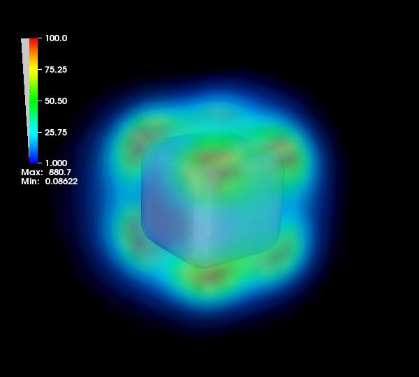
Caption: Increased intensity of the electric field near the silver nanoparticle surface in the excitation of plasmon resonance. Credit: Vladimir Bochenkov

Caption: Increased intensity of the electric field near the silver nanoparticle surface in the excitation of plasmon resonance. Credit: Vladimir Bochenkov

An Aug. 30, 2016 MSU press release on EurekAlert, which originated the news item, provides more information about the research,

The study is devoted to the protein corona — a layer of adsorbed protein molecules, which is formed on the surface of the silver nanoparticles during their contact with the biological environment, for example in blood. Protein crown masks nanoparticles and largely determines their fate: the speed of the elimination from the body, the ability to penetrate to a particular cell type, the distribution between the organs, etc.

According to the latest research, the protein corona consists of two layers: a rigid hard corona — protein molecules tightly bound with silver nanoparticles, and soft corona, consisting of weakly bound protein molecules in a dynamic equilibrium with the solution. Hitherto soft corona has been studied very little because of the experimental difficulties: the weakly bound nanoparticles separated from the protein solution easily desorbed (leave a particle remaining in the solution), leaving only the rigid corona on the nanoparticle surface.

The size of the studied silver nanoparticles was of 50-88 nm, and the diameter of the proteins that made up the crown — 3-7 nm. Scientists managed to study the silver nanoparticles with the protein corona in situ, not removing them from the biological environment. Due to the localized surface plasmon resonance used for probing the environment near the surface of the silver nanoparticles, the functions of the soft corona have been primarily investigated.

‘In the work we showed that the corona may affect the ability of the nanoparticles to dissolve to silver cations Ag+, which determine the toxic effect. In the absence of a soft corona (quickly sharing the medium protein layer with the environment) silver cations are associated with the sulfur-containing amino acids in serum medium, particularly cysteine and methionine, and precipitate as nanocrystals Ag2S in the hard corona,’ says Vladimir Bochenkov.

Ag2S (silver sulfide) famously easily forms on the silver surface even on the air in the presence of the hydrogen sulfide traces. Sulfur is also part of many biomolecules contained in the body, provoking the silver to react and be converted into sulfide. Forming of the nano-crystals Ag2S due to low solubility reduces the bioavailability of the Ag+ ions, reducing the toxicity of silver nanoparticles to null. With a sufficient amount of amino acid sulfur sources available for reaction, all the potentially toxic silver is converted into the nontoxic insoluble sulfide. Scientists have shown that what happens in the absence of a soft corona.

In the presence of a soft corona, the Ag2S silver sulfide nanocrystals are formed in smaller quantities or not formed at all. Scientists attribute this to the fact that the weakly bound protein molecules transfer the Ag+ ions from nanoparticles into the solution, thereby leaving the sulfide not crystallized. Thus, the soft corona proteins are ‘vehicles’ for the silver ions.

This effect, scientists believe, be taken into account when analyzing the stability of silver nanoparticles in a protein environment, and in interpreting the results of the toxicity studies. Studies of the cells viability of the immune system (J774 murine line macrophages) confirmed the reduction in cell toxicity of silver nanoparticles at the sulfidation (in the absence of a soft corona).

Vladimir Bochenkov’s challenge was to simulate the plasmon resonance spectra of the studied systems and to create the theoretical model that allowed quantitative determination of silver sulfide content in situ around nanoparticles, following the change in the absorption bands in the experimental spectra. Since the frequency of the plasmon resonance is sensitive to a change in dielectric constant near the nanoparticle surface, changes in the absorption spectra contain information about the amount of silver sulfide formed.

Knowledge of the mechanisms of formation and dynamics of the behavior of the protein corona, information about its composition and structure are extremely important for understanding the toxicity and hazards of nanoparticles for the human body. In prospect the protein corona formation can be used to deliver drugs in the body, including the treatment of cancer. For this purpose it will be enough to pick such a content of the protein corona, which enables silver nanoparticles penetrate only in the cancer cell and kill it.

Here’s a link to and a citation for the paper describing this fascinating work,

Dynamic protein coronas revealed as a modulator of silver nanoparticle sulphidation in vitro by Teodora Miclăuş, Christiane Beer, Jacques Chevallier, Carsten Scavenius, Vladimir E. Bochenkov, Jan J. Enghild, & Duncan S. Sutherland. Nature Communications 7,
Article number: 11770 doi:10.1038/ncomms11770 Published  09 June 2016

This paper is open access.

Human immune system and nanotoxicology in Québec (Canada)

At this point it’s starting to seem like there are thousands and thousands of nanotoxicology studies so the announcement of a new study based in Québec (Canada) didn’t immediately cause excitement  until I caught sight of the word ‘inflammation” which casts a newish light on the topic. From the Dec. 4, 2013 news item on Azonano,

… Professor Girard [Professor Denis Girard INRS–Institut Armand-Frappier Research Centre] will focus on the effects of NPs [nanoparticles] on human immune system cells (eosinophils) that play a key role in inflammation.

“Several studies on NPs have examined how tissues react in contact with these tiny foreign bodies,” said Girard. “Researchers have found that eosinophils flock to the contact site, but they have not examined the phenomenon in greater detail.” To further investigate why eosinophils come into contact with NPs and the role they play, protocols require expertise in both nanotoxicology and immunology, which is rare.

The Nov. 28, 2013 INRS [Institut national de la recherche scientifique] Université news release by Stéphanie Thibault, which originated the news item, delves into the issue of inflammatory responses,

According to Professor Girard, understanding the inflammatory response is currently a priority in the field of nanotoxicology. For a number of years, researchers have been observing links between exposure to NPs and asthmatic symptoms in some animals. Does the human body undergo similar inflammation upon contact with NPs? In the absence of any standards for workers, it’s best to take a closer look, insists Girard. “At this time, nanoparticles have not been properly identified and are often handled without protection. If they enter the body through the skin, respiratory tract, or even ingestion, we have no idea what happens next.” In his lab, a variety of approaches will help further understanding of how nanoparticles of different types and sizes interact. Cellular processes will be examined in detail.


At the rate at which NPs are being developed, Girard could be conducting systematic nanotoxicology studies for many years to come. “I will of course need the support of a strong team,” said Girard. “I already have one I am very proud of, and it will be expanded for the new project.” …

I gather there are going to be some jobs generated from this grant,

His research is being funded by Institut de recherche Robert-Sauvé en santé et en sécurité du travail (IRSST), which will award him a renewable $300,000 grant for the next three years.

… The IRSST grant will be used to hire staff and student researchers.

While I have heard of the IRSST before,, the INRS is new to me. Here’s more from the INRS English language homepage,

INRS (Institut national de la recherche scientifique) is one of Canada’s top universities in terms of research intensity (funding per faculty member). It brings together 150 professors, over 700 graduate and postgraduate students, and a hundred postdoctoral researchers at four centers in Montreal, Québec City, Laval, and Varennes. Conducting applied and fundamental research essential to the advancement of science in Quebec and around the world, our research teams plays a critical role in finding solutions to problems facing our society. Founded in 1969, INRS is one of the nine establishments that make up the Université du Québec network.


“The Institute is dedicated to fundamental and applied research, graduate studies, and the training of researchers. In keeping with its mission and objectives as a research university, the Institute specifically gears its activities towards Quebec’s economic, social, and cultural development, as well as the transfer of knowledge and technology stemming from all its fields of study.” INRS letters patent, 1999

There you have it.